Full Length Research Paper

ABSTRACT
The present study was aimed at investigating the effect of Cannabis sativa L. administration on neurobehavior and histology of the frontal cortex ofadult Wistar rats. Eighteen (18) adult Wistar rats (120 to 140 g) were divided into three (3) groups of six (6) animals per. Animals in group 1 (control) were given 1 ml/kg distilled water, while groups 2 and 3 were administered with 250 and 500 mg/kg of n-Butanol extract of Cannabis sativa L. respectively via the oral route, daily for 21 days. Motor function was assessed using Ladder rung walking while Haematoxylin and Eosin (H&E) and Cresyl Fast Violet (CFV) stains were used for histological studies of the cerebral cortex. A treatment of rats with 250 and 500 mg/kg showed decrease in foot fault scoring in group 2, and 3 when compared with the control. Also, cytoplasmic vacuolation, dissolution of nucleolus were observed in 250 and 500 mg/kg treatment groups when stained with Haematoxylin and Eosin (H&E) and chromatolysis was observed in 250 mg/kg and 500 mg/kg treatment group. It was concluded that Cannabis sativa L. administration results in motor impairment which could be due to degenerative changes in the cerebral cortex of adults Wistar rats.
Key words: Cannabis sativa L., n-butanol, cerebral cortex, Wistar rats, ladder rung walking.
INTRODUCTION
Cannabis among other names also known as marijuana, is a genus of flowering plant in the family Cannabaceae, originated in Central Asia and is now cultivated worldwide including in Africa, Europe, southern Asia, the Middle East, India, and the America (Elsohly and Slade, 2005). Cannabis is used more illicitly than as medicinal, although it may also be used for religious or spiritual purposes; this substance is majorly abuse by adolescence and young adult (Elsohly and Slade, 2005). The species Cannabis sativa L. was first classified by Carl Linnaeus in 1953 (Greg, 2005). Among 483 known compounds in the plant, the main psycho-active part of cannabis is delta-9-tetrahydrocannabinol (THC) including at least 65 other cannabinoids (Russo, 2002; Newton, 2013; Gloss, 2015). Cannabis is the preferred designation of the plant cannabis sativa, Cannabis indica and of minor significance, Cannabis ruderalis (Gloss, 2015).
Cannabis (marijuana) abuse has been in the increase and has become a source of concern globally affecting almost every nation for decades. The United Nations has found that cannabis is the most used illicit drug in the world and medical use of marijuana is also on the increase being legalized in some countries(Russo, 2016). In 2015, it was estimated that 255 million people used illicit drugs, such as cannabis, amphetamines, opiods, and cocaine, which translates into an annual prevalence of illicit drug use of 5.3%; cannabis is mainly used with 183 million users. Also, statistics obtained by the NDLEA has indicated that between 2014 and 2015, North Western Nigeria comprising Kaduna, Kano, Jigawa, Zamfara, and Kebbi had the highest number of people arrested in Nigeria with various causes related to cannabis (Shehu, 2017).
Unlike other substances, such as alcohol or tobacco, whose use may confer risk, no accepted standards exist to help guide individuals as they make choices regarding the issues of how to use cannabis safely and, in regard to therapeutic uses effectively. Despite extensive research on smoked Cannabis, little is known about the psychotropic effects of marijuana wax (similar to n-butanol extract), a high potency form of Cannabis that is gaining popularity (ElSohly et al., 2017).
Cannabis is the most used illicit drug in the world (WHO, 2010; Mbadugha et al., 2015). There are evidences that exposure to cannabis can lead to health challenges and consumption of cannabis has been reported to impair motor skills (Kano et al., 2009; Imam et al., 2017). However, there is paucity of information from literature on the effect of n-butanol extract of Cannabis sativa L. on cerebral cortex in adult Wistar rats, this study is therefore aimsed at investigating the effect of this n-butanol extract on motor activity of adult Wistar rats.
MATERIALS AND METHODS
Ethical approval
Ethical approval was obtained from Ahmadu Bello University ethical committee on animal use and care for the conduct of this research with approval number ABUCAUC/2018/046.
Experimental animals
Eighteen adult Wistar rats of both sexes (120 to 140) were obtained from the Department of Preventive Medicine, Faculty of Veterinary medicine, Ahmadu Bello University, Zaria, Kaduna, Nigeria.
The Wistar rats were transferred and housed in wired cages in the Animal House of the Department of Human Anatomy, Faculty of Medicine, Ahmadu Bello University, Zaria, and allowed to acclimatize for two (2) weeks before the start of the experiments.
All animals were given pelletized feed (Grand Cereals and Oil Mills Limited, Plateau State, Nigeria) and water ad libitum. Rats were weighed at the beginning, during and at the end of the study.
Plant material
Cannabis sativa L. was legally obtained from the National Drug Law Enforcement Agency (NDLEA), Kaduna State Command. The plant was identified and authenticated by Mal. Namadi Sunusi, a botanist at the Herbarium section of Botany Department, Faculty of Life Science, Ahmadu Bello University Zaria, and given a voucher number 2034.
Preparation of plant extract
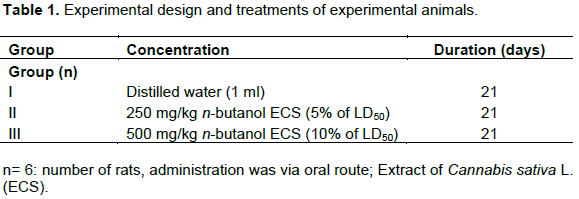
The dried plant of 500 g of Cannabis sativa was grounded and weighed which gave 400 g. The extract was obtained via soxhlet extraction technique in 3 L of ethanol and was evaporated using water bath to yield 27.55 g and was further fractionated using 300 ml of n-butanol and 200 ml of water to yield 17.45 and 3.75 g, respectively and was screened for the presence of alkaloids, saponins, tannins, and cardiac glycoside using standard methods (Trease and Evans, 1989; Sofowora, 1993). The extract was stored at room temperature until ready for use. The experimental groups were subjected to the different oral treatments once a day for twenty one (21) days after the LD50 of n-butanol extract of C. sativa L. was carried out based on Lorke’s method and it was found out to be greater than 5000 mg/kg body weight (Table 1).
Protocol
Horizontal ladder rung walking task paradigm consisting of side walls made of clear Plexiglas and metal rungs of 3 mm diameter which was inserted to create a floor with a distance of 1 cm between rungs was constructed locally using the method of Metz and Whishaw (2009). The side walls were 1 m long and 19 cm high from the height of the rungs (Plate 1). The ladder was raised 30 cm above the ground with a neutral start cage and a home cage at the end of the ladder. The elevation of the paradigm was unlikely to cause anxiety because the animals were habituated before the experiment. The width of the path was adjusted to the size of the animal; it was about 1 cm wider than an animal to prevent the animal from turning around.
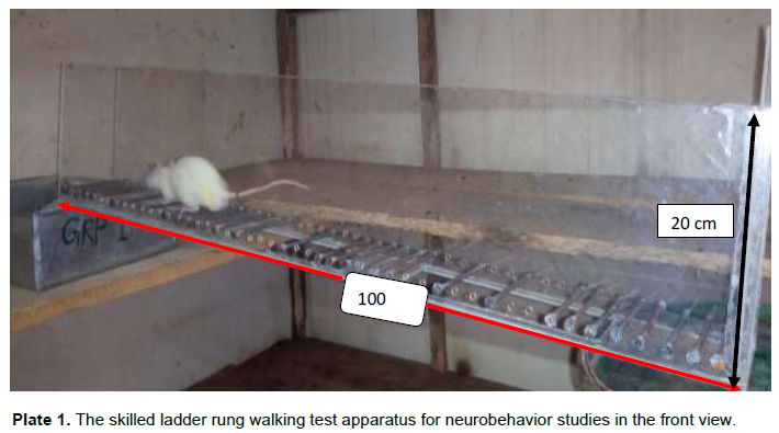
The difficulty of the task was modified by changing the position of the metal rungs. A regular pattern of the rungs allowed the animals to learn the pattern over several training sessions and to anticipate the position of the rungs. An irregular pattern was changed from trial to trial preventing the animal from learning the pattern. For the regular arrangement, the rungs were spaced at 2 cm intervals. For the irregular pattern, the distance of the rungs varied systematically from 1 to 5 cm. Five templates of irregular rung patterns were used, so that the same patterns will be applied to all animals to standardize the difficulty of the test and enhance comparability of the outcome (Metz and Whishaw, 2009).
Video recording
Canovision, Canon Inc. camera was placed slightly at a ventral angle, so that positions of all four limbs could be recorded simultaneously. The shutter speed was set at 500 to 2000 s. The video recordings were analyzed using frame-by-frame analysis at 30 f/s.
Behavioral training and test analysis
The animals were trained before the main experiment to cross the ladder from a neutral cage to reach their home cage; the home cage with littermates provided the positive support for walking. All animals crossed the ladder in the same direction. No further reinforcement was given to motivate the animals to cross the ladder. All animals were trained and tested five times per session.
Foot fault scoring
The qualitative evaluation of forelimb and hind limb placement was performed using a foot fault scoring system as described previously (Metz and Whishaw, 2002). Analysis was made by inspection of the video recordings frame-by-frame. Therefore, the last step before a gait interruption, such as a stop or a foot fault, and the first step after an interruption was not scored. Limb placement was scored in terms of limb placement on a rung and limb protrusion between rungs when a miss occurred. The types of paw placement on the rungs were rated using a seven (0, 1, 2, 3, 4, 5 and 6) category scale. Paw placement on the rung was assessed according to their position and errors that occurred in placement accuracy. (0) Total miss. 0 points were given when the limb completely missed a rung, that is, did not touch it, and a fall occurred. A fall will be defined as a limb deeply falling in-between rungs and body posture and balance were disturbed. (1) Deep slip. The limb was initially placed on a rung, then slipped off when weight-bearing and caused a fall. (2) Slight slip. The limb was placed on a rung, slipped off when weight bearing, but did not result in a fall nor interrupt the gait cycle. In this case, the animal was able to maintain balance and continue a coordinated gait. (3) Replacement. The limb was placed on a rung, but before it was weight bearing it was quickly lifted and placed on another rung. (4) Correction. The limb aimed for one rung, but was then placed on another rung without touching the first one. Alternatively, a score of 4 was recorded if a limb was placed on a rung and was quickly repositioned while remaining on the same rung. (5) Partial placement. The limb was placed on a rung with either wrist or digits of the forelimb or heel or toes of the hind limb. (6) Correct placement. The mid-portion of the palm of a limb was placed on the rung with full weight support. When different errors occurred at the same time, the lowest of the scores was recorded. For instance, if a foot was first placed on a rung and then placed on another one in the same step (score 3), and then slipped and fell in-between rungs (score 1), a score of 1 was recorded. When a fall occurred, only the limb initiating the error was rated and none of the other limbs was scored until the animal had repositioned all limbs. Error scores of five trials were averaged for analysis.
Foot placement accuracy analysis (number of errors)
The number of errors in each crossing will be counted. Errors will be determined based on the foot fault scoring system. An error will be defined as each limb placement that received a score of 0, 1 or 2 points, that is, an error represents any kind of foot slip or a total miss. The number of errors and the number of steps will be recorded for each limb separately. From these data, the mean number of errors per step will be calculated and averaged for five trials. The second quantitative parameter analyzed will be the average time needed to cross the entire length of the ladder task. Time measurement will start after the animal is placed on the ladder and began walking the entire length of it. The time an animal spent in a stop was not included in the measurement.
Histological and histochemical evaluation
The harvested brain was processed as outlined by Culling CFA (1974) for light microscopic studies using the routine Hematoxylin and Eosin (H&E) for general neural architecture and Cresyl Fast Violet stain for Nissl Substance demonstration under the light microscope. This was done in the Histopathology Department of Ahmadu Bello University Teaching Hospital, Shika.
Statistical analysis
The mean and standard error of mean (S.E.M.) were calculated for all values. Comparison between the control and experimental groups was done using one-way analysis of variance (ANOVA) with LSD Post-hoc Test. Differences were considered statistically significant at p<0.05.
RESULTS
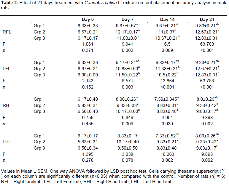
The effect of C. sativa L. extract (250 and 500 mg/kg) on ladder rung skilled walking after treatment of rats for 21 days is shown in Table 1. Treatment of rats with Cannabis sativa L. extract (250 and 500 mg/kg) produced significant (p<0.05) changes in ladder rung skilled walking values relative to their respective controls (Table 2).
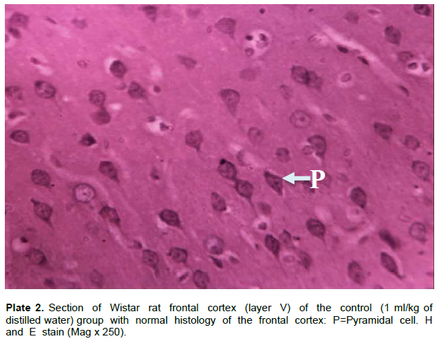
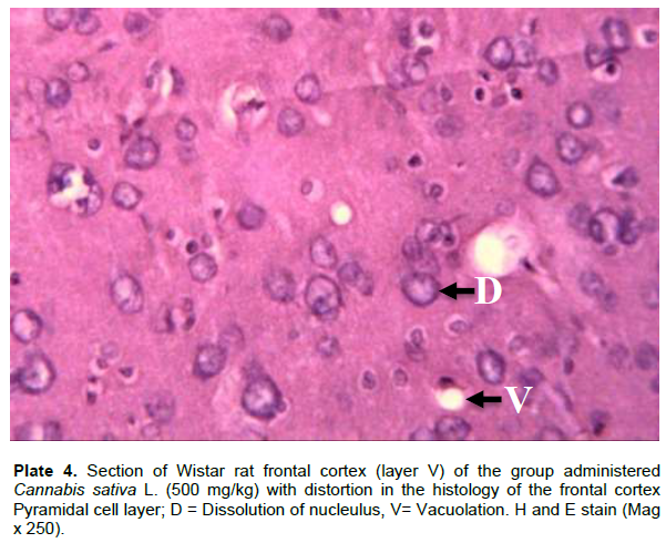
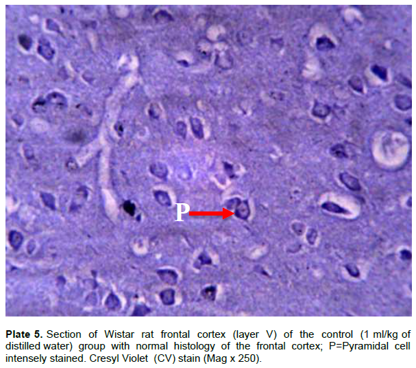
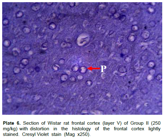
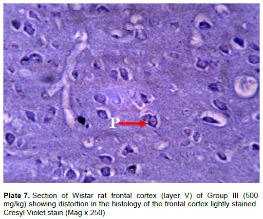
The effect of C. sativa L. extracts (250 and 500 mg/Kg) on the histology after treatment of rats for 21 days is shown in Plate 3 to 7, respectively. Treatment of rats with Cannabis sativa L. extract (250 and 500 mg/kg) on histology of Wistar rats caused significant distortion in the histoarchitecture of the frontal cortex as shown in the Plates of the treatment groups (Plate 2).

DISCUSSION
Horizontal rung ladder walking task with an irregular rung pattern has previously been described as a sensitive test to evaluate both forelimb and hind limb use in rats after lesions of the motor system (Metz and Whishaw, 2009). The result in this study showed forelimb and hind limb impairment in the treatment groups which could be as a result of motor impairment in the motor area of the cerebral cortex of the Wistar rats due to the extract administered since both animals and humans frontal region of the cerebral cortex contains high concentrations of CB1 receptors which the active compound in cannabis (Delta -9- tetrahydrocannabinol) binds, thus eliciting its effect (Iversen, 2003). Similar work was reported by Metz and Whishaw (2009) and Knieling et al. (2009) in which their data showed impaired forelimb and hind limb. Motor impairments, particularly in hindlimb use, were revealed by a reduced foot fault score and a higher error rate that was persistent for 21 days. Impairment of motor performance of the hind limbs predominates in this study which might be as a result of motor disabilities (Stroemer et al., 1995; Metz, 1998; Muir and Whishaw, 1999; Liebigt et al, 2012). The results revealed that the treatment groups (II and III) showed worse walking adaptability (lower skilled walking score) and longer duration of time to cross the ladder from the neutral cage to the home cage, when compared to the control group. Motor deficits are shown in ladder walking indicating that cannabis has a strong influence on the accuracy of limb placement for both affected limbs. This study also showed that, the high sensitivity of the ladder rung walking task is capable of detecting motor impairment in treated groups which is similar to a work which also reported high sensitivity of ladder rung walking task which aids the detection and observation of motor impairments in animal experiments (Metz and Whishaw, 2009). Other studies carried out using cannabis show that low dose (0.1 mg/kg) of cannabinoids increase motor activity, while high doses (1 mg/kg) decrease motor activity and produce catalepsy (Katsidoni et al., 2013). Another finding using different model by Ahmed (2018) indicate that embryos exposed to cannabis during a short-term but critical period of gastrulation showed alterations in motor neuronal morphology and also affect locomotion. Also, cannabis has been shown to have a deleterious effect on the brain altering corticostriatal connectivity and locomotor activity (Tomasâ€Roig et al., 2018) but the effect of cannabis on motor activity is dose-dependent (Boggs et al., 2018). The psychoactive compound found in cannabis (Delta-9-tetrahydrocannabinol) has also been observed to reduce motor activity in mice (Lloyd et al., 2018). A study by Kasten et al. (2019) reported a decrease in locomotor activity in rats administered 20 mg of cannabis sativa, this could be as a result of C/ sativa having a high amount of delta-9-tetrahydrocannabinol which binds to cannabinoid receptors and may interrupt well maintained inhibitory signaling regulated by endogenous cannabinoids (Svizenksa et al., 2008; Freund and Katona, 2007). However, other studies investigated the effects of cannabis smoke exposure on exploratory behavior. The studies showed that cannabis smoke increased locomotor activity when the rats were tested immediately after smoke exposure, but not when tested 4, 24 or 48 h after cannabis smoke exposure, the rapid action of cannabis by increasing locomotor activity might be because of the route of administration, other studies also revealed that smoke cannabis act faster for a short period of time. Another study similar to this work was also reported by Malyshevskaya et al. (2017); they observed behavioral changes after administration of Cannabis, including suppression of locomotor activity, extensor rigidity in hind limbs ataxia, impaired walking, and muscular jerks. All these changes observed could be a result of the presence of the psychoactive compounds in the plants. Okon et al. (2014) also reported a similar result to this study, the dose of 10 and 20 mg/kg body weight of ethanol extract of C. sativa L. for 28 days; they observed decrease locomotor activity compared with the control that received normal saline. Therefore, since a lower dose (10 and 20 mg/kg) of the extract is liable of causing motor impairment it is no doubt that 250 and 500 mg/kg of n-butanol extract of C. sativa L. would cause motor deficit as seen in the foot fault scoring. These studies therefore suggest that, n-butanol extract of C.sativa L. could have a high content of psychoactive compound thereby, causing motor deficit by binding with the cannabinoid receptors in the cerebral cortex and causing damage at the corticospinal tract linking the cerebral cortex and the spinal cord.
Histological assessments also verified the evidence of alterations in the structure of layer V (pyramidal neurons), degenerative alterations were detected as, cortical neuronal shrinkage and perineuronal vacuolations, necrosis and chromatolysis in the cerebral section of cannabis treated Wistar rats when compared with the cerebral section of the control group; it indicates treatment related neurotoxicity. The animals treated with cannabis extract especially in group two and three of H and E showed dissolution of nucleolus, vacuolation and pyknosis (Plate 3 and 4) while Cresyl Fast Violet stain showed distortion in the histology of the pyramidal cells of the frontal cortex lightly stained (Chromatolysis) (Plate 6 and Plate 7). Also, there are mark irregularities in the distributions of nissl granulation in the perikaryon and increased dark pyknotic nuclei, presence of dark neuron suggesting protein denaturation and neural degeneration as seen in group III of Cresyl Fast Violet stain. This agrees with the findings of previous studies on Cannabis who reported that Cannabis induce nervous tissue damage (Rapp et al., 2013; Jacobus et al., 2016; Orr et al., 2016). Administration of cannabis as reported by Imam et al. (2017) observed that, the pyramidal cells at the internal pyramidal cell layer show some degree of retraction of processes, vacuolation of the surrounding neuropil of the pyramidal cells, and hyperchromatic and shrunken perikarya which are also similar to this study. The histopathological changes detected in this study are similar to those recorded in the study of Solanke et al. (2016) and Imam et al. (2017) in which the animals treated with C. sativa showed neuro-degeneration of neurons and stroma. This could be as a result of the toxic effect of C. sativa which has been attributed to the tetrahydrocannabinol (Solanke et al., 2016). Another study by Odokuma EI and Ogbor-Omorie E (2015) showed that the histomorphologic changes induced by C. sativa in short- and long-term studies caused extensive cerebral gliosis in the brains of adult Wistar rats and concluded that there was both dose- and time-dependent toxic effects of Cannabis sativa L. on the experimental animals.
Another study by Adams et al. (2017) observed degeneration in the brain histology administered with 700 mg/kg methanol extract of C. sativa leaves for 21 days suggests that the extract possesses functional and structural toxicity in male rats. Findings from this study therefore support the assumption that consumption of the C. sativa may contribute to increasing incidence of brain damage. In treated male albino rats, Amaza et al. (2013) observed irregular shrunken cells with dense nuclei and cytoplasm. The cells are surrounded by irregular wide spaces which are also in line with this study. Amaza et al. (2013) also reported that administration of 500, 750 and 900 mg/kg of aqueous extract of C. sativa showed perivascular congestion as well as lymphatic infiltration in the cerebral cortex of adult Wistar rat. This study is also in agreement with the study conducted by Una et al., (1997) who also reported that there were changes in the neurons in selected brain regions most especially the cerebral cortex as to the administration of the same substance. Neurodegeneration is the progressive loss of structure or function of neurons. Also, is a process involved in both neuropathological conditions and brain aging (Arman, 2011; Kumar and Khanum, 2012). Also, this study is in consistency with the findings of Ebuehi and Abey (2016) who reported altered brain structure of neuronal and glial cells, as increased cellularity, hypertrophy of glial cells and hyperplasia in the brain following administration of Cannabis sativa compared to the control group. Nader et al. (2018) reported harmful effect of cannabis on the brain, that regular cannabis use is associated with cognitive changes, structural and functional alterations in adults brain which could affect brain function.
The results of this study suggest that cannabis use may be associated with altered brain structure, especially in particular regions rich in CB1 receptors like the cerebral cortex. Therefore, this altered architecture explains the lowered score of the foot fault task activity exhibited by the experimental rats.
CONCLUSION
In conclusion, this study has shown that C. sativa L. extract has some harmful effects on the cerebral cortex in male rats. However, the effect of this plant on human motor function and histology are still scarce; nevertheless, considering these findings in the animal model, it is recommended that abusing this substance should be strictly avoided.
CONFLICT OF INTERESTS
The authors have not declared any conflict of interests.
ACKNOWLEDGEMENT
The authors are grateful for the donation of C. sativa L. from National Drug Law Enforcement Agency (NDLEA) and support of the management of Ahmadu Bello University, Zaria, Nigeria.
REFERENCES
|
Adams MD, Okere OS, Tarfa FD, Daniel EE (2017). Toxicological Evaluation of Aqueous and Methanolic Leaf Extract of Cannabis Sativa in Liver and Brain of Male Rats. European Journal of Biomedical and Pharmaceutical Sciences 4(8):972-983. |
|
|
Ahmed KT, Amin MR, Shah P, Ali DW (2018). Motor neuron development in zebrafish is altered by brief (5-hr) exposures to THC (∆9-tetrahydrocannabinol) or CBD (cannabidiol) during gastrulation. Scientific Reports 8(1):10518. |
|
|
Amaza DS, Maidugu FA, Zirahei JV, Numan AI, Mari H (2013). The Effect of Cannabis Sativa Leaves Aqueous Extract on Cerebral Cortex in Albino Rats. Journal of Dental and Medical Sciences 6(2):53-58. |
|
|
Arman RH (2011). Histology of the Central Nervous System. Toxicologic Pathology 39(1):22 35. |
|
|
Boggs DL, Cortes-Briones JA, Surti T, Luddy C, Ranganathan M, Cahill JD, Skosnik PD (2018). The dose-dependent psychomotor effects of intravenous delta-9-tetrahydrocannabinol (Δ9-THC) in humans. Journal of Psychopharmacology 269881118799953. |
|
|
Culling CFA (1974). Handbook of Histopathological technique 2nd Ed Butterworth's, London. |
|
|
Ebuehi OAT, Abey NO (2016). Impact of Marijuana (Cannabis sativa) on Some Neurochemicals and Cognitive Function of Sprague-Dawley Rats. Research in Neuroscience 5(1):1-9. |
|
|
ElSohly MA, Radwan MM, Gul W, Chandra S, Galal A (2017) Phytochemistry of Cannabis sativa L. In Phytocannabinoids Springer, Cham. pp. 1-36. |
|
|
Elsohly MA, Slade D (2007). Chemical constituents of marijuana: the complex mixture of natural cannabinoids. Life Sciences 78(5):539-48. |
|
|
Freund TF, Katona I (2007). Perisomatic inhibition. Neuron 56:33-42. |
|
|
Gloss D (2015). An Overview of Products and Bias in Research. Neurotherapeutics 12(4):7314. |
|
|
Greg G (2005). The Cannabis Breeder's Bible. Human Psychopharmacology 22(3):135-48. |
|
|
Imam A, Ajao MS, Akinola OB, Ajibola MI, Ibrahim A, Amin A, Ali-Oluwafuyi A (2017). Repeated Acute Oral Exposure to Cannabis sativa Impaired Neurocognitive Behaviours and Cortico-hippocampal Architectonics in Wistar Rats Nigerian Journal of Physiological Sciences 31(2):153-159. |
|
|
Iversen L (2003). Cannabis and the Brain. A Journal of Neurology. 126(6):1252-1270. |
|
|
Jacobus J, Castro N, Squeglia LM, Meloy MJ, Brumback T, Huestis MA, Tapert SF (2016). Adolescent cortical thickness pre- and post marijuana and alcohol initiation. Neurotoxicology and Teratology 57:20-29. |
|
|
Kano M, Ohno-Shosaku T, Hashimotodani Y, Uchigashima M, Watanabe M (2009). Endocannabinoid-mediated control of synaptic transmission. Physiological Reviews 89:309-380. |
|
|
Kasten CR, Zhang Y, Boehm SL (2019). Acute Cannabinoids Produce Robust Anxiety Like and Locomotor Effects in Mice, but Long-Term Consequences Are Age- and Sex-Dependent. Frontiers in Behavioral Neuroscience 13(32). |
|
|
Katsidoni V, Kastellakis A, Panagis G (2013). Biphasic effects of Δ9- tetrahydrocannabinol on brain stimulation reward and motor activity. International Journal of Neuropsychopharmacology 16(10):2273-2284. |
|
|
Knieling M, Metz GA, Antonow-Schlorke I, Witte OW (2009). Enriched environment promotes efficiency of compensatory movements after cerebral ischemia in rats. Neuroscience 163(3):759-769. |
|
|
Kumar GP, Khanum F (2012). Neuroprotective potential of phytochemicals. Pharmacognosy Reviews 6(12):81-90. |
|
|
Liebigt NS, Schlegel J, Oberland OW, Witte CR, Keiner S (2012). Effects of rehabilitative training and anti-inflammatory treatment on functional recovery and cellular reorganization following stroke. Experimental Neurology 233:776-782. |
|
|
Lloyd D, Talmage D, Weickert CS, Karl T. (2018). Reduced type III neuregulin 1 expression does not modulate the behavioural sensitivity of mice to acute Δ9-tetrahydrocannabinol (D9 THC). Pharmacology Biochemistry and Behavior 170:64-70. |
|
|
Malyshevskaya O, Aritake K, Kaushik MK, Uchiyama N, Cherasse Y, Kikura-Hanajiri R, Urade Y (2017). Natural (∆9-THC) and synthetic (JWH-018) cannabinoids induce seizures by acting through the cannabinoid CB1 receptor. Scientific Reports 7(1):10516. doi: 10.1038/s41598-017-10447-2 |
|
|
Mbadugha CC, Ekandem GJ, Ekanem TB, Etuknwa BT (2015). Neurobehavioral and Immunohistochemical Assessment of the Cerebellum in Adult Male Albino Wistar Rats Following Cannabis Sativa Administration. Journal of Natural Sciences Research 5(16):82-89. |
|
|
Metz GA, Antonow-Schlorke I, Witte OW (2005). Motor improvements after focal cortical ischemia in adult rats are mediated by compensatory mechanisms, Behavioural Brain Research 162(1):71-82. |
|
|
Metz GA, Whishaw IQ (2002). Cortical and subcortical lesions impair skilled walking in the ladder rung walking test: a new task to evaluate fore- and hindlimb stepping, placing, and co-ordination. Journal of Neuroscience Methods 115(2):169-179. |
|
|
Metz GA, Whishaw IQ (2009). The Ladder Rung Walking Task: A Scoring System and its Practical Application. Journal of Visualized Experiments (28):1204. |
|
|
Metz GAS, Dietz V, Schwab, ME, Van de Meent H (1998). The effects of unilateral pyramidal tract section on hindlimb motor performance in the rat. Behavioural Brain Research 96(1-2):37-46. |
|
|
Muir GD, Whishaw IQ (1999). Complete locomotor recovery following corticospinal tract lesions: measurement of ground reaction forces during overground locomotion in rats. Behavioural Brain Research 103(1):45-53. |
|
|
Nader DA, Sanchez ZM (2018). Effects of regular cannabis use on neurocognition, brain structure, and function: a systematic review of findings in adults. The American Journal of Drug and Alcohol Abuse 44(1):4-18. |
|
|
Newton DE (2013). Marijuana: a reference handbook. Santa Barbara, Calif.: ABC-CLIO.7. |
|
|
Odokuma EI, Ogbor-Omorie E (2015). Histomorphologic effects of Cannabis sativa on the brains of adult Wistar rats. Annals of Bioanthropology 3:29-32. |
|
|
Okon VE, Obembe AO, Nna VU, Osim EE (2014). Long-Term Administration of Cannabis sativa on Locomotor and Exploratory Behavior in Mice. Research in Neuroscience 3(1):7-21. |
|
|
Orr JM, Paschall CJ, Banich MT (2016). Recreational marijuana use impacts white matter integrity and subcortical (but not cortical) morphometry. NeuroImage: Clinical 12:47-56. |
|
|
Rapp C, Walter A, Studerus E, Bugra H, Tamagni C, Rothlisberger M, Riecher-Rossler A (2013). Cannabis use and brain structural alterations of the cingulate cortex in early psychosis. Psychiatry Research 214(2):102-108. |
|
|
Russo EB (2016). Current therapeutic cannabis controversies and clinical trial design issues. Frontiers in Pharmacology 7:309 |
|
|
Shehu U (2017). Drug abuse taking frightening dimension in Northwest. |
|
|
Sofowora A (1993). Medicinal plants and Traditional medicine in Africa. Spectrum Books Ltd, Ibadan, Nigeria, P 289. |
|
|
Solanke AS, Ebuehi OAT, Adetona MO (2016). Toxicological Effect of Ethanolic Extract of Cannabis sativa on Brain Serotonin in Adult Wistar Rats. American Journal of Biochemistry 6(4):97-99. |
|
|
Stroemer RP, Kent TA, Hulsebosch CE, Rosenblum WI (1995). Neocortical neural sprouting, synaptogenesis, and behavioral recovery after neocortical infarction in rats. Stroke 26 (11):2135-2144. |
|
|
Svizenksa I, Dubovy P, Sulcova A (2008). Cannabinoid receptors 1 and 2 (CB1 and CB2), their distribution, ligands and functional involvement in nervous system structures - A short review. Pharmacology Biochemistry and Behavior 90:501-511. |
|
|
Tomasâ€Roig J, Piscitelli F, Gil V, Quintana E, Ramióâ€Torrentà LI, Antonio del Río J, Havemannâ€Reinecke U (2018). Effects of repeated longâ€term psychosocial stress and acute cannabinoid exposure on mouse corticostriatal circuitries: Implications for neuropsychiatric disorders. CNS Neuroscience and Therapeutics 24(6):528-538. |
|
|
Trease G, Evans W (1989). Pharmacognosy (11th ed., pp. 45-50), London: Bailliere. |
|
|
Una DM, Kelly AL, George ARC (1997). Neuroscience Biological psychiatry branch Bethseda Maryland Department of Neurology John Hopkins Medical Institution Baltimoore Maryland. |
|
|
World Health Organization (WHO) (2010). The health and social effects of nonmedical cannabis use. Geneva: World Health Organization. |
|
Copyright © 2024 Author(s) retain the copyright of this article.
This article is published under the terms of the Creative Commons Attribution License 4.0


