Full Length Research Paper

ABSTRACT
This study was to evaluate the effects of aqueous and ethanol fruit extracts of Phoenix dactylifera on the cerebellar cortex of Artesunate-Amodiaquine (AS–AQ) treated Wistar rats. Thirty-six adult male Wistar rats were randomly divided into nine groups (n=4). Group 1 served as the control; Group 2 received 2.86/8.75 mg/kg AS–AQ. Group 3 received 2.86/8.75 mg/kg AS–AQ + 100 mg/kg ascorbic acid; Groups 4, 5 and 6 received 2.86/8.75 mg/kg AS–AQ + 500 mg/kg aqueous extract of P. dactylifera (AEPD), 2.86/8.75 mg/kg AS–AQ + 1000 mg/kg AEPD and 2.86/8.75 mg/kg AS–AQ + 1500 mg/kg AEPD respectively. Groups 7, 8 and 9 received 2.86/8.75 mg/kg AS–AQ + 500 mg/kg ethanolic extract of P. dactylifera (EEPD), 2.86/8.75 mg/kg AS–AQ + 1000 mg/kg EEPD and 2.86/8.75 mg/kg AS–AQ + 1500 mg/kg EEPD respectively. All administration was orally done, and lasted for 28 days. Evidences of necrosis, chromatolysis and vacuolations were observed in the cerebellar cortex of the AS–AQ treated group. The histoarchitecture of the AEPD and EEPD treatment groups were preserved in a dose-dependent manner, and compared positively to the reference drug ascorbic acid treatment, with the AEPD-treated groups being better preserved. Amelioration of the severity of neurodegeneration by the extracts suggest that the extracts could have exerted their ameliorative effects by antioxidant activities ascribed to the phytochemicals present in the extracts.
Key words: Phoenix dactylifera L. cerebellum, Artesunate-Amodiaquine, Wistar rats.
INTRODUCTION
In 2017, a troubling shift in the trajectory of malaria disease was noted as data showed that less than half of countries with ongoing transmission were on track to reach critical targets for reductions in the death and disease caused by malaria. Progress appeared to have stalled and this worrying trend continues. Although there are some bright spots in the data, the overall decline in the global malaria burden has unquestionably leveled off.
And, in some countries and regions, reversals in the gains achieved have begun (WHO, 2001).
The broad collapse of preventive efforts, increasing parasite resistance and failure of single drug treatment of malaria in many endemic countries of Africa has led to a widespread promotion of Artemisinin–based combination therapies (ACTs) as a strategy for effective management of Plasmodium falciparum malaria (Trape, 1998; Lyda et al., 2007),
Artemisinin–based combination therapy for malaria is the simultaneous use of an Artemisinin and another blood schizonticidal drug where both have different biochemical targets in the parasites. This has exploited the synergistic and additive potential of the individual drugs Artemisinin kills majority of the parasites at the start of the treatment, while the slow–acting partner drug clears the remaining parasites (Oreagba, 2010).
Artesunate is a part of the artemisinin group of drugs used to treat malaria, especially Chloroquine–resistant malaria in Nigeria. It is the most widely used member of artemisinin derivatives (Nwanjo and Oze, 2007). It is used in combination with other antimalarial drugs such as Mefloquine (Looareesuwan et al., 1996; Nosten et al., 2000) or Amodiaquine (Adjuik et al., 2002). This is done so as to avoid development of resistance of the malaria parasite (Nosten et al., 2000).
Amodiaquine belongs to a class of drugs known as the 4–amino–quinoline compounds. The synthesis of this drug was first reported at the meeting of the American Chemical Society in 1946 and is in wide use mainly as an anti–malarial drug (Neftel et al., 1986; Misra et al., 1995). In Africa, Amodiaquine is significantly more effective than Chloroquine in parasite clearance and a faster clinical recovery (Schellenberg and Menendez, 2001). It is also more effective in areas of high Chloroquine resistance (Olliaro et al., 1996).
The cerebellum is a region of the brain that plays an important role in motor control (Wolf et al., 2009). Its movement–related functions are the most solidly established. The cerebellum does not initiate movement, but it contributes to coordination, precision, and accurate timing. It receives input from sensory systems of the spinal cord and from other parts of the brain, and integrates these inputs to fine–tune motor activity (Fine et al., 2002). The cerebellum is vulnerable to damage from a variety of sources such as developmental defects, degenerative diseases, infectious processes, chronic alcoholism, trauma and tumors (West, 1995). Cerebellar injuries have been reported to result from toxins, such as, antimalarial drugs (Agbon et al., 2014; Ajibade et al., 2012).
Medicinal plants continue to provide valuable therapeutic agents, both in modern and in traditional medicine (Krentz and Bailey, 2005) Traditional medicines are gaining importance and are now being studied to find the scientific basis of their therapeutic actions (Gupta and Briyal, 2004).
Phoenix dactylifera (Date palm) belongs to the Palmae (Aracaceae) family and is extensively cultivated for its edible fruit (Rani et al., 2007). Because of its high nutritional value, great yields, and its long life, the date palm has been mentioned as the “tree of life” (Augstburger et al., 2002).
The date palm is rich in phytochemicals like sterols, phenolics, carotenoids, procyanidins, anthocyanins, alkaloids, saponins, tannins, and flavonoids (Abdelhak et al., 2005; Abdul and Allaith, 2008). These natural compounds are known to function as free radical scavengers (Baliga et al., 2011) and restrain protein oxidation and iron–induced lipid peroxidation (Vayalil, 2002).
This study was aimed at evaluating the effects of aqueous and ethanol fruit extracts of P. dactylifera on the cerebellar cortex of Artesunate-Amodiaquine (AS–AQ) treated Wistar rats.
MATERIALS AND METHODS
Collection and identification of plant
Dried P. dactylifera fruits were obtained from a local Market in Zaria, Nigeria. It was identified and authenticated in the Herbarium Unit of the Department of Biological Sciences, Faculty of Life Sciences, Ahmadu Bello University, Zaria, Nigeria with the Voucher Specimen Number of 3252.
Extracts preparation and phytochemistry
Preparation of aqueous and ethanol fruit extracts of P. dactylifera was conducted in the Department of Pharmacognosy and Drug Development, Faculty of Pharmaceutical Sciences, Ahmadu Bello University, Zaria. The method of maceration as reported by Al–Qarawi et al. (2004) and Wahab et al. (2010) for the preparation of aqueous and ethanol P. dactylifera fruit extracts, respectively, was employed.
Phytochemical screening of aqueous and ethanol fruit extracts of P. dactylifera was conducted in the Department of Pharmacognosy and Drug Development, Faculty of Pharmaceutical Sciences, Ahmadu Bello University, Zaria. The method of Trease and Evans (2002) for phytochemical screening was adopted.
Experimental animals
For the purpose of this research, a total of thirty–six apparently healthy adult male Wistar rats weighing between 150 – 180 g were obtained and housed in the Animal House Center of the Department of Human Anatomy, Faculty of Basic Medical Sciences, Ahmadu Bello University, Zaria, and were acclimatized for two weeks prior to the start of the experiment.
All the rats were given food (rat chow – Vital Feeds) and water ad libitum. Rats were weighed on the day of commencement of the study (day 1), weekly during the study (days 8, 15, and 22), and on the day of sacrifice (day 29).
Drug
Camosunate manufactured by ADAMS Pharmaceutical (Anhui) Co., Ltd, China, was purchased. Each packet of Camosunate is made up of Artesunate and Amodiaquine tablets, with each made up of twelve blistered tablets each of Amodiaquine Hydrochloride USP equivalent to Amodiaquine (AQ) base (300 mg) and Artesunate (AS) (100 mg). The normal dosage of both drugs for a physiologic man per day is: 100 mg of AS and 306.2 mg of AQ. For an average weighted rat of 200 g, the dosages were calculated to be 2.86 mg/kg of AS and 8.75 mg/kg of AQ per day (Walker et al., 1984; Okafor, 2013).
Ascorbic acid was obtained and used for the experiment as standard antioxidant drug. The product is manufactured by Emzor Pharmaceutical Industries Ltd, Nigeria. Each of the blistered tablets contained an equivalent of Vitamin C (100 mg).
Experimental procedure
In this study, a total of thirty–six adult male Wistar rats (36) were obtained and randomly distributed into nine groups of four rats per group. Single dosage rate was used, and drugs and extracts administration was carried out once daily. Group 1 served as the control and was administered distilled water (1 ml/kg) while Groups 2 to 9 were the treatment groups and were all administered 2.86/8.75 mg/kg of Artesunate/Amodiaquine (AS/AQ). Group 2 received only 2.86/8.75 mg/kg of AS/AQ while Group 3 received 100 mg/kg of ascorbic acid (vitamin C) (Raghu et al., 2014) which was used as the standard drug. Groups 4, 5 and 6 received (in addition to the AS/AQ) 500 mg/kg (10% LD50), 1000 mg/kg (20% LD50) and 1500 mg/kg (30% LD50) of the aqueous extract of P. dactylifera respectively. Groups 7, 8 and 9 received 500 mg/kg (10% LD50), 1000 mg/kg (20% LD50) and 1500 mg/kg (30% LD50) of the ethanol extract of P. dactylifera respectively, while all administration was done orally and lasted for a period of 28 days.
Histological studies
At the end of the experiment, the rats were euthanized, and their brains carefully removed and processed histologically using Haematoxylin and Eosin (H&E) and Cresyl Fast Violet stains for light microscopic examination.
Data analysis
All data were presented as mean ± SEM and were statistically analyzed using Statistical Package for Social Science (SPSS) version 20 (IBM, Incorporation, NY). Comparisons between control values and those obtained in treated groups of rats were performed with one–way analysis of variance (ANOVA). A p–value < 0.05 was considered significant.
RESULTS
Phytochemical analysis
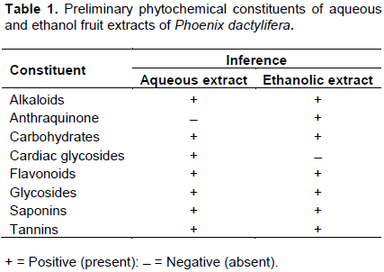
Phytochemical analysis of aqueous and ethanol fruit extracts of P. dactylifera (EFPD) produced positive reactions for secondary metabolites and negative for some as shown in Table 1.
Physical observation
Within the period of administration, the rats were observed to exhibit normal locomotor activity and liveliness with no obvious difference in activity levels between the control and treatment groups.
Similarly, the weights of the animals were found to have increased within the period of study but with no significant difference between the treatment groups and the control.
Neurobehavioral examination
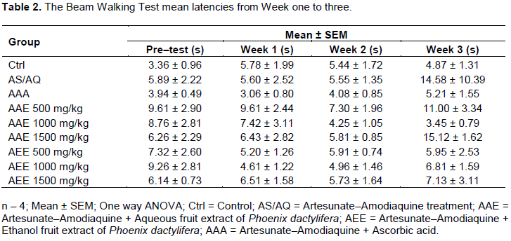
There was no significant difference in the latency time taken during the Beam Walking test. The time latencies are presented in Table 2.
Histological examination
Histological examination of sections of cerebellar cortex of rats, stained with routine histological (H&E) stain and Cresyl Fast Violet (CFV) stain revealed the following:
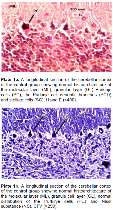
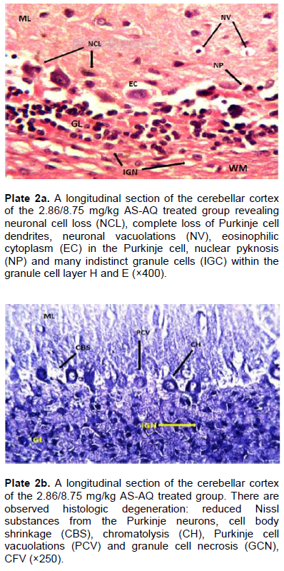
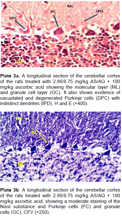
Plate 1a shows the histology of the cerebellar cortex of the control group showing the presence of the three distinct layers with normal distribution of neurons: the molecular layer, granule layer, and the Purkinje cell layer with its dendrites. Plate 1b reveals an even distribution of Nissl substance within the neurons. The AS–AQ treated group show histoarchitectural distortions and insults of the cerebellar cortex evidenced by condensed nuclei of Purkinje cells, bright eosinophilic cytoplasm, neuronal and cytoplasmic shrinkage, degeneration of Purkinje neuron dendrites, neuronal vacuolations within the overlying molecular layer, chromatolysis, neuronal cell loss, internal granule necrosis as well as cell body shrinkage and loss of Nissl substance (Plate 2a and b). The ascorbic acid treated group was observed to have ameliorated the histological insults incurred by the AS–AQ (Plate 3a and b). In addition, when compared with the control, it revealed a near–normal cerebellar section, with few vacuolations and degenerated Purkinje cells, but having a distinct dendritic circuitry.
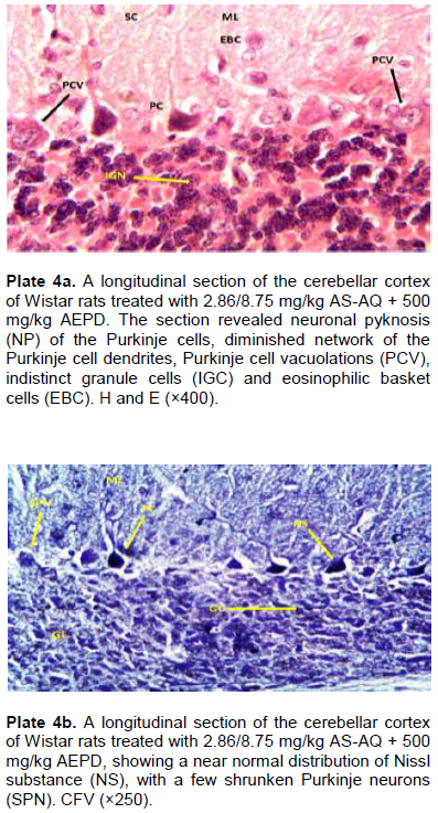
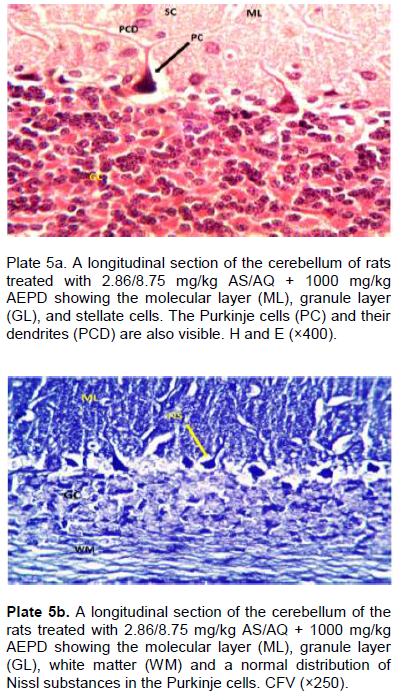
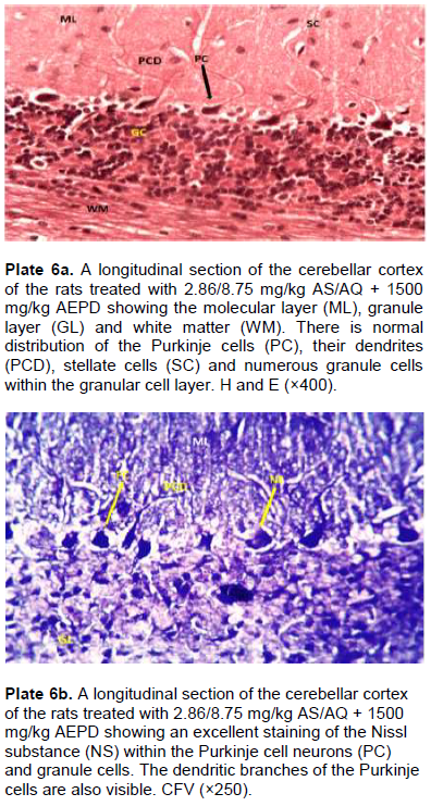
Treatment with 500 mg/kg of aqueous extract show a poor reparative response of the cerebellar cortex to histoarchitectural insults characterized by Purkinje and granule cells degeneration, pyknosis, neuronal swellings and vacuolations, though with few distinct Purkinje cell dendrites and moderate Nissl substance distribution (Plate 4a and b). The 1000 mg/kg and 1500 mg/kg aqueous extract treated groups (Plate 5a and b; 6a and b respectively) show a transition of progressively increased heterogeneous (medium to large sized) neuronal population in the three layers, with moderate to clearly distinct neuron dendrites in the molecular layer indicating no obvious degenerative changes. They reveal a normal appearance of distinct moderate to intensely stained Nissl substance.
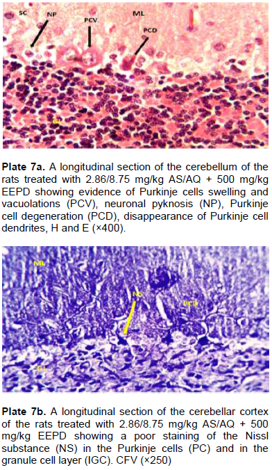
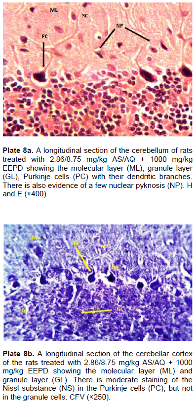
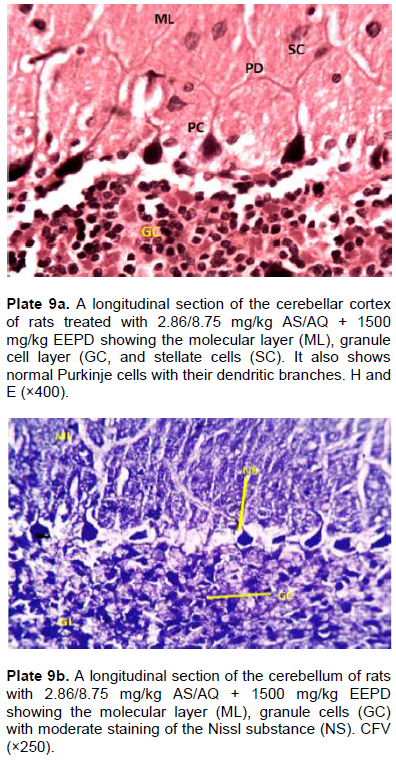
The histological sections of the ethanol extract–treated groups showed a similar ameliorative trend in the cerebellar cortex. At a dose of 500 mg/kg, the cerebellar sections revealed the presence of neuronal pyknosis, Purkinje cell swelling and vacuolations, eroded dendritic network of the Purkinje cells, poorly stained Nissl substance, and several indistinct granule cells (Plate 7a and b). There was a noticeable improvement in the cerebellar histology and Nissl substance staining of the groups administered higher doses of 1000 mg/kg (Plate 8a and b) and 1500 mg/kg (Plate 9a and b) respectively. However, nuclear pyknosis, chromatolysis, karyorrhexis, necrosis and vacuolations in the cerebellum of the 1000 mg/kg dose administered group was observed, whereas at the 1500 mg/kg dose, there was no obvious histological insults in the cerebellum.
DISCUSSION
The phytochemical results of both the aqueous and ethanol extracts indicated positive for secondary metabolites such as flavonoids, saponins and tannins. These natural compounds are known to function as free radical scavengers (Baliga et al., 2011) and also restrain protein oxidation and iron-induced lipid peroxidation (Vayalil, 2002). This lends credence to the reported phytochemical constituents in fruit extract of P. dactylifera L. as reported by Agbon et al. (2017).
Locomotor activity is considered as an index of alertness and a decrease in it is indicative of sedative activity (Lowry et al., 2005). Additionally, both extracts did not demonstrate any effect on the general behaviour or the muscle coordination, as indicated by the findings of the Beam Walking test. This is consistent with previous reports by Genovese et al. (1995, 1998) that show Artemisinin to have no overt behavioural signs of toxicity such as tremor of gait disturbances reflected on latency time. This suggests that dopaminergic neurotransmitter levels are unaltered by either AS–AQ, P. dactylifera or ascorbic acid, as reported by Doan et al. (2008), that animals with dopaminergic depletions display pronounced sensory and motor deficits, especially those in walking. Furthermore, the phytochemical alkaloid may have affected the central nervous system, including nerve cells of the brain which control many direct body functions and behavior, as well as interfere or compete with the action of serotonin in the brain (Pearson, 2001), causing a decreased firing in the brain, and thus, non–significant improvement in movement activities.
Neuronal injury may result in reversible or irreversible cell damage or cell death (Seilhean et al., 2004). Cell death has been reported to result from neuronal degeneration (Waters et al., 1994) and could result from necrosis, a pathological type of cell death that occurs from extrinsic insults to the cells, or after abnormal stresses such as chemical injury or toxin, thermal, traumatic and mechanical factors (Kumar et al., 2009).
Histoarchitectural distortions such as chromatolysis and necrosis when compared to the control as observed, is indicative of AS–AQ induced neurotoxicity. Central chromatolysis is a histopathologic change seen in the cell body of a neuron, where the chromatin and cell nucleus are pushed to the cell periphery in response to axonal injury (Holland, 1996); it is a morphologic manifestation of a reparative response to injury (Seilhean et al., 2004) and response is associated with increased protein synthesis to accommodate for axonal sprouting (Holland, 1996).
Necrosis is an indication of treatment–related toxicity (Kumar et al., 2009). It involves the cytoplasmic organelles and the cell membrane which ruptures, leading to cell death. Features include cytoplasmic shrinkage, intense eosinophilia (“red neurons”) (Mena et al., 2004; Seilhean et al., 2004), small, shrunken darkly stained (pyknotic) nucleus, or the eventual fragmentation of such shrunken nucleus (karyorrhexis). Degenerating neurons have a very poorly stained Nissl substance as a result of dissociation of ribosomes from the rough endoplasmic reticulum which occurs in the early stages of cell degeneration (Garman, 2011). These degenerative changes could have been induced by damage to mechanisms of DNA repairs (Hartwig et al., 1994).
The observed amelioration of histopathological degeneration in the cerebellum could equally be attributed to the free radical scavenging actions of the phytochemicals flavonoids, tannins and phenols as suggested by Ateeq et al. (2013).
Comparatively, the aqueous extract revealed a more potent ameliorative activity than the ethanol extract, evidenced by fewer neurodegenerative parameters at all the compared doses. At all doses, the AEPD ameliorated the neurohistopathologic effects of AS–AQ much better than the ethanol extract. The mechanism underlining this difference, however, is uncertain.
This study also supports the findings of Ibegbu et al. (2013) that ascorbic acid ameliorated induced degenerative changes in the brain of rats. Ascorbic acid is considered an important neuroprotective agent (Naseer et al., 2011) since it is potent, scavenging ROS production and can modulate glutamatergic, dopaminergic, cholinergic and GABAergic transmission and related behaviours (Fiona et al., 2009).
CONCLUSION
The findings of this study suggest that aqueous and ethanol fruit extracts of P. dactylifera have ameliorative properties against Artesunate–Amodiaquine–induced cerebellar damages which compare favourably with ascorbic acid, a standard antioxidant. This could be a function of some of the phytochemicals present in the extracts.
CONFLICT OF INTERESTS
The authors have not declared any conflict of interests.
ACKNOWLEDGEMENTS
The authors are grateful to the Department of Human Anatomy, Faculty of Basic Medical Sciences, Ahmadu Bello University, Zaria, Nigeria for providing the facilities to conduct this study.
REFERENCES
|
Abdelhak M, Guendez E, Eugene K, Kefalas P (2005). Phenolic profile and antioxidant activity of the Algerian ripe date palm fruit (Pheonix dactylifera). Food Chemistry 89:411-420. |
|
|
Abdul A, Allaith A (2008). Antioxidant activity of Bahraini date palm (Phoenix dactylifera L.) fruit of various cultivars. International Journal of Food Science and Technology 43:1033-1040. |
|
|
Agbon AN, Abubakar MG, Enemeli FU, Mahdi O, Bobb KA, Sule H, Yahaya MH, Okah CC (2017). Assessment of Ethanol Fruit extract of Phoenix dactylifera L. (Date Palm) on mecury chloride – induced cerebral and cerebellar alterations in Wistar rats. Journal of Anatomy Science 8(1):188-201. |
|
|
Agbon AN, Ingbian SD, Dahiru AU (2014). Preliminary Histological and Histochemical Studies on the Neuroprotective Effect of Aqueous Fruit Extract of Phoenix dactylifera L. (Date Palm) on Artesunate-induced Cerebellar Damage in Wistar rats. Sub-Saharan African Journal of Medical Practice 1(4):204-209. |
|
|
Al-Qarawi AA, Mousa HM, Ali BH, Abdel-Rahman H, El-Mougy SA (2004). Protective effect of extracts from Dates (Phoenix dactylifera) on carbon tetrachloride–induced hepatotoxicity in rats. The International Journal of Applied Research in Veterinary Medicine 2:176-180. |
|
|
Ateeq A, Sunil SD, Varun SK, Santosh SK (2013). Phoenix dactylifera Linn (Pind Kharjura): A review. International Journal of Research in Ayurveda and Pharmacy 4(3):447-451. |
|
|
Augstburger F, Berger J, Censkowsky U, Heid P, Milz J, Streit C (2002). Date Palm. Naturland. Germany. |
|
|
Baliga MS, Baliga BRV, Kandathil SM, Bhat HP, Vayalil PK (2011). A review of the chemistry and pharmacology of the date fruits (Phoenix dactylifera L.). Food Research International 44:1812-1822. |
|
|
Doan JB, Mevin KG, Whishaw IQ, Suchowersky O (2008). Bilateral impairments of skilled reach-to-eat in early Parkinson's disease patients presenting with unilateral asymmetrical symptoms. Behavioural Brain Research 194:207-213. |
|
|
Fiona E, Harrison J, May M (2009). Vitamin C function in the brain: vital role of the ascorbate transporter SVCT2. Free Radical Biology and Medicine 46:719-730. |
|
|
Garman RH (2011). Histology of the central nervous system. Toxicologic Pathology 39:22-35. |
|
|
Genovese RF, Newman DB, Petras JM, Brewer TG (1998). Behavioral and neural toxicity of arteether in rats. Pharmacology Biochemistry and Behavior 60(2):449-458. |
|
|
Genovese RF, Petras JM, Brewer TG (1995). Arteether neurotoxicity in the absence of deficits in behavioural performance in rats. A. Tropical Medicine and Parasitology 89(4):447-449. |
|
|
Hartwig A, Schwerdtle T (2002). Interactions by carcinogenic metal compounds with DNA repair processes: toxicological implications. Toxicology Letters 127(1):47-54. |
|
|
Holland GR (1996). Experimental trigeminal nerve injury. Critical Reviews in Oral Biology and Medicine 7(3):237–258. |
|
|
Ibegbu AO, Animoku AA, Ayuba M, Brosu D, Adamu SA, Akpulu P, Hamman WO, Umana UE, Musa SA (2013). Effect of Ascorbic Acid on Mercuric Chloride Induced Changes on the Cerebral Cortex of Wistar Rats. African Journal of Cellular Pathology, 1:23-29. |
|
|
Krentz AJ, Bailey CJ (2005). Oral antidiabetic agents: current role in type 2 diabetes mellitus. Drugs 65(3):385-411. |
|
|
Kumar S, Weaver VM (2009). Mechanics, malignancy, and metastasis: the force journey of a tumor cell. Cancer and Metastasis Reviews 28(1-2):113-127. |
|
|
Looareesuwan S, Wilairatana P, Andriel M (1996). Artesunate suppository for treatment of severe falciparum malaria in Thailand. Japanese Journal of Tropical Medicine and Hygiene 24(Suppl. 1):13-15. |
|
|
Lowry CA, Johnson PL, Hay–Schmidt A, Mikkelsen J, Shekhar A (2005). Modulation of anxiety circuits by serotonergic systems. Stress 8:233-246. |
|
|
Lyda O, Iveth G, Piero O, Walt RJ (2007). Artemisinin based combination therapy for uncomplicated falciparum malaria in Colombia. Malaria Journal 6:25-33. |
|
|
Mena H, Cadavid D, Rushing EJ (2004). Human cerebral infarct: a proposed histopathologic classification based on 137 cases. Acta neuropathologica, 108(6):524-530. |
|
|
Misra SP, Nandi J, Lai S (1995). Chloroquine versus amodiaquine in the treatment of plasmodium falciparum malaria. Journal of Postgraduate Medicine 50:40-44. |
|
|
Naseer MI, Ullah N, Ullah I, Koh PO, Lee HY, Park MS, Kim MO (2011). Vitamin C protects against ethanol and PTZ–induced apoptotic neurodegeneration in prenatal rat hippocampal neurons. Synapse 65:562-571. |
|
|
Neftel KA, Woodtly W, Schmid M, Frick PG, Fehr J (1986). Amodiaquine–induced agranulocytosis and liver disease. British Medical Journal, 292:721-723. |
|
|
Nosten F, Van Vugt M, Price R (2000). Effect of artemisinin–mefloquine combination on incidence of plasmodium falciparum malaria and mefloquine resistance in western Thailand: a prospective study. Lancet: 356:297-302. |
|
|
Nwanjo HU, Oze G (2007). Hepatotoxicity following administration of artesunate in male guinea pig. International Journal of Toxicology 1(4):7. |
|
|
Olliaro P, Nevill C, LeBras J, Ringwald P, Mussano P, Garner P (1996). Systematic review of amodiaquine treatment in uncomplicated malaria. Lancet 348:1196-1201. |
|
|
Oreagba IA (2010). Pharmacology of artemisinin – based combination therapies. Cement 363:9-17. |
|
|
Pearson W (2001). Pyrrolizidine Alkaloids in higher plants: hepatic veno–occlusive disease associated with chronic consumption. Journal of Nutraceuticals Functional and Medical Foods 3(1):87-96. |
|
|
Rani CI, Kalaiselvi T, Jegadeeswari V (2007). The date palm. |
|
|
Raghu J, Raghuveer CV, Mallikarjuna RC, Somayaji SN, Prakash BB (2014). Neuroprotective effect of ascorbic acid and Ginko biloba against Flourine caused neurotoxicity. Journal of Environmental Science, Toxicology and Food Technology 8(1):30-36. |
|
|
Schellenberg D, Menendez C (2001). Intermittent treatment for malaria and anaemia control at time of routine vaccinations in Tanzanian infants: a randomized trial, placebo–controlled trial. Lancet, 357:1471-1477. |
|
|
Seilhean D, De Girolami U, Gray F (2004). Basic pathology of the central nervous system. Manual basic neuropath. Butterworth, Heinemann, PA, pp.1-20. |
|
|
Trape JF (1998). Impact of chloroquine resistance of malaria mortality. Comptes rendu L'Academic des sciences, 321(111):689–697. |
|
|
Trease GE, Evans EC (1983). Pharmacognosy. 12th Edn., Bailliere Tindal, London P 185. |
|
|
Vayalil PK (2002). Antioxidant and Antimutagenic Properties of Aqueous Extract of Date Fruit (Phoenix dactylifera L. Arecaceae). Journal of Agricultural and Food Chemistry 50(3):610. |
|
|
Wahab AA, Mabrouk MA, Joro JM, Oluwatobi SE, Bauchi ZM, John AA (2010). Ethanolic extract of Phoenix dactylifera L. Prevents Lead Induced Hematotoxicity in Rats. Continental Journal of Biomedical Sciences 4:10-15. |
|
|
Waters CM, Moser W, Walkinshaw G, Mitchell IJ (1994). Death of neurons in the neonatal rodent and primate globus pallidus occurs by a mechanism of apoptosis. Neuroscience, 63(3):881-894. |
|
|
World Health Organization (WHO) (2001). Roll Back Malarial Technical Consultation: Antimalarial drug combination therapy. WHO, Geneva. WHO/CDS/RBM 2011/2 01/35 |
|
|
World Health Organization (WHO) (2001). WHO Policy Recommendation: Seasonal Malaria Chemoprevention (SMC) for Plasmodium falciparum malaria control in highly seasonal transmission areas of the Sahel sub–region in Africa. World Health Organization. |
|
Copyright © 2024 Author(s) retain the copyright of this article.
This article is published under the terms of the Creative Commons Attribution License 4.0


