Full Length Research Paper

ABSTRACT
The present study was carried out to investigate some biochemical alterations in layers experimentally infected with Pasteurella multocida. A total of 20 eighteen-week old ISA Brown layers were used in the experiment. The birds were randomly assigned to two groups (infected and control) of 10 layers each. To establish infection, each bird in the infected group was challenged by intra nasal (0.1 ml) and intramuscular (0.4 ml) administration of P. multocida inoculum containing 4.5 × 10⁸ CFU/ml. Meanwhile, birds in the control group were given clean drinking water and fed standard commercial layers mash ad libidum. All the experimental birds were monitored closely for clinical signs of fowl cholera. Blood samples were collected from both groups at day zero (Day 0), 2, 4, 7, 14, 21, 28, 35, 42, post-infection (pi) and used to assay some biochemical parameters. By day 5 post-inoculation (pi), all birds in the infected group manifested clinical signs typical of fowl cholera; weakness, ruffled feathers, sneezing, greenish-yellowish diarrhoea, decrease in feed and water consumption, weight loss, drop in egg production and mortality rate of (20%). However, there were significant increase in the plasma activities of aspartate amino transferase, alanine aminotransferase, alkaline phosphatase, and level of uric acid and significant hypoproteinaemia. The experimental P. multocida infection initiated hepatic, intestinal and renal dysfunctions.
Key words: Pasteurella multocida, cholera, ISA brown layers.
INTRODUCTION
Fowl cholera (avian pasteurellosis) is a contagious and economically important disease of poultry caused by a Gram negative, non-motile fermentative organism, Pasteurella multocida (Christensen and Bisgaard, 2000). Beside chickens, turkeys, ducks and geese, all other types of birds are also susceptible to the disease (Glisson et al., 2003). The disease can affect birds of any age, but it rarely occurs in commercial poultry of less than 8 weeks of age (Rimler and Rhoades, 1994). It causes devastating economic losses to the poultry industry through weight loss condemnations of carcasses and death world-wide (Aye et al., 2001). It has been reported to cause 1.8-21% mortality and a decline in egg production by 15-20% and resulted in shorter egg laying period leading to the infected flocks being culled at an earlier stage (Compi et al., 1990; Kempentanov et al., 2000). The disease remains a significant obstacle due to losses to commercial poultry production in most part of tropical Asia and Africa. It usually occurs as a fulminating disease with massive bacteraemia, high morbidity and mortality (OIE, 2008). Fowl cholera is one of the diseases known to affect and produce some pathological changes like oophoritis, regression of the ovary, serofibrinous pericarditis and catarrhal tracheitis with excessive mucus (Rosales, 2013).
In Nigeria, intensive large and medium scale poultry production has grown tremendously over the past two decades giving rise to numerous challenges (Hassan et al., 2006). However, family poultry production continues to make a significant contribution to the poverty alleviation and house hold food security in many developing countries (Alders and Pym, 2009; Gueye, 2012). The main challenge in raising chickens is the large economic losses due to various diseases prominent among which is fowl cholera (Davis-West, 1972).
Three clinical forms of the disease in poultry have been identified; namely, per- acute, acute and chronic forms. The per-acute form is associated with the most virulent and highly infectious organism; birds in good conditions are suddenly found dead with no premonitory signs. In the acute form, chickens will show anorexia, mucus discharge from the beak, high fever, loss of weight, drop in egg production, cyanosis of wattles and comb, green foetid diarrhoea (Rosales, 2013).The chronic form of the disease is associated with conjunctivitis, swollen wattles, tracheitis, lameness, and dyspnoea, swelling of joints and tendon sheaths of legs and wings, torticollis (Gustafson et al., 1998).
Gross lesions in chickens include petechial and ecchymotic haemorrhages on coronary fats of the heart, proventriculus, gizzard, peritoneum, intestines, and abdominal fats occur. The liver is frequently enlarged, congested streaked with multiple pinhead greyish necrotic foci and there is splenomegaly and congestion of ovarian follicles (Abdu, 2014). The mode of transmission of the disease can be by mechanical means through vectors, aerosol and ingestion of contaminated feed and water. Most farm animals may be carriers of P. multocida (Blackall, 2003).
MATERIALS AND METHODS
Study Area
This study was carried out in the Department of Veterinary Pathology, Faculty of Veterinary Medicine, Ahmadu Bello University Samaru, Zaria, which is located within the Northern Guinea Savannah Zone of Nigeria, between latitude 7° and 11° North and longitude 7° and 44°E; the average rainfall of this zone ranges from 1,000 to 1,250 mm and the average temperature ranges from 17 to 33° C (Sa’idu et al., 1994).
Experimental Birds and Housing
A total of 20 eighteen-week-old ISA Brown layers, immunized against all vaccinable endemic diseases other than fowl cholera, were acquired from a reputable farm that brooded poultry for research purposes at Kujama, Kaduna State. On arrival at the Poultry Research Unit of the Department of Pathology in the Faculty of Veterinary medicine, they were housed and managed intensively in pens that were thoroughly washed and sprayed with disinfectant. The birds were kept for 7 weeks to acclimatize to the new environment and handling conditions.
Source of bacterial organism
The challenge bacterium, P. multocida serotype A: 1, used in this study was provided by the Department of Bacteriology, National Veterinary Research Institute, Vom, Plateau State, Nigeria.
Sub-culture of organism
The bacterium on the slant was sub-cultured on blood agar and incubated at 37°C for 24 h. The resulting colonies that have similar characteristics with P. multocida sero-type A:1 were then subjected to biochemical tests (indole and sugar tests) according to standard procedures described by Glisson et al. (2008). Bacterial inoculum was prepared using McFarland Standards, which were turbidity standards used to approximate the concentration of bacteria in a liquid suspension as reviewed by Acharya (2016). In the laboratory McFarland turbidity Standards were prepared by mixing a 1% solution of anhydrous Barium chloride plus 1% solution of sulphuric acid to obtain a barium precipitate after which the volumes of the two reagents were adjusted to prepare standards of different turbidities that represent different concentrations of bacterium. The turbidity of a suspension of bacteria was visually compared using the standards.
Pre-inoculation bacteriological monitoring of experimental birds
Prior to commencement of the experiment, nasal swabs were collected from all the experimental birds and were used to inoculate an already prepared blood agar and MacConkey agar after which they were incubated at 37°C for 24 h. The growth (colonies) on the blood agar plates were then subjected to biochemical tests (indole and sugar tests) according to standard laboratory procedures described by Glisson et al. (2008).
Inoculation of birds with P. multocida
After reaching peak of egg production (80%) at 26 weeks old, the birds were assigned at a random into two groups (infected and control) of 10 layers each. several colonies from serotype A:1 were scooped from the blood agar and put into a single test-tube, containing 20 ml of 0.5% normal saline and mixed till when the turbidity was equivalent to 9 × 10⁸ CFU/ML which is standard 3 from McFarland standards. On the day of infection (Day 0) each of the birds in the infected group was challenged with dose of 0.5 ml of the inoculum containing 4.5 × 10⁸ CFU/ML of P. multocida. One tenth (0.1 ml) and 0.4 ml of the solution were administered intra-nasally (Arsov, 1965) and intra-muscularly (Amany and Abd-Alla, 1997) respectively to each bird using insulin syringe.
Determination of plasma biochemical parameters
Blood samples of 2.5 ml was collected from each of the birds in the infected and control groups via the brachial vein, using 5-ml syringe and 23 G needle, on day 0 2, 4, 7, 14, 21, 28, 35 and 42 pi. The blood was dispensed into EDTA sample bottles which were used for plasma biochemical analyses of the activities of aspartate aminotransferase (AST), alanine aminotransferase (ALT), alkaline phosphatase (ALP), levels of total proteins and uric acid at Chemical Pathology Laboratory, Ahmadu Bello University Teaching Hospital, Shika, Zaria.
Statistical analysis
All the data obtained were subjected to statistical analysis including the calculation of the means and standard error of the means. Data between groups were compared with Student t-test and values of P < 0.05 were considered significant using Graph Pad Prism Version 5.00 for Windows, Graph Pad Software, San Diego California USA.
RESULTS
Results of the bacteriological monitoring pre-infection showed that all the experimental birds in both groups tested negative to fowl cholera disease before the commencement of the experiment.
Clinical manifestation of fowl cholera disease in the infected layers
Following infection with P. multocida serotype A:1, birds in the infected group appeared clinically normal until day 5 pi when the birds started to show the typical clinical signs of fowl cholera, which included drop in egg production, reduced feed and water consumption, weakness, ruffled feathers, watery greenish-yellowish faeces and weakness, loss of weight and laboured breathing, and later followed included paleness and cyanosis of wattles and combs. During the experiment 20% mortality rate was recorded in the infected group on days 14 and 28 pi respectively, while two other birds that showed clinical signs of fowl cholera were selected and necropsied aseptically. Throughout the experiment period all the birds in the control group appeared apparently healthy.
Recovery of bacterial organism from infected birds
At necropsy, swabs were aseptically taken from the blood in the hearts, lungs and spleen of the birds in the infected group for the isolation of P. multocida starting from day 7 and throughout the period of experiment. Biochemical test carried in the laboratory revealed nitrate was reduced; indole and hydrogen sulphide were produced, while methyl red and Voges–Proskauer tests were negative.
Effect of P. multocida Infection on some biochemical parameters in layers
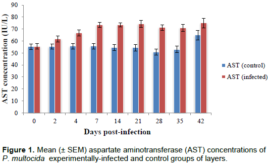
Mean plasma aspartate aminotransferase activity
The mean plasma aspartate amino transferase (AST) activity in the infected and control groups are as shown in Figure 1. Mean plasma AST activity in the infected group increased significantly (P < 0.05) from day 2 post-infection (pi) to reach a peak level (75.00 ± 3.89 IU/L) on day 42 pi when compared with that in the control group (65.83 ± 4.21 IU/L).
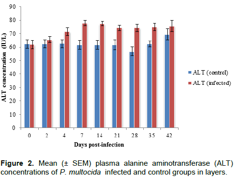
Mean plasma alanine aminotransferase activity.
The mean plasma alanine aminotransferase activity (ALT) in the infected and control groups are as shown in Figure 2. Progressive increase (P < 0.05) in mean ALT activity was observed in the infected group and this was significantly (P < 0.05) higher than that recorded in the control group beginning from day 4 with peak level (78.00 ± 1.89 IU/L) attained on day 7 pi. Thereafter, the mean ALT activity of the infected group remained relatively unchanged until termination of the experiment (day 42 pi).
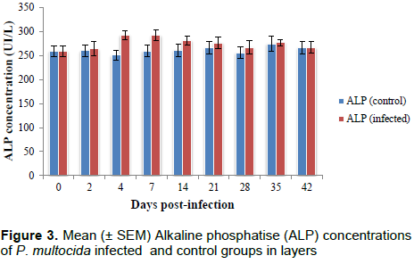
Mean plasma alkaline phosphatase activity
The mean plasma alkaline phosphatase activity (ALP) in the infected and control groups are shown in Figure 3. Mean plasma alkaline phosphatase activity showed no change in both the infected and control groups initially before it increased significantly to a highest level (4.293 ± 9.53 IU/L) on day 4 pi in the infected group of birds. On day 7 pi the level was still high in the infected before decreasing gradually from day 14 to 42 pi when no significant difference were observed between the two groups of birds.
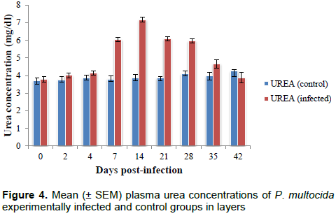
Mean plasma urea concentration
The mean plasma urea concentration in the infected and control groups are shown in Figure 4. The infection caused significant increase (P < 0.05) in mean plasma urea concentration in the infected group on day 7 pi with highest level (7.17 ± 0.55 mg/dl) reached on day 14 pi. It then decreased progressively until termination of the experiment on day 42 pi.
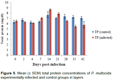
Plasma total protein concentration
The mean plasma total protein concentration in the infected and control groups are shown in Figure 5. Mean plasma total protein concentration significantly increase (P < 0.05) in the infected from day 7 pi to reach a highest level (8.70 ± 0.12 mg/dl) on day 14 pi which later dropped to a lowest level (5.50 ± 0.21 mg/dl) on day 35 pi until termination of the research on day 42 pi.
DISCUSSION
The clinical signs that manifested in the P. multocida-infected layers during the experiment included anorexia, weakness, loss of weight, greenish-yellowish diarrhoea, drop in egg production, pale wattles and combs which are in agreement with those previously reported by Christensen (2013). The observed significant (P < 0.05) increase in plasma activities of ALT, AST and ALP in the P. multocida-infected layers suggests hepatic, renal, and gastrointestinal lesions that had occurred consequent to possible effect of endotoxins that might have been released by the infecting organism or due to cytopathiec effect of the organism which are in line with the findings by Bokori and Karasi (1969). The significant (P < 0.05) increase in the mean plasma urea concentration noticed on days 7 to 28 pi in the P. multocida-infected layers compared to the control strongly suggests renal function impairment which may be due to the effect of P. multocida and/its endotoxin on the renal tubules or could be as a result of dehydration due to diarrhoea which is in line with the report of Harris (2000). In the same vein, the significant increase in the urea concentration in this study could be as a result of increased catabolism of tissue proteins in other to release energy consequent to anorexia that was earlier reported. This finding was similarly reported by Campbell (1998). The microscopic examination of the kidney in the P. multocida-infected layers showed degenerative and necrotic lesions of the renal tubules and which might have affected the renal excretion of uric acid which similarly reported by Harrison and Harrison (1986) and Amany (1997).
Total protein concentration is an important indicator of health status and production features of any organism because of its numerous roles in the physiology and diagnostic purposes (Geogieva et al., 2009). The initial significant increase (P < 0.05) in the mean total protein concentration in the P. multocida-infected group observed on days 7, 14 and 21 pi could be due to haemo-concentration that might have resulted from diarrhoea, while the subsequent decrease in the mean total plasma protein concentration on days 28, 35 and 42 pi following this rise may be due to malnutrition, that resulted from the infection-induced anorexia (Petersen et al., 2001), or could be as a result of combined effects of reduced hepatic synthesis or impaired renal reabsorption and consequent loss of protein in urine and diarrhoea which agrees with the findings by Campbell and Cole (1986) and Samia (2009).
CONCLUSION
In conclusion, experimental infection significant increase in plasma concentrations of AST, ALT, ALP, uric acid, and decrease in total protein thus, signified hepatic, intestinal and renal dysfunctions, due to cytopathiec effect caused by the infecting organism and/or its endotoxin.
CONFLICT OF INTERESTS
The authors have not declared any conflict of interests.
ACKNOWLEDGEMENT
Authors kindly appreciate the help of Hajia Salamatu, Mr Dodo and Mallam Yunusa Mohammed of the Department of Veterinary microbiology and Clinical Pathology Laboratories, Ahmadu Bello University, Zaria for their support.
REFERENCES
|
Abdu PA (2014). Manual of important poultry diseases in Nigeria.Third edition 5 and 6 ventures, Jos, Nigeria. Page 60. |
|
|
Acharya T (2016). |
|
|
Alders RG, Pym R (2009) Village poultry: still important to millions, eight thousand years after domestication. World's Poultry Science Journal 65:81-190. |
|
|
Amany A, Abd-Alla M (1997). Clinicopathological studies on the effect of Pasteurella multocida in chicken and ducks. Egypt Journalof Comparative Pathology and Clinical Pathology 10(2):149-159. |
|
|
Arsov R (1965).The portal of infection in fowl cholera.In: Diseases of poultry, 10th ed. (B.W.Calnek, H.J.Beard, L.R.McDougald and Y.M.Saif). Iowa State University press, Ames, Iowa, USA. pp. 208-219. |
|
|
Aye PP, Angerick EJ, Morishita TY, Harr BS (2001). Prevalence and characteristics of Pasteurella multocida in commercial turkeys. Avian Disease 45:182-190. |
|
|
Blackall PJ (2003). Fowl cholera an emerging disease in free range chickens. In Queensland poultry science symposium Gatton, Queensland. |
|
|
Bokori J, Karasi F (1969). Enzyme diagnostic studiesof blood from geese andducks, healthy and with liverdystrophy. Acta veterinaria Academiae Scientiarum Hungaricae. 19: 269-279. |
|
|
Campbell TW, Coles EH (1986). Avian clinical pathology. Veterinary clinical pathology 4:279-300. |
|
|
Christensen JP, Bisgaard M (2000). Fowl Cholera. Revised Scientific Technology. Office International Epizootics 19:626-637. |
|
|
Christensen JP (2013).Overview fowlcholera. |
|
|
Compi TW, Carpenter TE, Hirds DW, Snipes KP, Hirds DC (1990). Fowl cholera in California multipler breeder turkeys. Avian Disease 34:928-933. |
|
|
Davis-West KB (1972).Newcastle disease in Nigeria retrospection and anticipation. Bulletin Epizootics Africa 20:291-295. |
|
|
Georgieva TM, Zapryanova DS, Dishlyanova EV, Tanev SI, Georgiev I P, Andonova MI, Kanelov IN, Lazarov LV, Koleva PI (2009). Comparison of the results of serum total protein concentration measured by 3 methods: preliminary results. Turkish Journal of Veterinary and Animal Sciences 33(1):67-70. |
|
|
Glisson JR, Sandhu TS, Hofacre CL (2008). Pasteurellosis, Avibacteriosis, Gallibacteriosis, Riemerellosis, and Pseudotuberculosis. In: A Laboratory Manual for the Isolation, Identification, and Characterization of Avian Pathogens, Fifth Edition, Dufour-Zavala L., Swayne D.E., Glisson J.R., Pearson J.E., Reed W.M., Jackwood M.W. & Woolcock P.R., eds. American Association of Avian Pathologists, Athens, Georgia, USA. pp. 12-14. |
|
|
Glisson JR, Hofacre CL, Christensen JP (2003). Fowl cholera. In Diseases of poultry,pp 658-676.Edited by Y. M. Saif, H.J. Bames, J.R. Glisson, A.M. Fadly, L.R. McDougald and D.A Swayne. Ames: Iowa State University Press. |
|
|
Gueye EF (2012). Family poultry in developing countries. ISA Focus 7:5-6. |
|
|
Gustafson CR, Cooper GL, Charlton BR, Bickford AA (1998). - Pasteurella multocida infection involving cranial airspace es in White Leghorn chickens. Avian Disease 42:413-417. |
|
|
Harris DJ (2000). Clinical tests. In: Tully, T. N., Lawton, M. P. C. and Dorrestein, G. M. (Eds). Handbook of Avian Medicine. Butterworth Heinemann, Oxford. pp. 43-51. |
|
|
Harrison GJ, Harrison LR (1986). Clinical Avian Medicine and Surgery. W.B Saunders Company, Philadelphia, London Toronto. |
|
|
Hassan AA, Nwanta J, Mohammed A (2006).Profitability analysis of poultry egg production in Kaduna state, Nigeria. Nigeria Veterinary Journal 27:8-16. |
|
|
Kempentanove M, Kapetanove R, Suvajdzic L, Velhner M (2000). Cholera caused by Pasteurella in breeder flocks. Zivirnastro 35:211-213. |
|
|
Petersen KMD, Christensen JP, Permin A, Bisgaard M (2001). Virulence studies in domestic andfree living birds of a clone of Pasteurella multocidasppmultocida obtained from an out break of fowl cholera in wild birds. Avian Pathology 30:27-31. |
|
|
OIE Terrestrial Manual (2008). Fowl cholera 2(3):534-530. |
|
|
Rimler RB, Rhoades KR (1994). Hyaluronidase and chondroitinase activityof Pasteurella multocida serotype B:2 involved in hemorrhagic septicaemia. Veterinary Record 134:6768. |
|
|
Rosales AG (2013). Egg Bound or Impacted Oviducts in Poultry. |
|
|
Sa'idu LPA, Umoh JU, Abdullahi US (1994) Disease of Nigerian indigenous chickens. Bulletin of Animal Health and Production in Africa. 42:19-23. |
|
|
Samia MM (2009). Hematological biochemical immunological and pathological studies on pasteurellosis in chicken. Egypt Journal of Comparative Pathology and Clinical Pathology 22(2):195-209. |
|
Copyright © 2024 Author(s) retain the copyright of this article.
This article is published under the terms of the Creative Commons Attribution License 4.0


