Full Length Research Paper

ABSTRACT
The present study was conducted to determine egg production and weight in layers experimentally infected with Salmonella gallinarum. Twenty layers were used for the research. The layers were purchased at the age of 18 weeks from certified commercial poultry farm in Kujama farm, Kaduna State, Nigeria and housed in the Animal Research Unit of the Veterinary Teaching Hospital, Faculty of Veterinary Medicine, Ahmadu Bello University, Zaria, Kaduna State, Nigeria. The birds were examined to certify that they were disease free by collecting samples from the cloacal. The birds were assigned to two groups (infected and control) of ten layers each. The infected group was challenged with Salmonella gallinarum orally at the dose of 0.5 ml of 9 x 10⁸ CFU/ml. All the birds in the control group were orally given 0.5 ml of normal saline. After the infection, all the infected layers were closely observed for clinical signs of fowl typhoid. Percentage of egg production and body weight were measured from each group at days zero (Day 0), 2, 4, 7, 14, 21, 28, 35 and 42, post-infection (pi). By day seven post infection, all birds in the infected group showed clinical signs typical of fowl typhoid, namely, ruffled feathers, weakness, somnolence, greenish-yellow diarrhea, huddling together, decrease in feed and water consumption, and five of the layers died. There were, however, significant drop in egg production and loss of body weight in the S. gallinarum infected group.
Key words: Fowl typhoid, Salmonella, inoculum, layers, egg production, body weight.
INTRODUCTION
Salmonella species belong to the Family, Enterobacteriaceae. They are Gram negative, non-spore forming rods (Popoff et al., 2003). Fowl typhoid caused by Salmonella enterica serovar gallinarum in birds, is a severe systemic disease that affect both young and adult birds with macroscopic and microscopic lesions leading to massive economic losses due to high morbidity and mortality (Parmer and Davies, 2007).
Fowl typhoid (FT) has been discovered in many African countries which include Tanzania, Uganda (Okoj, 1993), Senegal (Arbelot et al., 1997), Nigeria (Sa’idu et al., 1994) and Morocco (Bouzoubaa et al., 1987). FT is a septicaemic infection affecting chicken and turkey mostly, but some natural infections in many other avian species has been studied (Wray et al., 1996; Shivaprasad, 1997). The outbreak of FT in young chicks may be due to vaccination against FT practiced by many farmers which result in vertical transmission of the infection (Jordan and Pattison, 1992; Roa, 2000). The control of FT through hygienic measures, together with some serological testing and slaughter of positive reactors, have resulted in the elimination of Salmonella gallinarum in many countries (Barrow, 1999). However, FT remains a leading disease of the poultry industry in many areas of the world (Okwori et al., 2013). Respiratory distress and depression is seen in acute FT and the clinical signs include greenish-yellow diarrhea, there may be enlargement and congestion of the liver, spleen and kidney. The liver may have pale multiple foci of 2 to 4 mm in diameter (Beyaz et al., 2010). In acute to subacute cases, there is multiple necrosis of the liver parenchyma with accumulation of fibrin and infiltration of heterophils mixed with a few lymphocytes and plasma cells can be seen in the liver (Kokosharov et al., 1997; Hossain et al., 2006).
In sub-acute outbreaks, sporadic mortality over a long period is experienced while in chronic cases, especially in cases where there are large nodules in the heart, the liver will have congestion with interstitial fibrosis. The spleen may have severe congestion or fibrin deposits and severe hyperplasia (Chishti et al., 1985). The transmission of S. gallinarum can be through faecal droppings of infected birds, bird carcasses and laid eggs. The infection could be introduced by importation of live infected chickens and hatched eggs. Mechanical spread may be by humans, wild birds, mammals, flies, ticks, feedsacks, etc (Steigh and Duguid, 1989).
Poultry production in Nigeria has witnessed a rapid growth to a well-established commercial enterprise. This increase in the production activity is greatly pronounced and has resulted in new challenges (Hassan et al., 2006). Poultry production is the most efficient and cost-effective way of increasing the availability of high-protein food, as eggs are known to provide the most perfectly balanced food containing all the essential amino acids, minerals and vitamins (FAO, 1987; Branckaert et al., 2000). Salmonellosis in poultry causes egg shell abnormalities including shell-less and infertile eggs with early embryonic mortality (Welish et al., 1997; Coufal et al., 2003). This study evaluated the determination of egg production and weight in layers experimentally infected with Salmonella gallinarum in Zaria, Kaduna State, Nigeria.
MATERIALS AND METHODS
Study area
This study was carried out in Zaria, Kaduna State, which is located within the Northern Guinea Savannah Zone of Nigeria, between latitude 7° and 11°N, and longitude 7° and 44°E; the average rainfall of this zone ranges from 1,000 to 1,250 mm, and the average temperature ranges from 17 to 33°C (Saʼidu et al., 1994).
Experimental chickens
Twenty eighteen-week-old hens were purchased from a commercial farm in Kuja, Kaduna State, Nigeria. These birds were vaccinated against other diseases but with the exception of fowl typhoid. On arrival, at the venue of the research, the birds were housed in the animal research unit of the Veterinary Teaching Hospital, Faculty of Veterinary Medicine, Ahmadu Bello University, Zaria, Kaduna State, Nigeria. The birds were kept for a period of four weeks to get used to the handling conditions they would be subjected to during the research. During this period, they were on layer mash (Hybrid®).
Experimental design
Allocation of chickens to experimental groups
At 22 weeks old, the hens were randomly allocated to two groups (infected and control) of 10 layers each. The control group of chickens was then moved to the research pen of Department of Veterinary Pathology as a precautionary measure against transmission of the experimental fowl typhoid disease to the control group. At this point, both groups were fed commercial layer mash (Hybrid feeds®) until termination of the experiment. Water was provided to the layers ad libitum, throughout the experimental period that lasted for 42 days.
Source of bacterial organism
S. gallinarum was obtained from the Department of Veterinary Microbiology, Ahmadu Bello University, Zaria, Kaduna State, Nigeria.
Bacteriological analysis
Cloacal swabs were collected from both infected and control groups of layers and dipped into a buffered peptone water for recovery of the S. gallinarum and subcultures were then made from each broth onto MacConkey agar. The agar plates were incubated aerobically at 37°C for 24 h using methods described by Wigley et al. (2001) and Parmer and Davies (2007).
Challenge bacteria
The challenge bacteria were collected from the Department of Veterinary Microbiology, Ahmadu Bello University, Zaria, Kaduna State, Nigeria. The bacteria from the slant were re-plated on MacConkey agar (MCA). The subcultured plates were then examined for their characteristic features, such as color, morphology using Gram’s stain (Gram negative). Some colonies were picked from the cultured plate and placed in sterile test tube with normal saline of 20 ml of 0.5% and turbidity equivalent to 9 x 10⁸ CFU/ML was obtained. Challenge of the layers was done orally using sterile syringes. The infected group was given a dose of 0.5 ml of 9 × 10⁸CFU/ML of the bacterium, but the control group were not infected with the organism, but received distilled water only.
Clinical observation
After challenge of the infected birds with the bacterial organism, the infected group was daily observed for typical signs of FT and findings were recorded.
Determination of body weight and egg production
Beginning from the day of infection (day 0) and throughout the experimental period, that lasted for 42 days, the live weights and egg production of the birds were recorded.
Bacteriological isolation
At necropsy, tissue samples of the liver, kidney, ovary and spleen were aseptically taken for isolation of S. gallinarum using standard laboratory methods (Wigley et al., 2001; Parmer and Davies, 2007).
Statistical analysis
Data obtained were expressed as ± SEM. Values were subjected to student T-test and values of P<0.05 were considered to be significant.
RESULTS
Clinical signs of fowl typhoid in the infected birds
All the birds in the control group appeared apparently healthy throughout the experiment. Following challenge with S. gallinarum, the birds appeared clinically normal until day 7 post challenge when the birds started passing greenish-yellow diarrhea, having depression and huddling, rough feathers, somnolence, reduction in feed and water consumption, decreased egg production and sudden death.
Bacterial recovery from infected birds
S. gallinarum organisms were isolated in some of the samples collected which include liver, kidney, spleen and ovary of the challenged birds. Biochemical test revealed indole negative, urea negative, catalase and citrate positive and it produces hydrogen sulphide (H2S) in triple sugar iron agar TSI.
Effect of S. enterica serovar gallinarum infection on egg production and body weight in the layers
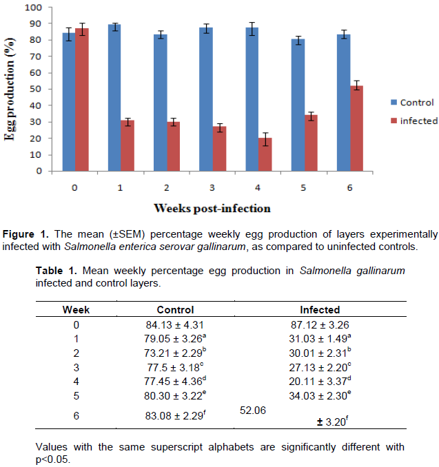
Mean weekly percentage egg production
The mean weekly percentage egg production in the S. enterica serovar gallinarum experimentally infected and control groups is presented in Figure 1. The mean weekly percentage egg production in the infected birds on week 0 pi (87.12 ± 3.26%) was not significantly different (P>0.05) from that of the control group (84.13 ± 4.31%). But by week 1 pi, a significant decrease (P< 0.05) in mean weekly percentage egg production was observed in the infected group (31.03 ± 1.49%) when compared with that of the control (89.05 ± 3.26%) with the infected group reaching its lowest value on week 4 pi (20.11 ± 3.37%).
Thereafter, a gradual rise was observed in the infected group on week 5 pi (34.03 ± 2.30%) until the end of the experiment on week 6 pi (52.06 ± 3.20%) Table 1.
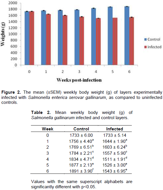
Mean weekly body weight
The mean body weights of the S. enterica serovar gallinarum experimentally infected and control groups are presented in Figure 2. The mean weekly body weight (g) of the infected birds on week 0 pi (1733 ± 5.14 g) and control (1733 ± 6.00 g) showed no significant difference (P>0.05). A significant decrease (P< 0.05) in mean weekly body weight was also observed on week 1 pi in the infected group (1644 ± 1.90 g) when compared with that of the control (1756 ± 4.40 g), with the infected group reaching its lowest value on week 4 (1511 ± 1.91g) post-infection. Following this, a gradual rise from its week 4 value was observed in the infected birds on week 5 pi (1526 ± 3.00g) till the termination of the experiment on week 6 pi (1543 ± 6.95 g) Table 2.
DISCUSSION
The clinical signs observed in the S. gallinarum-infected layers in this study, which included depression, ruffled feathers, huddling, loss of body weight, drop in egg production, somnolence and greenish-yellow diarrhoea were consistent with findings in previous reports (Shivaprasad, 2000; Freitas Neto et al., 2007; Ezema et al., 2009; Garcia et al., 2010). The 50% mortality in the layers recorded in this study was in the range (10 to 100%) reported previously (Shivaprasad, 1996; Uzzau et al., 2000; Oliveira et al., 2005; Paiva et al., 2009) in chickens. A significant (P<0.05) progressive drop in egg production was observed in the infected layers from 1th week pi and reaching its maximum drop on the 4th week pi. The significant drop in egg production in the S. gallinarum infected group recorded in this study was in the range of 50 to 70% reported previously by Shivaprasad (1997) and Ezema et al. (2009) in laying birds. The drop in egg production, which was recorded by week 4 pi showed that the disease progressed with increased severity. The drop in egg production observed in this study could be due to a number of factors. The factors known to cause drop in egg production in S. enterica serovar gallinarum-infected layers include decrease in feed and water consumption with consequent nutritional imbalances and possible impairment of renal and intestinal calcium absorption due to the infection-induced lesions in these systems (Ezema et al., 2009). The loss of body weight observed in the S. enterica serovar gallinarum infected birds was similarly reported by Ezema et al. (2009) in commercial layers afflicted by fowl typhoid and may be due to decrease in feed consumption which was supported by results of measurement of their feed consumption and intestinal disturbances evidenced by diarrhoea, which could have interfered with nutrients absorption as had been reported by Shah et al. (2013) in S. enterica serovar gallinarum infected broiler chickens.
CONCLUSION AND RECOMMENDATION
This study has shown that experimental infection of layers with S. enterica serovar gallinarum can cause significant reduction in egg production and weight loss. Therefore, those keeping layers should adhere to strict biosecurity measures as means of prevention and control of fowl typhoid in poultry farms, as this disease could lead to decrease in egg production, weight loss and other eggshell abnormalities.
CONFLICT OF INTERESTS
The authors have not declared any conflict of interests.
REFERENCES
|
Arbelot B, Dayon JF, Mamis D, Gneye JC, Tall, F, Samb H (1997). Sero-survey of Dominant avian disease in Senagal; Mycoplasmosis, Fowl Typhoid and Pullorum disease, Newcastle, Infectious Bursal and Infections Bronchitis disease. Revue d' Elevage et de Medicine veterinarier des Pays tropicaus 50:197-203. |
|
|
Barrow PA, Lowell MA, Murphy CK, Page K (1999). Salmonella infection in a commercial line of ducks;Experimental studies in virulence, intestinal colonization and immune protection. Epidemiology of Infection 123:12-132. |
|
|
Beyaz L, Atasever A, Aydin F, Gumusoy KS, Abay S (2010). Pathological and clinical findings and tissue distribution of Salmonella gallinarum infection in turkey poults. Turkish Journal of Veterinary and Animal Sciences 34:101-110. |
|
|
Bouzoubaa K, Nagarya KV, Newman JA, pomeraj BS (1987). Use of membrane proteins from Salmonella gallinarum for prevention of fowl typhoid infection in chickens. Avian Diseases. 1:699-704. |
|
|
Branckaert RDS, Gaviria I, Jallade J, Seiders RW (2000). Transfer of technology in poultry production for developing countries. FAO, Rome. |
|
|
Chishti MA, Khan MZ, Siddique M (1985). Incidence of salmonellosis in chicken in and around Faisalabad (Pakistan). Pakistan Veterinary Journal 5:79-82. |
|
|
Coufal CD, Chavez C, Knape KD, Carey JB (2003). Evaluating a method of ultraviolet sanitation of broiler hatching eggs. Poutry Science 5:754-759. |
|
|
Ezema WS, Onuoha E, Chah KF (2009). Observations on an outbreak of fowl typhoid in commercial laying birds in Udi, South Eastern Nigeria. Comparative Clinical Pathology 18(4):395-398. |
|
|
Food and Agricultural Organisation (1987). Termination Statement for Poultry development. Establishment of a poultry breeding farm, Bhutan. Rome, BHU/82/012. |
|
|
Freitas Neto OC, Arroyave W, Alessi AC, Fagliari JJ, Berchieri Jr A (2007). Infection of commercial laying hens with Salmonella gallinarum:Clinical, anatomopathological and haematological studies. Brazilian Journal of Poultry Science 9(2):133-141. |
|
|
Garcia KO, Santana AM, Freitas NOC, Berchieri Jr A, Fagliari JJ (2010). Experimental infection of commercial layers using a Salmonella enteric serovar Gallinarum strain: blood serum component and histopathological changes. Brazilian Journal of Veterinary Pathology, 3(2):111-117. |
|
|
Hassan AA, Nwanta J, Mohammed A (2006). Profitability analysis of poultry egg production in Kaduna state, Nigeria. Nigeria Veterinary Journal 27:8-16. |
|
|
Hossain MS, Chowdhury EH, Islam MM, Haider MG, Hossain MM (2006). Avian Salmonella infection: isolation and identification of organisms and histopathological study. Bangladesh Journal of Veterinary Medicine 4:7-12. |
|
|
Jordan FTW, Pattison M (1992). Poultry Disease 4th Edition. W.B. Sauder Company Ltd London pp.169-171. |
|
|
Kokosharov T, Hristov H, Belchev L (1997). Clinical, bacteriological and pathological studies on experimental fowl typhoid. Indian Veterinary Journal 74:547-549. |
|
|
Okoj L (1993). Diseases as important factors affecting increased poultry production in Uganda. Der Tropenlandwin, Zeitschrift in dentropen and Subtropen Jahrgag 94:S37-S44. |
|
|
Okwori AE J, Ogbe RJ, Chollom SC, Agada GOA, Ujah A, Okwori E, Adeyanju ON, Echeonwu GON (2013). Isolation Of Salmonella Gallinarum From Poultry Droppings in Jos Metropolis, Plateau State, Nigeria. IOSR Journal of Agriculture and Veterinary Science (IOSR-JASR) 5(2):14-44. |
|
|
Oliveira GH, Berchieri Jr. A, Fernandes AC (2005). Experimental infection of laying hens with Salmonella enterica serovar gallinarum. Brazilian Journal of Microbiology 36(1):51-56. |
|
|
Paiva JB de, Penha FRAC, Argüello YMS, da Silva MD, Gardin Y, Resende F, Berchieri Jr. A, Sesti L (2009). Efficacy of several Salmonella vaccination programs against experimental challenge with Salmonella gallinarum in commercial brown layer and broiler breeder hens. Brazilian Journal of Poultry Science 11(1):65-72. |
|
|
Parmer D, Davies R (2007). Fowl typhoid in small backyard laying flock. The Veterinary Record 160:348. |
|
|
Popoff MY, Bockemuhl J, Gheeshing LL (2003). Antigenic Formulas of the SalmonellaSerovars, 8th revision. WHO Collaborating Centre for Reference and Research on Salmonella. Supplement 2001 (No: 45) to the Kauffmann-White Scheme Research in Microbiology 154:173-174. |
|
|
Roa G (2000). A Comprehensive Textbook on Poultry Pathology. Medical Publisher Ltd pp. 7-10. |
|
|
Sa'idu L, Abdu PA, Umoh JU, Abdulahi US (1994). Disease of Nigerian indigenous chickens. Bulletin of Animals Health Production in Africa 42:19-23. |
|
|
Shah S, Kamil S, Darzi M, Mir M, Bhat S (2013). Haematological and some biochemical changes in experimental fowl typhoid infection in broiler chickens. Comparative Clinical Pathology. Academic Journal 22(1):83. |
|
|
Shivaprasad HL (1996). Pullorum Disease and Fowl Typhoid. Calneck BIN (ed). Disease of poultry. Tenth Edition. Iowa State University Press. pp. 82-96. |
|
|
Shivaprasad HL (1997). Pullorum disease and fowl typhoid. In B.W. Calnek., H.J. Barnes., C.W. Beard, L.R. McDougald and Y.M. Saif (Eds.), Disease of Poultry 10th edition. Ames, IA: Iowa State University Press. pp. 82- 96. |
|
|
Shivaprasad HL (2000). Fowl typhoid and Pullorum disease. Review Science 19(2):405-424. |
|
|
Steigh JD, Duguid JP (1989). Salmonella. In: 13th ed. Collee JG, Duguid JP, Fraser AG, Marmion BP editor. Practical Medical Microbiology. Volume 2:New York:Churchill Livingstone pp. 456-479. |
|
|
Uzzau S, Brown DJ, Wallis T, Rubino S, Leori G, Bernard S, Casadesus J, Platt DJ, Olsen JE (2000). Host adapted Serotypes of Salmonella enteric. Epidemology of Infection 125:229-255. |
|
|
Welish RD, Vanhooser SL, Dye LB, Neiman RW (1997). Salmonella infection in Ratites: Diagnosis, epidemiology and clinical significance. Veterinary Medicine 2:193-198. |
|
|
Wigley P, Berchieri Jr A, Page KL, Smith AL, Barrow PA (2001). Salmonella enterica serovar Pullorum persists in splenic macrophages and in the reproductive tract during persistent, disease-free carriage in chickens. Infection and Immunity 69(12):7873-7879. |
|
|
Wray C, Davies RH, Corkish JD (1996). Enterobacteriaceae. In F.T.W. Jordan and M. Pattison (Eds). Poultry Diseases 4th edition London; Saunders company Ltd. pp. 9-43. |
|
Copyright © 2024 Author(s) retain the copyright of this article.
This article is published under the terms of the Creative Commons Attribution License 4.0


