Full Length Research Paper

ABSTRACT
Mercury (Hg) is a poisonous substance associated with diseases, such as ataxia and Joubert syndrome. Therefore, it is important to find a way to disrupt the process of Hg poisoning in the cerebellum, by exploring the medicinal value of local herb such as Galinsoga parviflora (GP). This study examined the effects of aqueous leaf extract of GP in HgCl2-induced cerebellar toxicity in adult male mice. Twenty-five adult male mice of an average weight of 25 g were randomly divided into 5 groups (n=5). Animals in Group I received oral administration of 2 ml/kg distilled water for 6 weeks, Group II received 2.3 mg/kg of HgCl2 for 3 weeks, Group III received 2.3 mg/kg of HgCl2 for the first 3 weeks followed by oral administration of 800mg/kg of GP extract for the next 3 weeks, Group IV received 800mg/kg of GP extract for the first 3 weeks followed by 2.3 mg/kg of HgCl2 for the next three weeks, and Group V received 2.3 mg/kg of HgCl2 and 800 mg/kg of GP extract concurrently for three weeks. The test animals were subjected to beam walking tests during the experiment period, followed by euthanasia, perfusion fixation, and tissue sample collection for histological and histochemical analysis. Treatment with the extract of GP showed varying degrees of regeneration in the cerebellar Purkinje cells and myelin sheath of mice in HgCl2 induced toxicity, with corresponding improvements in balance and posture. Cerebellar HgCl2 exposure in the present study was neurotoxic; however, treatment with GP was of therapeutic value.
Key words: Mercury chloride, cerebellum, Galinsoga parviflora.
INTRODUCTION
Mercury(Hg) is the third most dangerous element on the earth, and humans are exposed to it in a various forms, including gaseous, organic, and inorganic forms (Aragão et al., 2018). Mercury is found in the environment, following natural processes and human activity (Brooks, 2011). Humans are exposed to Hg in a variety of ways, including eating Hg-contaminated food, coming into close contact with broken Hg-containing products, and working in mining industries, where Hg is used almost every day and workers are in constant contact with it (Bose-O'Reilly et al., 2010). Inhaling Hg-contaminated air is another effective way via which humans are exposed to Hg (Park and Zheng, 2012); Hg is taken into the bloodstream via the lungs and nervous system (Park and Zheng, 2012). Organic mercury accumulates in CNS-derived cells in the human body, converted to inorganic mercury (Aragão et al., 2018). Niigata Minamata disease and Hunter-Russell syndrome are all caused by methyl mercury, a byproduct of acetaldehyde production (Takahashi et al., 2017). Hg exposure has been known to cause serious health problems, affecting the nervous, immune, and digestive systems and organs such as brain, skin, eyes, lungs, and kidney (Genchi et al., 2017). Clinical signs of methyl chloride intoxication include cerebellar ataxia, sensory and auditory abnormalities, and restriction of visual fields, depending on the location of the lesions (Takahashi et al., 2017). Hg exposure is associated with the following conditions such as behavior changes, fatigue, tremor, headaches, cognitive loss hallucination, incoordination and death (Genchi et al., 2017). Health conditions associated with Hg exposure contribute to the general global and local disease burden. Most of burden from Hg exposure is notable among the developing countries (Budnik and Cateleyn, 2019). In the developing countries, effects of Hg exposure are often not discovered early, therefore making the management of Hg exposure a very difficult task (Bose-O’Reilly et al., 2010).
Cerebellum is an important part of the central nervous system and controls all muscular movements and coordination (Gallucci et al., 2003); made up of the middle vermis and two lateral hemispheres (halves) (Izawa et al., 2012). The cerebellum has three lobes which are anterior, flocculonodular, and posterior lobes (Lunnon et al., 2016). Also, the cerebellar consists of the cerebellar cortex, and an embedded medullary center of white matter (Mihailoff and Haines, 2018). There are layers within the cerebellar cortex which are; outer (molecular layer), Purkinje and the inner layer (granular layer) (Koeppen et al., 2012). The Purkinje cells are the most important neurons in the cerebellar cortex (Jelliffe et al., 2012). The cerebellar cortex is highly vulnerable to the effects of toxic metals, including Hg (Gandhi et al., 2000). Injury or damage to the cells of the cerebellum may lead to conditions like; ataxia, Joubert syndrome, and many others (Jelliffe et al., 2012). Treatment of clinical conditions associated with Hg exposure is also considered to be expensive and not readily available among different populations of the developing world. Therefore, it is necessary to explore other options or approaches, such as the use of local herbs such as Galinsoga parviflora (GP) especially in the management of Hg exposure.
GP also known as Gallant Soldier, Kofume (Luganda), and Mpunika or Empunika (Runyankore), is an Asteraceae plant that is widely consumed as a vegetable around the world (Ali et al., 2017). These plants are found in almost every part of the world including North America, Europe, Asia, and Africa (Bazylko et al., 2012). According to research, GP contains flavonoids, aromatic esters, caffeic acid derivatives, diterpenoids, alkaloids, and derivatives, as well as vitamin C and phenolic acid (Bazylko et al., 2012). Plants that contain alkaloids have shown to have potential therapeutic effects against a variety of neurodegenerative diseases, including Alzheimer's disease (AD), Huntington disease (HD), Parkinson's disease (PD), Epilepsy, Schizophrenia, and stroke (Hussain et al., 2018). GP has been reported to have analgesic and anti-inflammatory effects, including free radical scavenging activity, reduction of hyaluronidase activity, and inhibition of IL-6 production, all of which can help to reduce the activation of an inflammatory response (Studzi?ska-Sroka et al., 2018). In addition, earlier study has shown that the herbs of GP are an attractive source of antioxidant-rich preparations (Bazylko et al., 2012). The aim of the present studies was to assess behavioral, histological, and histochemical changes in the cerebellar cortex following treatment with GP in Hg induced cerebellar damage.
MATERIALS AND METHODS
Ethical approval
Ethical approval was obtained from the Research and Ethics Committee of Kampala International University-Western Campus, and registered as KIU-2021-14. The study adhered to the international and national ethical regulations outlining use of animals in scientific experiments.
Experimental design
GP leaves (Mbr-10007-2) was collected from a garden in Bushenyi district of Uganda. The leaves were dried at room temperature, powdered with a grinding machine, and extracted. Sixteen grams (16g) of powdered leaves was dissolved in 300 ml of distilled water and allowed to rest for three days, often shaking until the authors extracted all plant elements. They then filtered the extract using filter paper (Dr wax) and solidified in an oven at 70°C for three days (Bazylko et al., 2012). The Mercuric chloride (May and Bakers, England) was purchased from a reputable chemical store in Ishaka town, Western Uganda.
Experimental animals
Twenty-five (25) adult male mice weighing 20-35 g were obtained from the Kampala International University Western Campus (KIU-WC) animal facility for this investigation. All mice were placed in a well-ventilated plastic cage and given free access to food and water for two weeks to acclimatize. These animals were further divided into five (5) groups based on their weight (n = 5). Animals in Group (I-V) received oral administration of 2 ml/kg distilled water for 6 weeks, 2.3 mg/kg (Franciscato et al., 2011) of HgCl2 for 3 weeks only, 2.3 mg/kg of HgCl2 for the first 3 weeks followed by oral administration of 800 mg/kg (Yadav and Tangpu, 2008) of GP extract for the next 3 weeks, 800 mg/kg of GP extract for the first 3 weeks followed by 2.3 mg/kg of HgCl2 for the next three weeks, and 2.3 mg/kg of HgCl2 and 800 mg/kg of GP extract concurrently for three weeks, respectively.
Beam walking test
This test was conducted using the method outlined by Luong et al. (2011). The beam walking test can be used to examine fine motor coordination and balance. This is helpful for detecting modest deficiencies in motor skills and balances that other motor test, such as the Rotarod (Loung et al., 2011). The beam apparatus consisted of two poles supporting one-meter beams with a flat surface of 12 mm width that rested 50 cm above the tabletop. The endpoint was represented by a black box put at the end of the beam. The mouse was moved to the room with the beam device around 10 min before the training. The mouse was positioned in the middle of the beam, facing one of the ends, with care. The mouse was permitted to walk to the darkroom's end, and the time it took to get there was recorded. Each mouse was put through three trials through the design that causes the animal to become conscious that a goal box could be reached (Magaji et al., 2012). The timer began when the nose of the mouse entered the center of the beam and ended when the animal reached the end.
Histological and histochemical study
The brain was transcardially perfused using 4% paraformaldehyde fixatives and fixed tissues were processed in a procedure described by (Okesina and Ajao, 2019), for Hematoxylin and Eosin (H and E) Staining to understand the cytoarchitecture of the cerebellum after the various treatments. Subsequently, myelin staining was done as de Also, myelin staning was done as describe by Jouihan (2012).
Statistical analysis
Statistical analysis was performed using SPSS version 25. Data obtained from this study were analyzed using ANOVA to compare mean across the groups followed by Tukey post-hoc test. Statistical significance of p < 0.05* (95% C.I) was used. All results were expressed as mean ± SD or SEM.
RESULTS
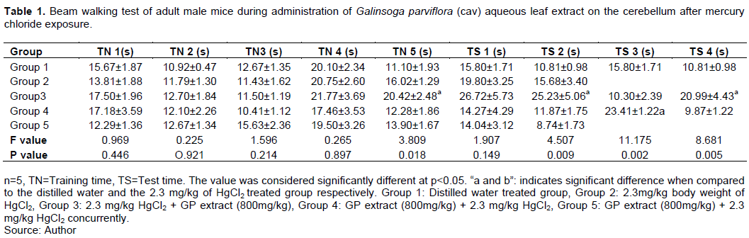
Beam walking test
When compared to control group, the group given 2.3 mg/kg of HgCl2 followed by oral administration of 800 mg/kg of GP extract, showed remarkable increase in the time taken to transit the beam to the dark area, notably at the beginning of the test period, but a significant decrease in time at the end of the test time (P < 0.05). However, there was a decrease in time during test time when compared to group 2 animals which were given 2.3 mg/kg of HgCl2. Also, when compared to the control group, group 4 animals which were given 800 mg/kg of GP extract for the first three weeks followed by 2.3 mg/kg of HgCl2 orally for the next three weeks, showed an increase in the time taken to transit the beam at the beginning of the test and a significant reduction in time at the end of the test time (P < 0.05). During the test period, Group 5 animals which were concurrently given 2.3 mg/kg of HgCl2 and 800mg/kg of GP extract for three weeks showed a reduction in time when compared to control group (distilled water treated group) and the 2.3 mg/kg of HgCl2 treated group (Table 1).
Histological observation
Normal histo-architecture featuring Purkinje layers, molecular layer (ML), Purkinje cell layer (PL), and granular cell layer (GL) were visible in the photomicrograph of the cerebellum of the adult male mice treated with distilled water (control) (Figure 1A). In the Purkinje cell layer of the cerebellar cortex of group 2, histological examinations based on H and E revealed indications of pyknosis following three weeks of oral administration of 2.3 mg/kg HgCl2 (Figure 1B). In group 3, the section revealed rather normal Purkinje cells with marked reduction of pyknotic cells in mice given 800 mg/kg of GP extract orally for three weeks after receiving 2.3 mg/kg HgCl2 for three weeks (Figure 1C). In the pyramidal cell layer of the cerebellar cortex of group 4 mice, both pyknosis and normal cells were observed in mice given 800mg/kg of GP extract prior to 3 weeks oral administration of 2.3 mg/kg of HgCl2 (Figure 1D). Group 5 demonstrated a more normal pyramidal cell (with an intact nucleus) (Figure 1E).

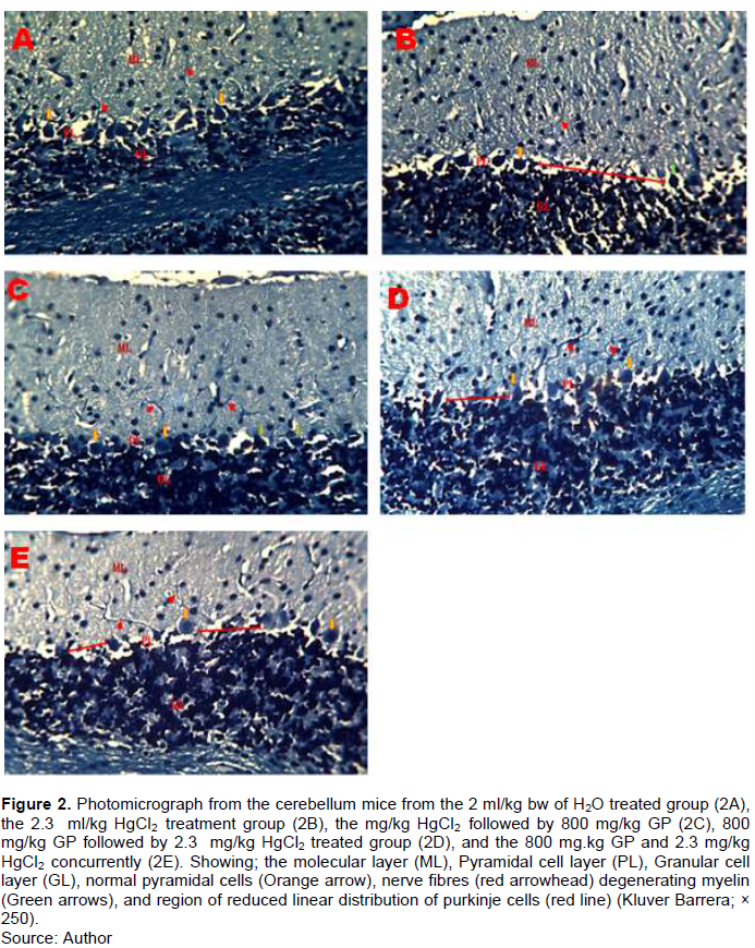
The photomicrograph of the cerebellum of adult male mice treated with distilled water group 1 (normal control), revealed relatively normal cyto-architecture; having normal myelination (Figure 2A). Section of the cerebellum from the 2.3 mg/kg HgCl2 treated group revealed degeneration of myelin sheath and a region of reduced linear distribution of Purkinje cells (Figure 2B). Section of the cerebellar cortex from Group 3 animals given 800 mg/kg of GP extract orally for three weeks after receiving 2.3 mg/kg HgCl2 for three weeks revealed regions with degenerating myelination (Figure 2C). Sections of the cerebellar cortex from the group 4 animals given 800mg/kg of GP extract prior to 3 weeks oral administration of 2.3 mg/kg of HgCl2 revealed a region of reduced linear distribution of Purkinje cells (Figure 2D). In group 5, the section of the cerebellum shows a region of reduced linear distribution of Purkinje cells (Figure 2E)
DISCUSSION
The beam walking test was employed in this study to examine fine motor coordination, and the time it took the mice to get to the darkroom. The present showed that the administration of Hg was neurotoxic and affected motor coordination among the test animals, as indicated by the increase in the time taken to transit the beam to the dark box; this could possibly be attributed to a cortical impact lesion. the authors observation align with Buccafusco et al. (2009) findings, who reported earlier that mice with cortical impact lesions frequently exhibits contralateral slippage on the beam in an earlier investigation. The poor fine motor coordination and balance seen in the beam walk test suggest that CNS lesions may have caused motor impairments as a result of mercury damage to the CNS. On the other hand, treatment with GP was associated with improved coordination in all the treatment groups. This finding may support the previous report on the possible anti-inflammatory effects, free radical scavenging activity, reduction of hyaluronidase activity, and inhibition of IL-6 production associated with GP (Studzi?ska-Sroka et al., 2018).
Hg exposure was associated with degenerative alterations in the Purkinje layer of the cerebellar cortex, as evident in the depletion of Purkinje cells population and the presence of pyknotic Purkinje cells. The finding may support the previous report by Ibegbu et al. (2014) revealed degenerative and necrotic changes in the Purkinje cells of the Purkinje cell layer of the cerebellar cortex following aluminum chloride exposure. Pyknosis seen in cerebellar cortex in this study is a sign of neuronal death, with its consequent effects on fine motor coordination. Alteration of fine motor function, balance, posture, and motor learning are often affected in cerebellar injury (Gray’s Anatomy, 2008). Damage to the Purkinje cells in the cerebellum can obstruct fine motor performance and learning (Gandhi et al., 2000); this could have happened as a result of chromatin condensation, which causes cell death (Bianchi and Manfredi, 2004). Because the cerebellum possesses a blood brain barrier (BBB) that is susceptible to mercury intoxication (Takahashi et al., 2017). Methyl mercury has the potential to cause damage to the BBB through the production of RECA-1 and extravasation of endogenous IgG (Takahashi et al., 2017). Our findings are in line with those of (Olivier et al., 2021), who found glial cell multiplication and granular cell loss beneath the Purkinje cell layer. Furthermore, our findings using a histochemical stain (Kluver-Barrera) demonstrated myelin component depletion in the Purkinje layer. This could be due to oligodendroglia cell loss or disruption, as well as an immune-mediated response (Duncan and Radcliff, 2016). The processes of immunotoxicity, on the other hand, are claimed to be depending on the amount of Hg exposed to and the type of Hg (Eagles-Smith et al., 2018). Higher doses can cause the production of cytokine signals, whilst smaller doses only cause the production of cytokine signals without affecting cell numbers (Eagles-Smith et al., 2018).
To test the curative therapeutic potential of GP extract, mice were administered 800 mg/kg orally after being exposed to HgCl2. When compared to the control group, the histoarchitecture of the cerebellar cortex revealed a reduction in Purkinje cells, with the myelin and Nissl staining suggesting possible recovery. Plants containing alkaloids have potential therapeutic effects against a variety of neurodegenerative diseases, according to Hussain et al. (2018), who studied the function of plant-derived alkaloids and their processes in neurodegenerative diseases. The presence of phytochemicals found in GP, such as alkaloid, carbohydrate, flavonoid, polyphenol, and glycoside, could explain the observed therapeutic potential of GP. This shows that the presence of such phytochemicals could be responsible for the observed recovery of cells.
In comparison to the control group, further investigation into the preventative impact of GP revealed a dramatic and considerable loss of cells in the Purkinje layer. The Purkinje layer has few cells as a result of cell death. A postmortem analysis found a drop in Purkinje cell numbers in the brains of patients with essential tremor who did not have Lewy bodies, according to Axelrad et al., (2008). This shows that a decrease in the linear distribution of Purkinje cells in the cerebellum may be caused by cell death or Lewy bodies. When compared to the control group, histochemical demonstration with Kluver-Barrera revealed a considerable reduction in the content of the myelin sheath in the Purkinje layer. Due to its improved histoarchitecture and lower number of pyknotic nuclei, the therapeutic treatment to Hg poisoning appears to be better when compared to group three. This could be because the GP extract boosted the immunological response produced by early HgCl2 exposure. To confirm that GP has a neuromodulatory effect, more study is needed in this area. Future study may need to concentrate on figuring out how GP works and investigating the impact of the phytochemicals involved in the process. The molecular interplay between the neuron-glia triad may aid in elucidating the therapeutic role of GP extract.
Concurrent administration of HgCl2 and GP extract in group 5 revealed normal histoarchitecture of the cerebellar cortex when compared to the control group. However, histochemical demonstration of the myelin sheath revealed reduction in the content of the Purkinje cells which seems to be recovering. Overall, myelin sheath degeneration occurs when a neuron dies or if its axons have been severed, the myelin sheath surrounding the degenerating axon breaks up and is phagocytosed (Burnett and Zager, 2004). The conduction of signals in the affected nerves is hampered by myelin degradation. As a result, the reduction in conduction ability causes deficiency in sensation, movement, cognition, or other functions depending on which nerves are involved (Swanson, 2017). Degradation of myelin in cerebellar cortex neurons, as found in the study, may have disrupted the conduction of electrical signals in neurons, impairing normal cerebellar cortex functioning. The pyknosis and degenerating myelin seen in histomorphological investigations of the cerebellum could be implicated in the motor deficiencies seen in neurobehavioural studies.
CONCLUSION
The study revealed that Hg exposure in the cerebellum of mice caused pyknosis, reduced myelination in the axons, loss of neuromuscular function, loss of balance, and coordination. However, the administration of GP was of therapeutic value against Hg induced cerebellar toxicity in adult male mice.
CONFLICT OF INTERESTS
The authors have not declared any conflict of interests.
REFERENCES
|
Ali S, Zameer S, Yaqoob M (2017). Ethnobotanical, phytochemical and pharmacologicalproperties of Galinsoga parviflora (Asteraceae): A review. Tropical Journal of Pharmaceutical Research 16(12). |
|
|
Aragão WA B, Teixeira FB, Fagundes NCF, Fernandes RM, Fernandes LMP, da Silva MCF, Amado LL, Sagica FES, Oliveira EHC, Crespo-Lopez ME, Maia CSF, Lima RR (2018). Hippocampal Dysfunction Provoked by Mercury Chloride Exposure: Evaluation of Cognitive Impairment, Oxidative Stress, Tissue Injury and Nature of Cell Death. Oxidative Medicine and Cellular Longevity 2018:1-11. |
|
|
Axelrad JE, Louis, ED, Honig LS, Flores I, Ross GW, Pahwa R, Vonsattel JPG (2008). Reduced Purkinje cell number in essential tremor: a postmortem study. Archives of Neurology 65(1):101-107. |
|
|
Bazylko A, Stolarczyk M, Derwi?ska M, Kiss AK (2012). Determination of antioxidant activity of extracts and fractions obtained from Galinsoga parviflora and Galinsoga quadriradiata, and a qualitative study of the most active fractions using TLC and HPLC methods. Natural Product Research 26(17):1584-1593. |
|
|
Bianchi ME, Manfredi A (2004). Chromatin and cell death. Biochimica et Biophysica Acta (BBA)-Gene Structure and Expression 1677(1-3):181-186. |
|
|
Brooks N (2011). Human responses to climatically-driven landscape change and resource scarcity: Learning from the past and planning for the future. In Landscapes and Societies: Selected Cases.https://doi.org/10.1007/978-90-481-9413-1_4 |
|
|
Bose-O'Reilly S, McCarty KM, Steckling N, Lettmeier B (2010). Mercury exposure and children's health. Current problems in Pediatric and Adolescent Health Care 40(8):186-215. |
|
|
Buccafusco JJ, Beach JW, Terry AV (2009). Desensitization of nicotinic acetylcholine receptors as a strategy for drug development. Journal of Pharmacology and Experimental Therapeutics 328(2): 364-370. |
|
|
Budnik LT, Casteleyn L (2019). Mercury pollution in modern times and its socio-medical consequences. Science of the Total Environment 65(4):720-734. |
|
|
Burnett MG, Zager EL. (2004). Pathophysiology of peripheral nerve injury: a brief review. Neurosurgical Focus 16(5):1-7. |
|
|
Duncan ID, Radcliff AB (2016). Inherited and acquired disorders of myelin: The underlying myelin pathology. Experimental Neurology 283:452-475. |
|
|
Eagles-Smith CA, Silbergeld EK, Basu N, Bustamante P, Diaz-Barriga F, Hopkins W A, Kidd KA, Nyland JF (2018). Modulators of mercury risk to wildlife and humans in the context of rapid global change. Ambio 47(2):170-197. |
|
|
Franciscato C, Moraes-Silva L, Duarte FA, Oliveira CS, Ineu RP, Flores EMM, Pereira ME (2011). Delayed biochemical changes induced by mercury intoxication are prevented by zinc pre-exposure. Ecotoxicology and Environmental Safety 74(3):480-486. |
|
|
Gallucci M, Iannessi F, Puglielli E, Splendiani A, Russo R (2003). Embriologia e genetica dello sviluppo cerebellare. Rivista di Neuroradiologia 16(3):349-357. |
|
|
Gandhi CC, Kelly RM, Wiley RG, Walsh TJ (2000) Impaired acquisition of a Morris water maze task following selective destruction of cerebellar purkinje cells with OX7-saporin. Behavioural Brain Research 109(1):37-47. |
|
|
Gray's Anatomy 2008. (40th ed.), Chapter 20. P 297. ISBN 978-0-8089-2371-8 |
|
|
Genchi G, Sinicropi MS, Carocc, A, Lauria G, Catalano A (2017). Mercury exposure and heart diseases. International Journal of Environmental Research and Public Health 14(1):74. |
|
|
Hussain G, Rasul A, Anwar H, Aziz N, Razzaq A, Wei W, Li X (2018). Role of plant derived alkaloids and their mechanism in neurodegenerative disorders. International Journal of Biological Sciences 14(3):341. |
|
|
Ibegbu AO, Animoku AA, Ayuba M, Brosu D, Adamu SA, Akpulu P, Musa SA (2014). The effect of ascorbic acid on mercury-induced changes on the histomorphology of the cerebellum of adult wistar rats. African Journal of Cellular Pathology 3(9):9-15. |
|
|
Izawa J, Criscimagna-Hemminger SE, Shadmehr R (2012). Cerebellar contributions to reach adaptation and learning sensory consequences of action. Journal of Neuroscience 32(12):4230-4239. |
|
|
Jelliffe SE, White WA, Jelliffe SE, White WA (2012). Cerebellar syndromes. In Diseases of the nervous system: A text-book of neurology and psychiatry. |
|
|
Jouihan H (2012). Iron-Prussian blue reaction-Mallory's method. Bioprotocol 2(13):3-6. |
|
|
Koeppen AH, Ramirez RL, Yu D, Collins SE, Qian J, Parsons PJ, Yang KX, Chen Z, Mazurkiewicz JE, Feustel PJ (2012). Friedreich's ataxia causes redistribution of iron, copper, and zinc in the dentate nucleus. The Cerebellum 11(4):845-860. |
|
|
Lunnon K, Hannon E, Smith RG, Dempster E, Wong C, Burrage J, Troakes C, Al-Sarraj S, Kepa A, Schalkwyk L, Mill J (2016). Variation in 5-hydroxymethylcytosine across human cortex and cerebellum. Genome biology 17(1):1-15. |
|
|
Luong TN, Carlisle HJ, Southwell A, Patterson PH (2011). Assessment of Motor Balance and Coordination in Mice using the Balance Beam. Journal of Visualized Experiments 49:2376. |
|
|
Magaji MG, Yaro AH, Musa AM, Anuka JA, Abdu-Aguye I, Hussaini IM (2012). Central depressant activity of butanol fraction of Securinega virosa root bark in mice. Journal of Ethnopharmacology 141(1):128-133. |
|
|
Mihailoff GA, Haines DE (2018). The Pons and Cerebellum. In Fundamental Neuroscience for Basic and Clinical Applications: Fifth Edition. |
|
|
Okesina AO, Ajao MS (2019). Progesterone reversed the trimethyltin-induced injury on the histo-architectural integrity of the hippocampus of adult male wistar rat. Journal of the Neuroscience Society of Nigeria 10(1):29-35. |
|
|
Olivier C, Oliver L, Lalier L, Vallette FM (2021). Drug resistance in glioblastoma: the two faces of oxidative stress. Frontiers in Molecular Biosciences 7:468. |
|
|
Park JD, Zheng W (2012). Human exposure and health effects of inorganic and elemental mercury. Journal of Preventive Medicine and Public Health. Journal of Preventive Medicine and Public Health 45(6):344. |
|
|
Studzi?ska-Sroka E, Dudek-Makuch M, Chanaj-Kaczmarek J, Czepulis N, Korybalska K, Rutkowski R, ?uczak J, Grabowska K, Bylka W, Witowski J (2018). Anti-inflammatory Activity and Phytochemical Profile of Galinsoga Parviflora Cav. Molecules 23(9):2133. |
|
|
Swanson JW (2017). Demyelinating Disease; What Can You Do About It. Mayo Foundation for Medical Education and Research (MFMER). |
|
|
Takahashi T, Fujimura M, Koyama M, Kanazawa M, Usuki F, Nishizawa M, Shimohata T (2017). Methylmercury Causes Blood-Brain Barrier Damage in Rats via Upregulation of Vascular Endothelial Growth Factor Expression. Plos One 12(1):e0170623. |
|
|
Yadav AK, Tangpu V (2008). Therapeutic efficacy of Bidens pilosa L. var. radiata and Galinsoga parviflora Cav. in experimentally induced diarrhoea in mice. Recent Progress in Medicinal Plants 23:35-36. |
|
Copyright © 2024 Author(s) retain the copyright of this article.
This article is published under the terms of the Creative Commons Attribution License 4.0


