Full Length Research Paper
ABSTRACT
The essential oils and defatted ethanolic extracts isolated from Cupressus sempervirens L. leaves collected from two geographically distinct regions in Lebanon; Beit El-Dein (mountain) and Jbeil (sea-side) were analysed for their chemical composition by GC/MS and qualitative phytochemical screening, respectively. These were then tested for their in-vitro antioxidant and antibacterial activities. Fourteen and eighteen compounds were identified in Beit El-Dein and Jbeil samples, respectively with α-pinene (45.69-20.97%) and δ-3-carene (24.55-16.7%) representing the major constituents. C. sempervirens L. ethanolic extracts were able to reduce the stable radical 1,1-Diphenyl-2-picrylhydrazyl (DPPH) reaching 50% reduction with IC50 values equal to 113.17 and 155.75 μg/ml, respectively. These results positively correlated with the high phenol (77.4-66.4 mg gallic acid equivalent/g dry weight) and flavonoid (3.8-2.8 mg rutin equivalent/g dry weight) contents in both samples, respectively. Regarding the in-vitro antibacterial activity, essential oils exhibited a stronger potential mainly towards the Gram-positive bacteria Staphylococcus aureus compared with the ethanolic extracts as this was revealed by higher diameters of inhibition zone ranging between 7 and 12.3 mm and minimal inhibitory concentrations equal to 15.62 μl/ml. The results obtained from this study show that the Lebanese Cypress may be used as a good source of natural food preservatives and as drugs for various ailments.
Key words: Lebanese Cupressus sempervirens L., essential oil and ethanolic extract, antioxidant, antibacterial activities.
INTRODUCTION
Since ancient times, human societies have exploited medicinal plants to recover from a wide array of ailments. The active components present in their different parts have been proven to have direct and indirect therapeutic effects on living organisms; thus, being suggested as candidates for drugs development (Jamshidi-Kia et al., 2018). The use of natural products from plants in health care, food, agriculture and industry is regaining interest in the recent years as problems associated with the usage of synthetic drugs and chemicals in various fields arise (Mazari et al., 2010; Boukhris et al., 2012). This turning to rely back on the surrounding environment occurred due to the fact that ingredients from natural sources are much safer with negligible drawbacks on the health and environment (Mazari et al., 2010; Boukhris et al., 2012). On the other hand, the development of microbial strains with resistance to many commercial antibiotics is a growing concern nowadays and necessitates the finding of new, safe, viable, and effective alternatives. Therefore, reducing or eliminating the unfavorable effects of synthesized chemicals is a worldwide demand today; and the use of plant-derived extracts or essential oils in pharmaceutical industry and food preservation is a new approach (Mazari et al., 2010).
The genus Cupressus has an immense importance in traditional medicine. It is comprised of twelve species with a limited distribution in the northern hemisphere mainly in North America, the Mediterranean region, and subtropical Asia at high altitudes (Nouri et al., 2015). Cupressus sempervirens L., usually known as “Cypress”, is a monoecious (Ahmad et al., 2006), medium-sized, evergreen tree up to 35 m that is native to the eastern Mediterranean region (Elansary et al., 2012; Fayed, 2015). In general, it is widely cultivated as a windbreak species to protect fields (Ibrahim et al., 2009; Selim et al., 2014). Since the middle ages, Cypress has been used in traditional medicine to treat cold, flu, foul throats, rheumatism (Mazari et al., 2010), coughs, and bronchitis (Ahmad et al., 2006; Fayed, 2015). Several papers have reported the antiseptic, astringent, antispasmodic, deodorant, balsamic, sedative, vasoconstrictive, anti-inflammatory, antioxidant, antimicrobial, and antiviral activities of its extracts and essential oils (Elansary et al., 2012; Fayed, 2015) thus, rendering Cypress as a potential source of new drugs and natural antimicrobial compounds (Fayed, 2015). In addition to its medicinal and therapeutic applications, C. sempervirens is widely used in cosmetics particularly the use of their essential oils in perfumes and soap-making (Nehdi, 2013; Selim et al., 2014).
Analysis of the chemical composition and the biological activities of C. sempervirens essential oils and extracts has been investigated in several countries including Egypt (Elansary et al., 2012; Fayed, 2015; Ibrahim et al., 2009), Tunisia (Boukris et al., 2012), Iran (Ahmad et al., 2006), Saudi Arabia (Selim et al., 2014), and Algeria (Mazari et al., 2010). However, and to our knowledge, no such studies have been previously done on the Lebanese Cypress even though it is an endemic species and is included among the 128 medicinal plants in Lebanon (Deeb et al., 2013).
In the current study, we aim to assay, for the first time, the chemical composition as well as some biological potentials of C. sempervirens L. leaf extracts obtained from two topographically distinct regions in Lebanon. A comparative evaluation of the results obtained between the corresponding locations and with respect to other countries is done as well, in an attempt to apply these products as natural antimicrobial and antioxidant agents in various fields.
MATERIALS AND METHODS
The leaves of C. sempervirens L. were collected in April 2019 from Beit El-Dein at the mountain (33° 41′ 21″ N 35° 34′ 38″ E) and Jbeil at the sea-side (34° 07’ 37” N 35° 39’ 46” E) in Mount Lebanon Governorate. These sites characterized by the presence of Lebanese Cypress differ in terms of their altitude, annual temperature range, and rainfall level (DÄʾirat al-Ará¹£Äd al-JawwÄ«yah al-LubnÄnÄ«yah, 1977). Triplicates of samples were collected from each region. Leaves were washed thoroughly to remove dust and impurities. Some were stored at -20°C to avoid any change in their chemical composition until used (Ahmad et al., 2006); while others were shade-dried in the laboratory at ambient temperature (25-30°C) for three weeks.
Isolation of essential oils
The fresh leaf samples (165 g) were cut into small pieces and were subjected to steam-distillation in 700 ml of distilled water using Clevenger-type apparatus for 4 h. Hydro-distillation was carried out in triplicates. The essential oils obtained were collected and dried over anhydrous sodium sulfate and stored in sealed vials at 4°C prior to further analysis. The yield percentage obtained was calculated based on the fresh weight of the samples taken (v/w %) (Elansary et al., 2012).
Gas chromatography-mass spectrometry (GC-MS) analysis
The analysis of the essential oils dissolved in hexane was performed using a Shimadzu (QP2010) GC-MS equipped with an Agilent Technologies capillary DB-5MS column (30 m length, 0.25 mm diameter, 0.25 μm film thickness), and coupled to a mass selective detector. The carrier gas was He used at 2 ml/min flow rate. The oven temperature program was set as follows: 5 min hold at 60°C ramped from 60 to 280°C at a rate of 3°C/min and 5 min hold at 280°C. The injection volume was 1 μl. The chromatogram was equipped with a split/split less injector system used in the split mode. The MS detector was operated in electron impact (EI) ionization mode with an ionizing energy of 70eV scanning from m/z 50-650. Identification of the compounds was done by matching their mass spectra with the National Institute of Standards and Technology (NIST) library data. The component relative abundance was obtained via peak area integration from the GC peaks.
Isolation of non-volatile components
The shade-dried leaves of each replicate were powdered separately. From each sample, 30 g of powder was defatted with 500 ml petroleum ether (BP: 40-60°C) using Soxhlet apparatus for 6 h (Ahmad et al., 2006). The chemical components of the defatted powders were extracted by maceration with 70% ethanol (300 ml) twice, 24 h each. The ethanolic extracts were concentrated at reduced pressure using a rotary evaporator at 40°C and then stored at 4°C for further analysis.
Phytochemical screening of non-volatile portion
The preliminary screening of the ethanolic extract was done as a qualitative analysis for the detection of phenols, alkaloids, tannins, saponins, flavonoids, terpenoids, resins (Farhan et al., 2012), steroids, phlobatannins (Kavit et al., 2013), glycosides (Shah, 2015), and quinones (Gopinath et al., 2012). These standard tests were performed before concentrating the ethanolic extract via the rotary evaporator.
Estimation of total phenol content of ethanolic extracts
The total phenol content (TPC) of the plant’s ethanolic extract was estimated following the Folin-Ciocalteu principle described by Shah (2015) (Not included in Reference List) with some modifications. To 0.5 ml of each of the ethanolic extracts (1 mg/ml), 0.5 ml of Folin-Ciocalteu’s reagent dissolved in water (1:10 v/v) was added and the mixture was incubated in the dark for 5 min. Thereafter, 1.5 ml of saturated sodium carbonate (Na2CO3 7%w/v) were added and the reaction mixture was incubated in the dark for 30 min.
Absorbance was measured at 765 nm using a UV-Vis spectrophotometer against a blank prepared from 0.5 ml of 70% ethanol with 0.5 ml of Folin-Ciocalteu’s reagent and 1.5 ml of sodium carbonate. A standard curve was generated using several dilutions with known concentrations of gallic acid (y=0.0224x, R2=0.9907 where R2 represents the coefficient of determination). The concentrations of phenols in the tested samples were determined from the calibration plots and the phenol content was expressed as mg gallic acid equivalent of phenol/g of sample according to the following equation:

Where GAE represents gallic acid equivalent in μg/ml; ‘V’ represents the extract volume in ml; ‘D’ represents the dilution factor; and ‘m’ represents the weight of the pure plant extract in g. The tests were carried out as triplicates.
Estimation of total flavonoid content of ethanolic extracts
The total flavonoid content (TFC) of the ethanolic extracts was estimated following the aluminum chloride principle described by Shah (2015) with some modifications. To 0.5 ml of the ethanolic extracts (1 mg/ml), 1 ml of 2% AlCl3 solution (w/v) was added. The mixture was vigorously shaken and incubated for 30 min at room temperature. The absorbance was determined at 415 nm using a UV-Vis spectrophotometer against a blank prepared by mixing 0.5 ml of 70% ethanol with 1ml of AlCl3. A standard curve was generated using several dilutions with known concentrations of rutin (y=0.0318x, R2=0.9952). The concentrations of flavonoids were obtained from the calibration plot and the total flavonoid content was expressed as mg rutin equivalent/ g of sample according to the following equation:

Where RE represents rutin equivalent in μg/ml; ‘V’ represents sample volume in ml; ‘D’ represents the dilution factor; and ‘m’ represents the weight of the plant extracts in g. The tests were carried out as triplicates.
In-vitro antioxidant activities of ethanolic extracts
DPPH radical-scavenging assay
The free radical scavenging activity of the ethanolic extracts was determined using the 1,1-diphenyl-2-picryl-hydrazyl (DPPH) method according to Farhan et al. (2012) with some modifications. From a stock solution of 0.15 mM DPPH dissolved in ethanol, 1 ml was taken and added to 1ml of extract solution dissolved in ethanol at various concentrations (100, 200, 500, 700 and 1000 μg/ml). The reaction mixtures were mixed well then allowed to react in the darkness at room temperature for 30 minutes. As a blank, 2 ml of ethanol was used. A negative control was prepared by mixing 1 ml of ethanol with 1 ml of 0.15 mM DPPH. Ascorbic acid was used as a positive control. The antioxidant capacity to scavenge free DPPH radicals was determined according to the following equation:

Where Ablank and Asample are the absorbances of the control containing all reagents except the test compound and the sample with the test compound, respectively. The extract concentration providing 50% inhibition known as IC50 μg/ml was calculated from the graph showing the scavenging activity percentage as a function of the extracts’ concentrations. The tests were carried out in triplicates.
In-vitro antimicrobial activity of essential oils and ethanolic extracts
Bacterial strains
The antimicrobial assays of essential oils and ethanolic extracts were tested against three bacterial strains including the Gram-positive Staphylococcus aureus (ATCC25923); and the Gram-negative Pseudomonas aeruginosa (ATCC27853) and Escherichia coli (ATCC35218). These bacterial strains were cultivated in their appropriate medium.
Disc diffusion method
The disc diffusion method was carried out as a preliminary antimicrobial assay of the essential oils and ethanolic extracts as described by Mazari et al. (2010) with slight modifications. Suspensions from the tested bacteria (100 μl) taken at their exponential phase of growth with an optical density (O.D) equal to 0.406 at 550 nm after an 18 h pre-culture in nutrient broth was spread on Muller Hinton Agar (MHA) plates. Filter paper discs (6 mm in diameter) dipped with 15 μl of the essential oil dissolved in DMSO and 10 μl of ethanolic extracts (10 mg/ml) dissolved in 10% DMSO (v/v) were placed on the previously inoculated agar plates. The DMSO solvent was used as a negative control in addition to growth and sterility controls with and without bacterial strains, respectively. Standard antibiotic assay discs were used as positive controls (10 μg of Streptomycin). Both treatment and control groups were inoculated at 37°C for 24 h. The diameters of the inhibition zones (IZ) formed were measured in millimeters. Tests were carried out in triplicate.
Minimum inhibitory concentration (MIC) and minimum bactericidal concentration (MBC)
The broth micro-well dilution method was used to determine the MIC and MBC of the essential oils and ethanolic extracts as described by Selim et al. (2014) with modifications. This test was done using 96-microwell plates for the strains that showed susceptibility in the disc diffusion assay. The wells were prepared by dispensing 95 μl of nutrient broth and 5 μl of the inoculum into each well. Again, the inoculum of each bacterium was taken at the exponential phase of growth from an 18 h pre-culture in nutrient broth with an optical density (O.D) of 0.399 at 550 nm. Stock solutions of the essential oils and ethanolic extracts were initially prepared at a concentration of 250 μl/ml in DMSO (v/v) and 250 μg/ml in 10% DMSO (w/v), respectively. 100 μl aliquot from the stock solution and their corresponding serial dilutions were transferred into five constitutive wells, thus adjusting the final volume in each well to 200 μl. Sterility and growth controls were used as negative control. Ampicillin was used as positive control and dilution with DMSO was also done to confirm that it has no contributions to the antibacterial activity observed. Thereafter, the micro-wells were covered and incubated at 37°C for 24 h. Inhibition of growth was determined by clear solutions or a definite decrease in the turbidity. To determine the MBC, 5 μl was taken from the wells showing no visible growth and cultured on nutrient agar at 37°C for 24 h. The MIC is defined as the lowest concentration where no microbial growth is observed. The MBC is an extension of the MIC and defined as the lowest concentration where no viable microorganisms appear in the culture. Tests were carried out in triplicates.
Statistical analysis
The results of percentage (%) yield of essential oils, TPC, TFC, and percent SA were expressed as means of the triplicates. The SPSS software was used to find correlations between the results obtained and the geographical distribution of Lebanese Cypress by applying the bivariate correlation analysis.
RESULTS AND DISCUSSION
Chemical composition of the essential oils
The essential oils obtained by hydro-distillation from the leaves of C. sempervirens L. had a pale-yellow color with strong odor. The yields (relative to material fresh weight) from Beit El-Dein and Jbeil samples were 0.121 and 0.242%, respectively. These results were similar to the ones obtained by other studies done on C. sempervirens L. in Algeria (Mazari et al., 2010), and Egypt (Ibrahim et al., 2009).
The chemical composition of the essential oils was unraveled by GC/MS analysis. As shown in Table 1, 14 and 18 compounds representing 99.8 and 84.19% were identified in Beit El-Dein and Jbeil samples, respectively. Both oils were predominantly composed of monoterpene hydrocarbons (89.03-63.76%) where α-pinene (45.69-20.97%) and δ-3-carene (24.55-16.7%) represented the major components. Cedrol was the third major compound (8.1%) in Beit El-Dein while α-cubebene covered the third highest percentage (8.4%) in Jbeil samples.
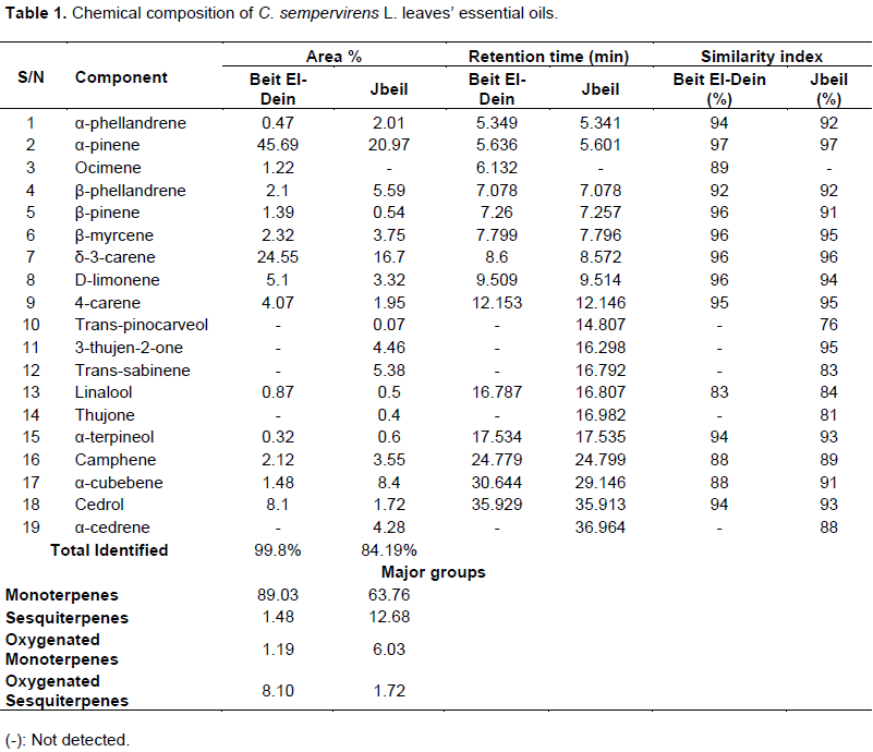
Many reports have been available on the chemical composition of essential oils obtained from C. sempervirens L. leaves.. The dominance of α-pinene and δ-3-carene was also observed in Tunisia (37.14 and 19.64%, respectively) (Boukhris et al., 2012), Saudi Arabia (48.6 and 22.1%, respectively) (Selim et al., 2014), and Egypt (29.21 and 18.92%, respectively) (Fayed, 2015). Furthermore, α-pinene (60.5%), cedrol (8.3%), myrcene (3.9%) and β-pinene (2.9%) were reported in the essential oil of C. sempervirens L. leaves in Algeria while δ-3-carene only accounted for 0.2% (Mazari et al., 2010). On the other hand, the chemical composition of essential oil extracted from Cypress leaves in Egypt showed the predominance of cedrol (23.68%), followed by ocimene (24%), α-pinene (21.15%) and α-cadinene (2.18%) (Ibrahim et al., 2009).
In general, such compositional differences have been proven to be mainly due to geographic, climatic, harvest season, and soil conditions (Fayed, 2015);in which all of these factors can cause fluctuations in the presence and in the relative abundance of the essential oil’s volatile constituents.
Phytochemical screening for the non-volatile portion of the ethanolic extracts
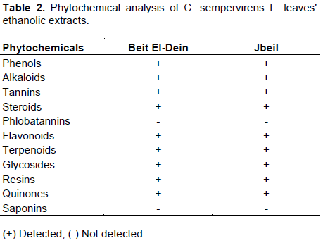
The presence of non-volatile components in the defatted ethanolic extracts of Cypress leaves from Beit El-Dein and Jbeil was investigated after maceration with 70% ethanol and prior to concentrating the extracts via the rotary evaporator. The extracts were examined qualitatively for the presence of phenols, alkaloids, terpenoids, flavonoids, saponins, tannins, steroids, phlobatannins, resins, quinones, and glycosides. As it can be seen from Table 2, similar results were obtained for both extracts. With the exception of saponins and phlobatannins, all other compounds were present in the corresponding extracts regardless the site from which the leaves were collected. These results are in accordance with other studies in which phenols (Ibrahim and El-seedi, 2007), flavonoids, tannins (Ahmad et al., 2006), alkaloids, and terpenoids (Preedy and Watson, 2014)were present in the defatted ethanolic extracts of C. sempervirens L. leaves. The presence of various phytochemicals in the tested plant renders it a good source of new drugs as most of these compounds have been reported to have therapeutic properties including anti-constipation, antifungal, and antibacterial activities (Shah, 2015).
Estimation of total phenol content in the ethanolic extracts
Phenolic compounds possess several health benefits as their antioxidant, anti-inflammatory, anticarcinogenic, and other biological activities render them capable to protect against oxidative stress and hence several diseases (Kabera et al., 2014). Due to their importance, the total phenol content (TPC) in the ethanolic extracts of C. sempervirens L. leaves was estimated according to Folin-Ciocalteu’s reagent method. The standard plot of gallic acid (y=0.0224x, R2=0.9907) was used, and the phenol content in the extracts was found to be 77.39 ±1.17 mg of gallic acid equivalent/g of sample dry weight and 66.43 ±1.06 mg of gallic acid equivalent/g of sample dry weight in Beit El-Dein and Jbeil samples, respectively. The high phenol content in both samples gives C. sempervirens L. a chance to be traced for the extraction of its valuable phytochemicals and development of several drugs.
Estimation of total flavonoid content in the ethanolic extracts
Flavonoids represent one of the most diverse and probably the most important group of natural phenols. Several biological activities including anti-allergic, anticancer, antioxidant, antiviral, and anti-inflammatory ones have been attributed to them (Kabera et al., 2014). Using the Aluminum Complexation Reaction method, the total flavonoid content (TFC) in the ethanolic extracts of C. sempervirens L. leaves was determined. The standard plot of rutin was used (y=0.0318x, R2=0.9952) and the values obtained were equal to 3.80 ±0.45 mg of rutin equivalent/g of sample dry weight and 2.83 ±0.70 mg of rutin equivalent/g of sample dry weight in Beit El-Dein and Jbeil samples, respectively.
The ability of flavonoids to relief high fever, eczema, asthma, sinusitis, and mainly heart diseases have been illustrated by several epidemiological studies (Kabera et al., 2014)thus making C. sempervirens L. able to participate in the treatment and prevention of such ailments with its moderate flavonoid content in its leaves.
In-vitro antioxidant activity of ethanolic extracts
DPPH-radical scavenging potential
The antioxidant capacity of C. sempervirens L. leaves’ ethanolic extracts was determined by applying the DPPH assay. In fact, the extracts of both Beit El-Dein and Jbeil samples remarkably reduced the DPPH free-radicals and were capable to transform its stable, purple color into yellow-colored DPPH-H upon electron abstraction form the antioxidant with an efficiency of IC50 respectively (Sarikurkcu et al., 2009).
The degree of discoloration; hence the decrease in the absorbance at 517 nm; indicates the free-radical scavenging potential of the samples tested. As it could be revealed form the results obtained, the free-radical scavenging potential of the samples tested exhibit a dose-dependent increase. The weakest DPPH scavenging activity was observed at the lowest concentration (100 μg/ml) with a value of 43.55 and 33.78% for Beit El-Dein and Jbeil samples, respectively. This activity increased gradually till reaching 95.05 and 93.56% at the highest concentration (1000 μg/ml) as mentioned, respectively. The ethanolic extracts of Beit El-Dein and Jbeil samples were able to reduce DPPH reaching 50% reduction with IC50 = 113.17 and 155.75 μg/ml, respectively. All the samples tested showed a lower DPPH radical scavenging activity when compared to ascorbic acid (IC50 = 4.1 μg/ml).
Many reports concerning the antioxidant activity of phenolic acids and flavonoids can be found in the literature. In addition, (Ibrahim and El-seedi, 2007)showed that the methanolic extract obtained from the leaves of C. sempervirens L. grown in Egypt has a high antioxidant activity with DPPH scavenging potential ranging from 95.80-99.75% for different isolated phenolic compounds. A significant amount of phenols and flavonoids have been detected in Lebanese Cypress extracts as revealed by TPC and TFC tests done. Therefore, these can be considered as the potential compounds contributing for the high antioxidant activity of C. sempervirens L. ethanolic leaves extracts. A positive correlation exists between the phenol and flavonoid content on one hand; and the antioxidant capacity on the other. The TPC and TFC of Beit El-Dein samples were relatively higher than those of Jbeil samples, which can attribute for its higher antioxidant capacity at different concentrations in one way or another.
Consequently, C. sempervirens L. leaves’ extract can act as a potential source of natural antioxidants used in food, pharmaceutical, and cosmetic industries where synthetic antioxidants being currently used have been found to possess adverse health effects (Miguel, 2010).
In-vitro antibacterial activity of the essential oils and ethanolic extracts
The in-vitro antibacterial activity of the essential oils and ethanolic extracts was evaluated against three bacterial strains using disc diffusion and micro-well dilution assays. The diameter of inhibitions zones (IZ) and the minimal inhibitory and minimal bactericidal concentrations (MICs and MBCs respectively) are represented in Tables 3 and 4.
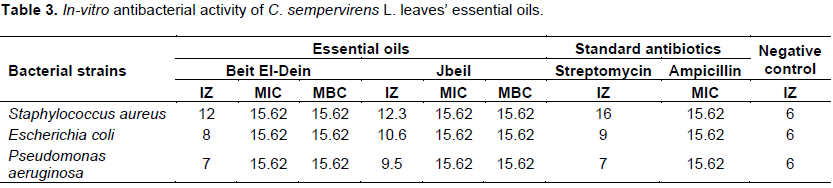
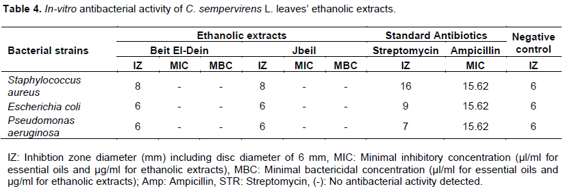
The results obtained show that the essential oils inhibited the growth of the bacterial strains tested with a diameter of inhibition zone ranging from 7-12 mm for Beit El-Dein and from 9.5-12.3 mm, for Jbeil samples. In both cases, S. aureus showed the highest susceptibility with a larger diameter of inhibition zone (12-12.3 mm for Beit El-Dein and Jbeil samples, respectively) and the lowest MIC (15.62 μg/ml). P. aeruginosa showed the highest resistance in both samples with lowest diameters of inhibition zone (7-9.5 mm, respectively). The results obtained regarding relative bacteria susceptibility are consistent with other studies (Ahmad et al., 2006; Mazari et al., 2010; Selim et al., 2014; Boukhris et al., 2012). The ethanolic extracts showed a much lesser antibacterial activity compared to the essential oils with an effect only on S. aureus with a diameter of inhibition zone equal to 8 mm in both Beit El-Dein and Jbeil samples. However, the methanolic extract of C. sempervirens L. leaves showed a mild yet a higher antibacterial activity compared to that of the essential oil in a study done in Egypt (Selim et al., 2014).
The antibacterial activity of both the essential oils and ethanolic extracts was more pronounced towards Gram-positive than Gram-negative bacteria. The results obtained regarding the essential oil are in accordance with other studies (Ahmad et al., 2006; Mazari et al., 2010; Selim et al., 2014; Boukhris et al., 2012). Generally, the higher resistance observed in the Gram-negative bacteria could be related to the presence of an outer phospholipidic membrane that is almost impermeable to lipophilic compounds. As a result, the absence of this barrier in the case of Gram-positive bacteria renders them much more susceptible due to the direct contact between the components of the essential oil with their phospholipidic bilayer cell membrane (Selim et al., 2014; Boukhris et al., 2012). The synergistic effect of the essential oil’s various constituents where α-pinene, β-phellandrene, and cedrol seem to play the major role in the antibacterial activity of the essential oils (Mazari et al., 2010; Selim et al., 2014). On the other hand, the antibacterial activity of the ethanolic extracts could be associated with phenolic compounds capable of forming complexes with extracellular and soluble proteins, in addition to bacterial cell wall peptides and membrane-bounded enzymes hence disrupting the latter (Cowan, 1999).
CONCLUSION
This work has reported for the first time the chemical composition as well as the antioxidant and antibacterial potentials of essential oils and ethanolic extracts obtained from the leaves of the Lebanese C. sempervirens L. As the study revealed, these can be used as natural therapeutic antioxidant and antibacterial agents to prevent or slow down diseases related to oxidative stresses and microbial growth. These promising results obtained by the in-vitro analysis motivate us to further investigate the tested activities of essential oils and ethanolic extracts in-vivo as such studies provide more convincing evidences and confirmations for their corresponding biological potentials taking into consideration any possible toxicity. Also, other studies including antifungal, anticancerous and insecticidal effects can be done. Comparing the in-vitro antioxidant potential of the essential oils to that of the ethanolic extracts could be suggested in future studies. However, the present findings could be considered as useful data which can in turn provide the Lebanese Cypress a potential pharmaceutical role.
CONFLICT OF INTERESTS
The authors have not declared any conflict of interests.
REFERENCES
|
Ahmad S, Asili J, Rahimizadeh M, Fazly-bazzaz BS (2006). Chemical and antimicrobial studies of Cupressus sempervirens L . and C . horizentalis Mill . essential oils. Iranian Journal of Pharmaceutical Sciences 2(2):103-108. |
|
|
Boukhris M, Regane G, Yangui T, Sayadi S, Bouaziz M, (2012). Chemical composition and biological potential of essential oil from Tunisian Cupressus sempervirens L. Journal of Arid Land Studies 332:329-332. |
|
|
Cowan MM (1999). Plant products as antimicrobial agents. Clinical Microbiology Reviews 12(4):564-582. |
|
|
DÄʾirat al-Ará¹£Äd al-JawwÄ«yah al-LubnÄnÄ«yah (1977). Atlas climatique du Liban. Publié par le service météorologique du Liban avec l'aide de l'observatoire de Ksara. 2éme Edition. |
|
|
Deeb T, Knio K, Shinwari ZK, Kreydiyyeh S, Baydoun E (2013). Survey of medicinal plants currently used by herbalists in Lebanon. Pakistan Journal of Botany 45(2):543-555. |
|
|
Elansary HO, Salem MZM, Ashmawy NA, Yacout MM (2012). Chemical composition ,antibacterial and antioxidant activities of leaves essential oils from Syzygium cumini L., Cupressus sempervirens L. and Lantana camara L. from Egypt. Journal of Agricultural Sciences 4(10):144-152. |
|
|
Farhan H, Rammal H, Hijazi A, Badran B (2012). Preliminary phytochemical screening and extraction of polyphenol from stems and leaves of a Lebanese plant Malva parviflora L. International Journal of Current Pharmaceutical Research 4(1):1-5. |
|
|
Fayed SA (2015). Chemical composition, antioxidant, anticancer properties and toxicity evaluation of leaf essential oil of Cupressus sempervirens. Notulae Botanicae Horti Agrobotanici Cluj-Napoca 43(2):320-326. |
|
|
Gopinath SM, Rakesh CK, Murthy TPN, Dayananda KS (2012). Preliminary phytochemical evaluation of leaf extracts of Gymnema. International Journal of Pharmacognosy and Phytochemical Research 4(3):109-111. |
|
|
Ibrahim NA, Mohammed MMD, El-kom S (2009). Chloroform extract and essential oil of Cupressus sempervirens. Chemistry of Natural Compounds 45(3):265-268. |
|
|
Ibrahim NA, El-seedi HR (2007). Phytochemical investigation and hepatoprotective activity of Cupressus sempervirens L . leaves growing in Egypt. Natural Product Research: Formerly Natural Product Letters 21(10) 37-41. |
|
|
Jamshidi-Kia F, Lorigooini Z, Amini-Khoei H (2018). Medicinal plants: past history and future perspective. Journal of Herbmed Pharmacology 7(1):1-7. |
|
|
Kabera JN, Semana E, Mussa AR, He X (2014). Plant secondary metabolites: biosynthesis, classification, function and pharmacological properties. Journal of Pharmacy and Pharmacology 2:377-392. |
|
|
Kavit M, Patel BN, Jain BK (2013). Phytochemical analysis of leaf extract of Phyllanthus fraternus. Research Journal of Recent Sciences 2:12-15 |
|
|
Mazari K, Bendimerad N, Bekhechi C, Fernandez X (2010). Chemical composition and antimicrobial activity of essential oils isolated from Algerian Juniperus phoenicea L. and Cupressus sempervirens L. Journal of Medicinal Plant Research 4(10):959-964. |
|
|
Miguel MG (2010). Antioxidant activity of medicinal and aromatic plants: A review. Flavour and Fragnance Journal, pp. 291-312. |
|
|
Nehdi IA (2013). Cupressus sempervirens var . horizentalis seed oil : Chemical composition, physicochemical characteristics and utilizations. Industrial Crops and Products 41:381-385. |
|
|
Nouri A, Dhifi W, Bellili S, Ghazghazi H, Aouadhi C, Chérif A, Hammami M, Mnif W (2015). Chemical composition, antioxidant potential, and antibacterial activity of essential oil cones of Tunisian Cupressus sempervirens. Hindawi Publishing Corporation. Journal of Chemistry Volume 2015, Article ID 538929, 8p. |
|
|
Preedy VR, Watson RR (2014). The Mediterranean diet: an evidence-based approach. 1st Edition. Academic Press. |
|
|
Sarikurkcu C, Arisoy K, Tepe B, Cakir A, Abali G, Mete E (2009). Studies on the antioxidant activity of essential oil and different solvent extracts of Vitex agnus castus L . fruits from Turkey. Food and Chemical Toxicology 47(10):2479-2483. |
|
|
Selim SA, Adam ME, Hassan SM, Albalawi AR (2014). Chemical composition , antimicrobial and antibiofilm activity of the essential oil and methanol extract of the Mediterranean Cypress (Cupressus sempervirens L.). BMC Complementary and Alternative Medicine 14(1):1-8. |
|
|
Shah RK (2015). Qualitative phytochemical analysis and estimation of total phenols and flavonoids in leaf extract of Sarcochlamys Pulcherrima Wedd.. Global Journal of Bioscience and Biotechnology 4(1):81-84. |
|
Copyright © 2024 Author(s) retain the copyright of this article.
This article is published under the terms of the Creative Commons Attribution License 4.0


