Full Length Research Paper
ABSTRACT
Punica granatum, specifically the fruit, has a long ethno medical history and is a phytochemical reservoir of great medicinal value. The phytochemistry and pharmacological actions of all P. granatum components suggest a wide range of clinical applications. The aim of the present study is to investigate the anticancer potential of aqueous extract of P. granatum (AEPG) in experimental models. The chemical composition of the AEPG was assessed by HPLC-DAD. In vivo antitumor activity was assessed in sarcoma 180 bearing mice. To evaluate the toxicological aspects related to the AEPG treatment, hematological, biochemical, histopathological and morphological analyses of treated animals were performed. Gallic acid, punicalagin α, punicalagin β, and ellagic acid were identified as the major phytochemical compounds of the extract. AEPG and 5-fluorouracil (5-FU) induced significant inhibition of tumor growth when compared with saline (p < 0.05). The percentage of apoptotic cells was significantly increased in 5-FU (p < 0.01) and AEPG treated groups (p < 0.01). No significant difference was observed between 5-FU and the three doses of AEPG. 5-FU induced toxic effects, such as decrease of body weight, splenic atrophy, and leukopenia, but these effects were not found in AEPG treated groups. The results provide evidence that AEPG exhibits comparable antitumor effects as 5-FU in a murine model, likely the result of increased apoptotic rate, but with no remarkable side effects presented by 5-FU.
Key words: Cancer, chemotherapy, Punica granatum, sarcoma 180.
INTRODUCTION
Cancer is a disease characterized by uncontrolled multiplication of subtly modified normal human cells (Mubeen et al., 2012). An exceptionally difficult problem in cancer treatment is multidrug resistance, when cancer cells lose their sensitivity to multiple structurally different chemotherapeutics, leading to the search for alternative treatments, such a medicinal plants (Amaral et al., 2015). Plants have a long history of use in the treatment of cancer and the interest in nature as a source of potential chemotherapeutic agents continues. The present day research and development tailored towards the discovery of new antiproliferative agents from natural products have been buoyed by improvement in the science and technology of anticancer drug discovery (Akindele et al., 2015).
Punica granatum is a deciduous tree belonging to the family Punicaceae and contains hydrolysable tannins as major active chemical constituents, that is, punicalagin, punicalin, gallic acid, ellagic acid, and ellagic acid derivative (Akhtar et al., 2015; Sing et al., 2018). There is extensive literature about bioactive compounds from its fruit (pomegranate), showing important biologic activities, such as healing (Zekavat et al., 2016), antimicrobial (Mohammad et al., 2016), chemopreventive (Bishayee et al., 2011) and antitumor effects (Panth et al., 2017).
Several studies have demonstrated that natural compounds such as tannins have a wide variety of biologic functions that are mainly related to modulation of carcinogenesis and antiproliferative effects, such as antioxidant and proapoptotic activities (Dilkmen et al., 2011). Furthermore, these compounds are generally safe, with low toxicity, and receive general acceptance (Fresco et al., 2006).
Apoptosis plays an important role in elimination of tumor cells by chemotherapeutic agents (Hassan et al., 2014). According to Dai and Mumper (2010) apoptosis-inducing compounds are expected to be ideal anticancer drugs due to their ability to promote DNA damage in tumor cells, which are then rapidly recognized by macrophages and removed without inducing an inflammatory response.
In vitro studies have already described the positive results of treatment with P. granatum extract, with apoptosis in many cell lines, such as colon cells-SW620, HT-29, and HCT-116 (Joseph et al., 2013), prostate cells-DU145, PC3, mouse prostate cancer cell TRAMP-C1 (Deng et al., 2017; Deng et al., 2018), lung cells-A549, H1299 (Li et al., 2016), and breast cells-MCF-7 (Shirode et al., 2014; Chen et al., 2015). However, in these models, the systemic effect of these compounds could not be assessed.
The aim of this study was to evaluate the antitumor activity of the aqueous extract of P. granatum (AEPG) in mice transplanted with sarcoma 180. Hematological, biochemical, histopathological and morphological analyses of the tumor and the organs, including liver, spleen and kidney, were performed to evaluate the toxicological aspects of the treatment.
MATERIALS AND METHODS
Plant
Fruits of P. granatum were collected in Petrolina, PE, Brazil (09°23’34”S, 40°30’28”W) in September 2011. Samples were identified and a voucher specimen (ASE 20881) has been deposited in the herbarium of the Department of Biology, Federal University of Sergipe, São Cristóvão, Sergipe, Brazil.
Extraction procedure and sample preparation
The fruits were washed with tap water and pulp samples were obtained. The peel was dried at 55°C and uniformly powdered. The extraction was carried out by dynamic maceration using boiling water 1:100 (w/v) as a solvent for 2 h. The suspension was filtered and the solvent was removed in a circulating air stove at 50 ± 5°C for 48 to 72 h. The percentage of extraction yield was 63.4%, calculated in terms of dry weight.
For HPLC analysis, the crude extract of P. granatum (AEPG) was solubilized in a mixture of water: methanol (1:1 v/v) (1 mg/mL), filtered through a 0.45 µm membrane (Millipore, Merck-Billerica, MA, USA) and an aliquot of 10 μL was injected into the chromatographic system.
Apparatus and chromatographic conditions
The HPLC analyses were performed on a Shimadzu liquid chromatograph (Tokyo, Japan), equipped with a LC-6AD pump, an SPD-M20A diode array detector (DAD), and operated with the LC Solution data station software (Shimadzu, Tokyo, Japan). The water used in experiments was obtained with the Millipore (São Paulo, Brazil) Milli-Q purification system. Analysis was carried out on the analytical C18 Luna column (250 × 4.6 mm, 5 µm, Phenomenex, Torrance, CA, USA) with the following conditions: flow rate 1 mL/min and mobile phase consisting of 0.1% aqueous phosphoric acid (v/v, A) and acetonitrile (B). The gradient program was: 1 to 5% B at 0 to 5 min, 5 to 8% B at 5 to 10 min, 8% B at 10 to 16 min, 8 to 25% B at 16 to 22 min, 25 to 90% B at 22 to 27 min, 90 to 1% B at 27 to 33 min. The chromatogram was monitored at 260 nm. Quantification was achieved using the linear calibration curves of gallic acid (1 to 10 µg/mL) and ellagic acid (1 to 5 µg/mL) standards.
In vivo antitumoural assay
Animals
Sixty Swiss mice (male, 20 ± 2 g) were obtained from the central biotery of the Tiradents University (Aracaju, Brazil). The animals were housed in cages with free access to food and water. All animals were maintained under controlled temperature (25 ± 2°C) and relative humidity (50 ± 5%), with a 12h:12 h light-dark cycle (lights on at 6:00 a.m.). The experiments were conducted after approval of the protocols by the Institutional Ethics Committee (021113) of the Tiradents University (Aracaju, Brazil) and were carried out in accordance with the current guidelines for the care of laboratory animals.
Determination of the effect of the AEPG on the growth of solid tumors in mice
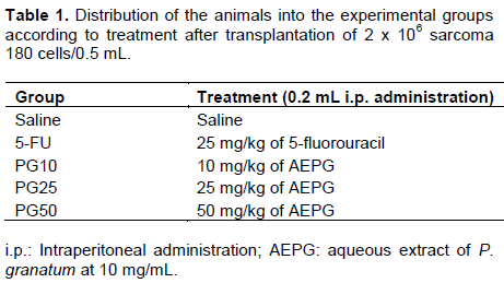
The in vivo antitumor effect was evaluated using sarcoma 180 ascites tumor cells according to the method described by Bezerra et al. (2006). Ten-day-old sarcoma 180 ascites tumor cells (2 × 10⁶ cells per 500 µL) were implanted subcutaneously into the left hind groin of the experimental mice. One day after inoculation, the AEPG was suspended in saline (vehicle) at final concentration of 10 mg/mL and administered intraperitoneally (10, 25 and 50 mg/kg) once a day for seven consecutive days. The negative control was injected with saline solution and the positive control was injected with 5-fluorouracil (5-FU, purity > 99%; Sigma Chemical Co., 25 mg/kg). At the beginning of the experiment, the mice were divided into five groups (n = 12 animals/group) shown in Table 1. Body weight and food and water intakes were measured daily over the time course of the experiment. On the 8th day, peripheral blood samples were collected from the orbital plexus of the mice while under light ether anesthesia and submitted to further hematological and biochemical analyses. The animals were then sacrificed in a CO2 chamber. The tumors, livers, spleens, and kidneys were excised, weighed, and examined for morphology. Then, they were fixed in 10% formaldehyde for histological analysis. The inhibition ratio (%) was calculated by the following formula: inhibition ratio (%) = ((A – B) / A) × 100, where A is the average tumor weight of the vehicle group and B is the average tumor weight of the treated group.
Systemic toxicology analysis
Determination of the effect of the AEPG on body and organ weight: Body weights were determined at the start and on the last day of treatment, and the animals were observed for signs of abnormalities throughout the study.
Tumor, livers, kidneys and spleens were dissected, weighed and observed for any signs of gross lesions or color changes and hemorrhages.
Determination of the effect of the APEG on biochemical parameters: After fasting for 6 to 8 h, the animals were submitted to blood collection from the orbital plexus for biochemical analysis (urea and creatinine to investigate any renal function alterations; AST and ALT as liver parameter). The analysis was carried out in semi-automatic equipment (Bioplus 200®), using enzymatic colourimetric kits (Labtest®).
Determination of the effect of the APEG on hematological parameters: After fasting for 6 to 8 h, the animals were submitted to blood collection from the orbital plexus for hematological analyses. To determine hematological parameter, an automated blood cell counter was used (Sysmex America, Inc., USA). The total count as well as differential counts of leukocytes, including eosinophils, lymphocytes, neutrophils and monocytes was performed using optical light microscopy after staining with Pappenheim's method.
Histopathology and morphological observations
The tumors, spleens, liver and kidneys were fixed in 10% formaldehyde (pH 7.4), dehydrated in alcohol, and diaphanized in xylene and paraffin-embedded.
Subsequently, 7 μm thick histological sections were obtained and stained with hematoxylin and eosin. Histological analyses were performed under light microscopy.
Terminal deoxyuridine nick-end labeling (TUNEL) staining
The number of apoptotic cells was assessed by the TUNEL technique described by Woodside et al. (2003). Histological sections (7 μm thick, n = 3) were obtained from the paraffin-embedded tissue and incubated using an in situ cell death detection kit, POD (Roche Diagnostics, Indianapolis, IN, USA). At first, the sections were deparaffinized in xylene (three changes at 3 min intervals with air-drying in between each change for better section adherence), rehydrated in graded alcohol (99, 95 and 70%) for 3 min each, and washed with deionized water. Then, the samples were treated with proteinase K (20 μl/ml in PBS) to digest the proteins, and endogenous peroxidase activity was quenched with 2% H2O2 in PBS for 10 min at room temperature. Thereafter, sections were washed with 50 μl PBS buffer, diluted TdT enzyme solution was applied, and the sections were incubated at 37°C in a humidified chamber for 1 h. After incubation, the sections were washed again with PBS buffer. Subsequently, 50 μl of antidigoxygenin peroxidase was added, and the sections were incubated in a humidified chamber for 30 min at room temperature. Once more, the sections were washed with PBS, and diaminobenzidine (DAB)-hydrogen peroxide was used for color development. For negative controls, the TdT enzyme was replaced with PBS on one section on each slide and was processed in parallel. Counterstaining of nuclei was performed with 2% Meyer’s hematoxylin and mounted for examination. Apoptotic cells were identified as cells with brown-stained nuclei or as apoptotic bodies (fragments of apoptotic cells engulfed by neighboring cells). The number of TUNEL-positive cells was determined in 1000 counted cells, and the apoptosis ratio (AR) was calculated according to the following equation: AR (%) = (TnP/1000) × 100, where AR is the apoptosis ratio and TnP is the number of TUNEL-positive cells.
Statistical analysis
Results are expressed as mean ± standard error of the mean (SEM) or as a percentage of the saline group. Subsequently, data were assessed by one-way analysis of variance (ANOVA) followed by post hoc Tukey-Kramer multiple comparison test. The values at p < 0.05 were considered to be statistically significant.
RESULTS
HPLC-DAD analysis
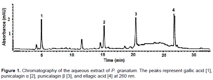
The HPLC-DAD analysis of the AEPG revealed the presence of four major peaks (Figure 1) which were identified according to retention times, UV and comparison with authentic samples as gallic acid [1], punicalagin α [2], punicalagin β [3], and ellagic acid [4]. The ellagic and gallic acids were quantified using external standard method, which presented concentrations of gallic acid and ellagic acid in the sample were 32.24 and 41.67 mg/g, respectively.
In vivo anti-tumor activity of the AEPG
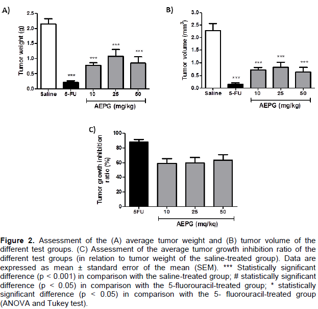
The effects of the AEPG on mice transplanted with sarcoma 180 tumors cells are as shown in Figure 2. As shown in Figure 2A and B, the tumor weight and volume of 5-FU-treated animals were significantly lower than those of the saline group (p < 0.001). Similarly, intraperitoneal administration of AEPG (10, 25, and 50 mg/kg) also reduced significantly the average weight and volume of the tumors (p < 0.001). The reductions of tumor weight and volume obtained with the treatment with AEPG at 10 and 50 mg/kg were statistically similar to that obtained with 5-FU (p > 0.05). Moreover, there was no significant difference in the average tumor weight and volume between the AEPG-treated groups (p > 0.05). No significant difference in the tumor growth IR was observed between 5-FU and treatment with 10 and 50 mg/kg AEPG (p > 0.05) (Figure 2C). However, the IR for 25 mg/kg AEPG was significantly lower than that for 5-FU (p < 0.05). Assessment of the average tumor weight and tumor growth IR indicated that the response of sarcoma 180 growth to treatment with AEPG was not dose-dependent.
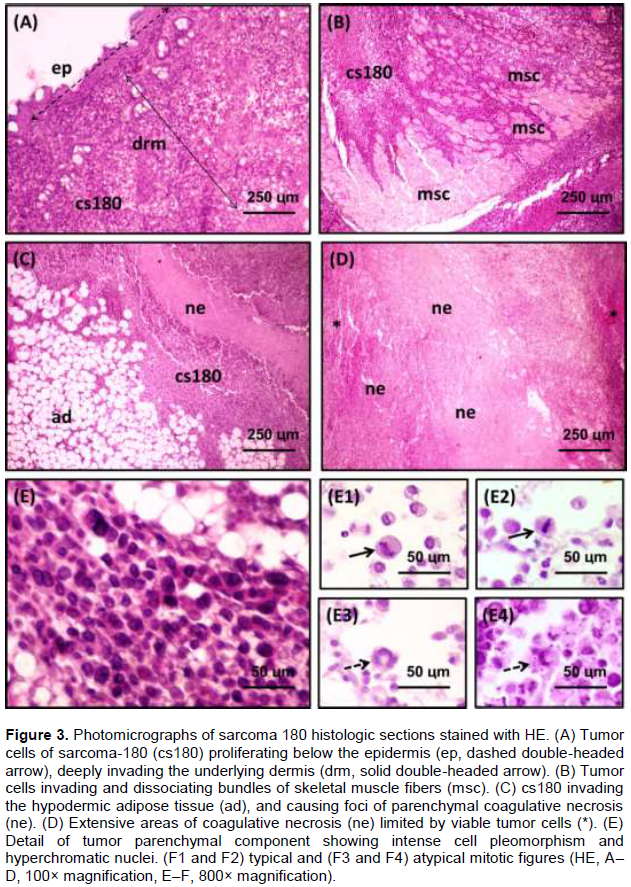
Post-mortem analysis of the tumors revealed similar histopathologic features in all groups (Figure 3). The tumors were characterized by neoplastic sheets of small polygonal and ovoid cells compactly arranged in some areas but loosely disposed in others. Tumor cells often invaded and dissociated lobules of adipose tissue and striated skeletal muscle bundles. Most of the tumor cells exhibited strongly eosinophilic cytoplasm and round-shaped hyperchromatic and moderately pleomorphic nuclei, but sometimes the nuclear chromatin was disperse and presented prominent nucleoli. Typical and atypical mitotic figures were often found (2 to 3 mitoses/histologic field at 400× magnification) among the neoplastic parenchyma. In addition, extensive areas of coagulative necrosis and a mild to moderate inflammatory response composed of lymphocytes andneutrophils were also observed. Vascular and perineural invasion were rare histologic findings in all groups, regardless of the treatment applied to the animals.
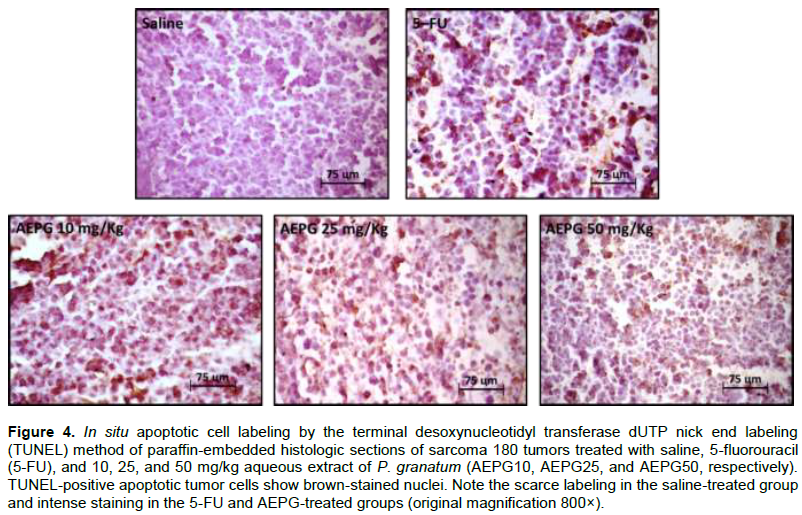
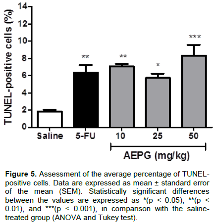
Apoptotic cell death was also detected using TUNEL assay. Positive labeling was identified by brownish color of the nucleus, regardless of the intensity of the staining (Figure 4). TUNEL-positive cell count varied considerably in all groups (Figure 5), but the average percentage was significantly increased in the groups treated with 5-FU (p < 0.01) and AEPG at the doses of 10 mg/kg (p < 0.01), 25 mg/kg (p < 0.05), and 50 mg/kg (p < 0.001). No significant difference was observed either between 5-FU and the three doses of AEPG or between the groups treated with AEPG.
Systemic toxicological evaluation
No behavioral changes were observed in the animals treated either with AEPG or 5-FU, and no remarkable changes in the intake of food and water were observed, regardless of treatment. Figure 6A shows the body weight of the animals at the beginning and end of the experiment. No significant differences in body weight between the groups were observed at the beginning of the experiment (p > 0.05), but at the eighth day, the body weight of the mice treated with 5-FU was significantly lower than that of the other groups (p < 0.05). Furthermore, as shown in Figure 6B, the average percentage of weight loss (in relation to the initial body weight) was significantly greater in the group treated with 5-FU than in the others (p < 0.05).
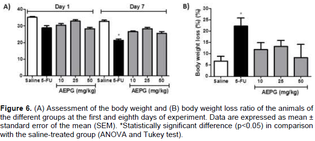
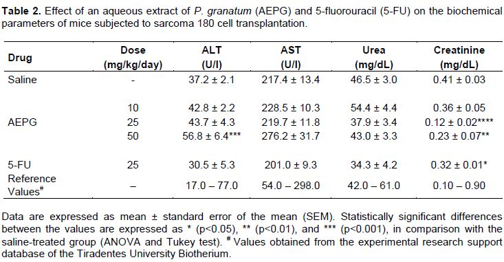
Table 2 shows the results of the biochemical parameters. Serum levels of aspartate aminotransferase (ALT) significantly increased in the group treated with 50 mg/kg AEPG in comparison with the saline-treated group (p < 0.001). Creatinine levels were significantly decreased in the groups treated with 25 mg/kg AEPG (p < 0.001) and 50 mg/kg AEPG (p < 0.01), as well as in the 5-FU-treated group (p < 0.05), again in comparison with the group treated with saline. Despite the significant differences found in these data, the values were within the normal interval established by the experimental research support database of the Biotery of the Tiradentes University.
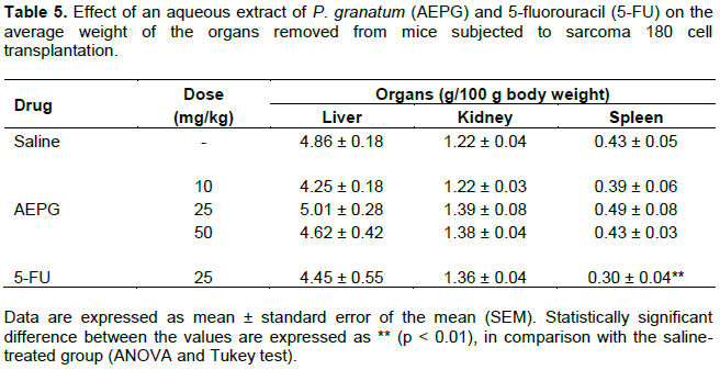
Analysis of the hematologic parameters revealed no significant changes in the erythrogram data (Table 3, p > 0.05). However, as shown in Table 4, treatment with 5-FU induced a significant decrease in the total leukocyte count (p < 0.01) and relative neutrophil count (p < 0.001).
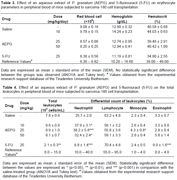
In addition, a relative increase in the differential count of eosinophils was observed (p < 0.001). In contrast, the administration of AEPG induced an increased neutrophil count at the three doses of 10 mg/kg (p < 0.05), 25 mg/kg (p < 0.001), and 50 mg/kg (p < 0.05), without causing expressive leukocytosis.
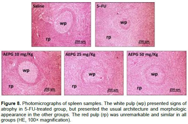
Pathological examination of the organs removed on a gross basis revealed that livers and spleens of the 5-FU-treated group showed an opaque surface tissue that was not present in the other groups, but no gross difference was detected between the experimental groups regarding the shape and consistency. Furthermore, a significant reduction of the average weight of the spleen in relation to that of the saline group was observed in the 5-FU-treated animals (p < 0.05), but not in the AEPG groups (p > 0.05) (Table 5).
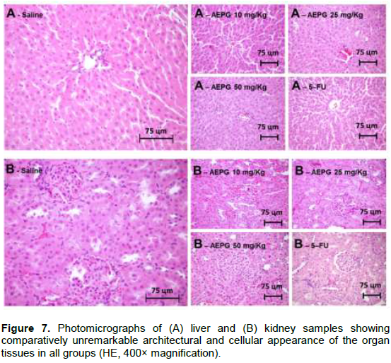
Histologic analysis revealed that the architectural and cellular appearances of the organ tissues were comparatively unremarkable in all groups (Figure 7), except for the spleen samples from the 5-FU-treated group, which displayed atrophy of the white pulp (Figure 8).
DISCUSSION
Natural products play a relevant role in cancer therapy, with a substantial number of natural anticancer agents (Rayan et al., 2017; Blowman et al., 2018; Cui et al., 2018). Furthermore, because cancer treatments have become more aggressive during the last 20 years, the need for new methods to manage adverse and/or side effects of such therapy has become apparent (Redd et al., 2001).
In this study, treatment with an aqueous extract of P. granatum at doses of 10, 25, and 50 mg/kg significantly inhibited the growth of sarcoma 180 tumors in mice, but not in a dose-dependent manner. These data suggest antitumor activity of the extract, which is similar to the effects of 5-FU, a chemotherapeutic agent widely used in experimental models (Mousinho et al., 2011). Therefore, this study provides data indicating the promise of AEPG in anticancer therapy, which is consistent with previous in vitro research (Jayakumar and Haridass, 2012; Joseph et al., 2013). It is possible that such antitumor effects are related to the chemical compounds present in the extract. It has been demonstrated that phytochemical constituents found in P. granatum present cytotoxic effects on tumor cell lines, particularly tannic compounds such as ellagic acid (Zhao et al., 2013; Zahin et al., 2014; Zhang et al., 2014), gallic acid (Liang et al., 2012; Locatelli et al., 2013) and punicalagin (Zahin et al., 2014). As reported by Qu et al. (2012), high contents of these tannic compounds were found in the AEPG using the HPLC-MS method, suggesting that ellagic and gallic acids may play an important role in the inhibition of murine sarcoma 180 growth. The reason why the antitumor activity was not dose-dependent is not fully understood, but it is possible that all receptors activated by the chemical compounds present in the EAPG are already saturated at 25 mg/kg, and therefore increasing it provided no additional biological effect. However, further investigations are necessary to clarify the precise biochemical mechanisms underlying these biological effects. In addition, as the effect is not dose-dependent, and therefore does not require dose adjustments to body weight.
Since tissue homeostasis is the result of the balance between proliferation and cell death, the apoptotic rate plays a key role in tumor formation and progression (Evan and Vousden, 2001; Fulda, 2009).
Apoptosis is the process of highly controlled programmed cell death triggered by intrinsic biochemical signaling pathways. The mechanisms of apoptosis involve an energy-dependent cascade of molecular events that includes activation of cysteine proteases such as interleukin-1β-converting enzyme (ICE), Fas signaling, cell cycle interfaces, stress responses, the B-cell lymphoma 2 family, and the tumor suppressor gene p53.
This process leads to the cleavage of caspase-3 and results in DNA fragmentation, degradation of cytoskeletal and nuclear proteins, cross-linking of proteins, formation of apoptotic bodies, expression of ligands for phagocytic cell receptors, and finally uptake by phagocytic cells, with no secondary inflammation (Elmore, 2007).
Thus, to explore the antitumor effect of AEPG on sarcoma 180 tumor in the present study, apoptosis was detected by in situ TUNEL staining. It was found that treatment with AEPG significantly increased the number of TUNEL-positive cells in comparison with saline control. The increased number of TUNEL-positive cells observed in AEPG-treated groups was statistically comparable to that resulting from 5-FU treatment. Thus, the results strongly suggest that the antitumor effects of the extract are related to increased apoptosis-mediated tumor cell death. It is possible that the major tannic compounds present in the extract, such as ellagic and gallic acids, are involved in the proapoptotic effects of AEPG. It has been demonstrated that ellagic acid is able to stimulate apoptosis in poorly differentiated MIAPaCa-2 and moderately differentiated PANC-1 human pancreatic carcinoma cell lines, as a response of inhibition of the transcription factor NF-κB. The decrease in NF-κB leads to activation of the mitochondrial proapoptotic pathway, resulting in cytochrome C release and caspase activation (Edder Kaoui et al., 2008).
In addition, gallic acid has been shown to induce apoptosis of HL-60 human promyelocytic leukemia cells (Yeah et al., 2011) and A375.S2 human melanoma cells (Lo et al., 2010) through caspase-dependent and -independent pathways. However, further investigations are necessary in order to clarify the precise mechanisms underlying the proapoptotic effect of AEPG on tumor cells.
One of the most important challenges regarding chemotherapy against cancer are the minimization of the adverse/side effects of the drugs. As previously demonstrated by Gonzaga et al. (2009), treatment with 5-FU promotes a variety of undesirable adverse effects, such as body weight loss, severe myelosuppression, and spleen atrophy.
Herein, no remarkable changes were observed in the total leukocyte count of the AEPG-treated groups, indicating no suppressive effects on peripheral blood white cells. In fact, AEPG induced the increase of neutrophils, suggesting that the extract might exert a possible stimulatory effect on the bone marrow. In addition, although significant differences were observed in biochemical parameters of liver and renal function in the AEPG-treated groups, all the serum values remained within the physiologic reference range, suggesting that those changes might be considered irrelevant. The fact that the weight, gross appearance, and histologic features of the organs in the AEPG treated groups were unremarkable and comparable to those of the saline group seems to support the hypothesis that the biochemical changes were not severe enough to cause real functional damage. Thus, as these hematologic, biochemical, and gross/histologic parameters have been used to assess the toxicity of P. granatum fruit extracts (Vidal et al., 2003), the present data seem to point to the safety of AEPG at the studied doses.
CONCLUSION
This study demonstrated that intraperitoneal admini-stration of AEPG inhibited the growth of transplanted sarcoma 180 cells in a murine model, and that the antitumor effects were likely related to increased apoptosis rates. In addition, the use of the extract was proven to be safe, with none of the adverse/side effects associated with the use of chemotherapeutics.
CONFLICT OF INTERESTS
The authors have not declared any conflict of interests.
ACKNOWLEDGMENTS
The authors thank the Research and Technology Innovation Support Foundation of Sergipe, Brazil (FAPITEC/SE) that supported this research. RLCAJ, SMT, and FFP are recipients of CNPq productivity grants. TSB received scholarship from Brazilian Coordination for Improvement of Higher Education Staff (CAPES) for the financial support.
REFERENCES
|
Akhtar S, Ismail T, Fraternale D, Sestili P (2015). Pomegranate peel and peel extracts: Chemistry and food features. Food Chemistry 174: 417–425. |
|
|
Akindele AJ, Wani ZA, Sharma S, Mahajan G, Satti NK, Adeyemi OO, Mondhe DM, Saxena AK (2015). In vitro and in vivo anticancer activity of root extracts of Sansevieria liberica Gerome and Labroy (Agavaceae). Evidence Based Complementary and Alternative Medicine. |
|
|
Amaral RG, Fonseca CS, Silva TKM, Andrade LN, França ME, Barbosa-Filho JM, Sousa DP, Moraes MO, Pessoa CO, Carvalho AA, Thomazzi SM (2015). Evaluation of the cytotoxic and antitumour effects of the essential oil from Mentha x villosa and its main compound, rotundifolone. Journal of Pharmacy and Pharmacology 67(8):1100-1106 |
|
|
Bezerra DP, Castro FO, Alves APNN, Pessoa C, Moraes MO, Silveira ER, Lima MAS, Elmiro FJM, Costa-Lotufo LV (2006). In vivo growth-inhibition of sarcoma 180 by piplartine and piperine, two alkaloid amides from Piper. Brazilian Journal of Medical and Biological Research 39(6):801-807. |
|
|
Blowman K, Magalhães M, Lemos MFL, Cabral C, Pires IM (2018). Anticancer Properties of Essential Oils and Other Natural Products. Evidence-Based Complementary and Alternative Medicine. |
|
|
Chen HS, Bai MH, Zhang T, Li GD, Liu M (2015). Ellagic acid induces cell cycle arrest and apoptosis through TGF-beta/Smad3 signaling pathway in human breast cancer MCF-7 cells. International Journal of Oncology 46(4):1730-1738. |
|
|
Cui Q, Yang DH, Chen ZS (2018). Special Issue: Natural Products: Anticancer and Beyond. Molecules 23:23. |
|
|
Dai J, Mumper RJ (2010). Plant Phenolics: Extraction, Analysis and Their Antioxidant and Anticancer Properties. Molecules 15(10):7313-7352. |
|
|
Deng Y, Li Y, Yang F, Zeng A, Yang S, Luo Y, Zhang Y, Xie Y, Ye T, Xia Y, Yin W (2017). The extract from Punica granatum (pomegranate) peel induces apoptosis and impairs metastasis in prostate cancer cells. Biomedicine and Pharmacotherapy 93:976-984. |
|
|
Deng YL, Li YL, Zheng TT, Hu MX, Ye TH, Xie YM, Yin WY (2018). The Extract from Punica Granatum (Pomegranate) Leaves Promotes Apoptosis and Impairs Metastasis in Prostate Cancer Cells. Sichuan da xue xue bao. Yi xue ban= Journal of Sichuan University. Medical science edition 48(1):8-12. |
|
|
Edderkaoui M, Odinokova I, Ohno I, Gukovsky I, Go VL, Pandol SJ, Gukovskaya AS (2008). Ellagic acid induces apoptosis through inhibition of nuclear factor kB in pancreatic cancer cells. World Journal of Gastroenterology: WJG 14(23):3672. |
|
|
Elmore S (2007). Apoptosis: a review of programmed cell death. Toxicologic Pathology 35(4):495-516. |
|
|
Evan GI, Vousden KH (2001). Proliferation, cell cycle and apoptosis in cancer. Nature 411(6835):342. |
|
|
Fresco P, Borges F, Diniz C, Marques MP (2006). New insights on the anticancer properties of dietary polyphenols. Medicinal Research Reviews 26(6):747-766. |
|
|
Fulda S (2009). Apoptosis pathway and their therapeutic explotation in pancreatic cancer. Journal of Cellular and Molecular Medicine 13(7):1221-1227. |
|
|
Gonzaga ML, Bezerra DP, Alves AP, de Alencar NM, Mesquita Rde O, Lima MW, Soares Sde A, Pessoa C, de Moraes MO, Costa-Lotufo LV (2009). In vivo growth-inhibition of Sarcoma 180 by an (1→4)-glucan– b-(1→6)-glucan-protein complex polysaccharide obtained from Agaricus blazei Murill. Journal of Natural Medicines 63(1):32-40. |
|
|
Hassan M, Watari H, AbuAlmaaty A, Ohba Y, Sakuragi N (2014). Apoptosis and Molecular Targeting Therapy in Cancer. BioMed Research International 2014. |
|
|
Jayakumar S, Haridass S, Krishnamurthy V (2012). Anticancer activity of Punica granatum rind extracts against human lung cancer cell line. Asian Journal of Pharmaceutical and Clinical Research 5(2):204-210. |
|
|
Joseph MM, Aravind SR, George SK, Varghese S, Sreelekha TT (2013). A galactomannan polysaccharide from Punica granatum imparts in vitro and in vivo anticancer activity. Carbohydrate Polymers 98(2):1466–1475. |
|
|
Li Y, Yang F, Zheng W, Hu M, Wang J, Ma S, Deng Y, Luo Y, Ye T, Yin W (2016). Punica granatum (pomegranate) leaves extract induces apoptosis through mitochondrial intrinsic pathway and inhibits migration and invasion in non-small cell lung cancer in vitro. Biomedicine & Pharmacotherapy 80: 227-235. |
|
|
Liang CZ, Zhang X, Li H, Tao LJ, Yang ZR, Zhou XP, Shi ZL, Tao HM (2012). Gallic acid induces the apoptosis of human osteosarcoma cells in vitro and in vivo via the regulation of mitogen-activated protein kinase pathways. Cancer Biotherapy and Radiopharmaceuticals 27(10):701-710. |
|
|
Lo C, Lai TY, Yang JH, Yang JS, Ma YS, Weng SW, Chen YY, Lin JG, Chung JG (2010). Gallic acid induces apoptosis in A375.S2 human melanoma cells through caspase-dependent and -independent pathways. International Journal of Oncology 37(2):377-385. |
|
|
Locatelli C, Filippin-Monteiro FB, Creczynski-Pasa TB (2013). Alkyl esters of gallic acid as anticancer agents: a review. European Journal of Medicinal Chemistry 60:233-239. |
|
|
Lotufo LV, Pessoa C, de Matos MP, Ramos MV, Moraes MO (2011). Antitumor effect of laticifer proteins of Himatanthus drasticus (Mart.) Plumel – Apocynaceae. Journal of Ethnopharmacology 137(1):421-426. |
|
|
Mohammad GJ, Al-Jassani MJ, Hameed IH (2016). Anti-bacterial, antifungal activity and chemical analysis of punica grantanum (Pomegranate peel) using GC–MS and FTIR spectroscopy. International Journal of Pharmacognosy and Phytochemical Research 8(3):480-494. |
|
|
Mousinho KC, Oliveira CC, Ferreira JR, Carvalho AA, Magalhães HI, Bezerra DP, Alves AP, Costa-Mubeen M, Kini SG (2012). A review on the design and development of EGFR tyrosine kinase inhibitors in cancer therapy. International Journal of Therapeutic Applications 5:29-37. |
|
|
Panth N, Manandhar B, Paudel KR (2017). Anticancer Activity of Punica granatum (Pomegranate): A Review. Phytotherapy Research 31(4):568-578. |
|
|
Qu W, Breksa III P, Pan Z, Ma H (2012). Quantitative determination of polyphenols constituent in pomergranate products. Food Chemistry 132(3):1585-1591. |
|
|
Rayan A, Raiyn J, Falah M (2017). Nature is the best source of anticancer drugs: Indexing natural products for their anticancer bioactivity. PLoS One 12(11):e0187925. |
|
|
Redd WH, Montgomery GH, DuHamel KN (2001). Behavioral Intervention for cancer treatment side effects. JNCI: Journal of the National Cancer Institute 93(11):810-823. |
|
|
Shirode AB, Kovvuru P, Chittur SV, Henning SM, Heber D, Reliene R (2014). Antiproliferative effects of pomegranate extraction MCF-7 breast cancer cells are associated with reduced DNA repair gene expression and induction of double strand breaks. Molecular Carcinogenesis 53(6):458-470. |
|
|
Singh B, Singh JP, Kaur A, Singh N (2018). Phenolic compounds as beneficial phytochemicals in pomegranate (Punica granatum L.) peel: A review. Food Chemistry 261:75-86. |
|
|
Vidal A, Fallarero A, Pe-a BR, Medina ME, Gra B, Rivera F, Gutierrez Y, Vuorela PM (2003). Studies on the toxicity of Punica granatum L. (Punicaceae) whole fruit extracts. Journal of Ethnopharmacology 89(2-3):295-300. |
|
|
Woodside KJ, Spies M, Wu XW, Song J, Quadeer SS, Daller JA, Wolf SE (2003). Decreased lymphocyte apoptosis by anti-tumor necrosis factor antibody in Peyer's patches after severe burn. Shock 20(1):70-73. |
|
|
Zahin M, Ahmad I, Gupta RC, Aqil F (2014). Punicalagin and ellagic acid demonstrate antimutagenic activity and inhibition of benzo[a]pyrene induced DNA adducts. BioMed Research International. |
|
|
Zekavat O, Amanat A, Karami M, Paydar S, Gramizadeh B, Zareian-Jahromi M (2016). Wound Healing Studies Using Punica granatum Peel: An Animal Experimental Study. Advances in Skin & Wound Care 29(5):217-225. |
|
|
Zhang H, Guo ZJ, Xu WM, You XJ, Han L, Han YX, Dai LJ (2014). Antitumor effect and mechanism of an ellagic acid derivative on the HepG2 human hepatocellular carcinoma cell line. Oncology Letters 7(2):525-530. |
|
|
Zhao M, Tang SN, Marsh JL, Shankar S, Srivastava RK (2013). Ellagic acid inhibits human pancreatic cancer growth in Balb c nude mice. Cancer Letters 337(2):210-217. |
|
Copyright © 2024 Author(s) retain the copyright of this article.
This article is published under the terms of the Creative Commons Attribution License 4.0


