Full Length Research Paper

ABSTRACT
The study aimed to evaluate cytotoxicity and antitumor activity of perillaldehyde 1,2-epoxide (PE), a p-menthane monoterpene derivative against four human tumor cell lines ovarian cancer (OVCAR-8), colon carcinoma (HCT-116), glioblastoma (SF-295) and leukemia (HL-60) using the colorimetric MTT assay. PE showed a high degree of inhibition of cell proliferation (GI = 95.66 to 99.71%) and IC50 16.14 μM (± 1.86), 23.61 μM (± 1.13), 21.99 μM (± 2.64) and, 9.70 μM (± 1.01) against tumor cells, respectively. Then, in vivo antitumor activity of the PE was assessed in sarcoma 180-bearing mice. Tumor growth inhibition rates were 33.4, 56.4 and 66.6% at doses of 100 and 200 mg/kg/day for the PE and 25 mg/kg/day for 5-FU intraperitoneal treatments, respectively. Toxicological effects related to the spleen, kidneys, liver, and hematological were investigated in mice submitted to treatment. Furthermore, histopathological analyses of these organs were absent of any morphological changes in the animals treated with PE. The viability of HL-60 cells was affected by perillaldehyde 1, 2-epoxide after an exposure period of 72 h when analyzed by trypan blue exclusion. PE reduced the number of viable cells associated with an increase in non-viable cells, which contributes to the increased number of dead cells in the morphological analysis. The incorporation of ethidium bromide/acridine orange, the treated cells suggests cytotoxicity via apoptosis and necrosis. So on the results, we conclude that PE presents cytotoxic and antitumoral activity through apoptotic and necrotic processes.
Key words: Essential oils, p-menthanes, natural products, cytotoxicity, antitumor activity, sarcoma 180.
INTRODUCTION
Cancer is the second most common cause of death in the world, surpassed only by cardiovascular disease and is also known as malignant tumor characterized by abnormal growth and proliferation of cells (Amaral et al., 2015; Polu et al., 2015). It is a frightful disease because the patient suffers pain, disfigurement, and loss of many physiological processes. Cancer may be uncontrollable and incurable and may occur at any time at any age in any part of the body (Umadevi et al., 2013).
Natural products, especially constituents of essential oils from medicinal plants have been successful in treating various disorders in traditional medicine (Chinta et al., 2015; Sousa, 2015). Compounds of natural origin have provided new and potential leads to cancer chemotherapy, and many of them are the drug of choice in cancer treatment (Polu et al., 2015). Natural source products have offered many useful anticancer agents in current use, such as the plant-derived drugs vinblastine, irinotecan, topotecan, etoposide, and paclitaxel (Bhanot et al., 2011; Qurishi et al., 2011).
Sources are still available in abundance and offer the best possibilities of finding substances of therapeutic interest (Butler, 2008). Although there are several cancer therapies, an ideal anticancer drug has not been discovered, and numerous side effects limit treatment. However, research into new drugs has revealed a variety of new chemical structures and potent biological activities
that are of interest in the context of cancer treatment (Carvalho et al., 2015).
In fact, several compounds derived from natural products are in phase clinical trials mainly for cancer treatment, such as the monoterpene S-(-)-perillyl alcohol which shows cytotoxic and antitumor activity in various experimental models (Andrade et al., 2015; Andrade et al., 2016). Perillyl alcohol, a naturally occurring monoterpene found in essential oils of peppermint and lavender has been widely researched it is useful agents against a variety of human tumor cell lines (Andrade et al., 2015; Chen et al., 2015; Garcia et al., 2015).
A study performed by Andrade and collaborators (Andrade et al., 2015)was demonstrated the cytotoxicity of seventeen analogous compounds of perillyl alcohol having featured (-)-8,9-perillaldehyde epoxide, (-)-perillaldehyde, (+)-limonene 1,2-epoxide and, (-)-8-hydroxycarvotanacetone.
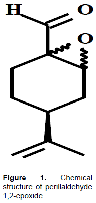
In this context, the development of new derived compounds from natural plants and their analogues for anticancer and antitumor activities, and for this, the significant challenge is to synthesize, isolate and characterize novel derivatives based on bioactivity and mechanisms of action of these compounds. However, there is a continuing need for research and development of new anticancer drugs, drug combinations and chemotherapy strategies, by methodical and scientific exploration of an enormous pool of synthetic, biological and natural products (Umadevi et al., 2013). Therefore, considering the perillyl alcohol anticancer bioactivity, this study aimed to evaluate its chemical analogue, perillaldehyde 1,2-epoxide, on antitumor activity, toxicology effects and cytotoxicity mechanism (Figure 1).
MATERIALS AND METHODS
Preparation of perillaldehyde 1,2-epoxide
A solution of perylaldehyde (4.5 g; 30 mmols) in MeOH (60 ml) was added to a solution of H2O2 (30%, 15.3 ml, 150 mmols). Then an aqueous solution KOH 6 mol/L (5.0 ml, 30 mmol) was added slowly, drop by drop, and kept at 0°C (ice bath), as described by Tantanak and collaborators (Tantanak et al., 1998).
The reaction medium remained under stirring for 4 h at the same temperature. Subsequently, it was removed from the ice bath, and an aqueous phase was extracted with CH2Cl2 (50 ml), and the combined organic phases washed twice with distilled water (50 ml) and water stripped with anhydrous Na2SO4. Then the material obtained was concentrated on a rotary evaporator, and the product purified by silica gel column chromatography (hexane/ethyl acetate 9:1). Perillaldehyde 1,2-epoxide was obtained with a 71.6% (4.77 mmol) yield. Perillaldehyde 1,2-epoxide is a p-menthane cyclic compound that contains an epoxide ring. It has a molecular formula of C10H14O2, molar volume of 143.1 ± 3.0 cm3/mol, surface tension of 29.6 ± 3.0 dyn/cm, and density of 1.161 ± 0.06 g/cm3.
The compound perillaldehyde 1,2-epoxide was analyzed by infrared, 1H and 13C NMR. The 1H- and 13C-NMR measurements were obtained with a Mercury-Varian spectrometer (Palo Alto, CA, USA) operating at 200 MHz (for 1H), and 50 MHz (for 13C). The infrared spectra were recorded on a Bomen Michelson model 102 FTIR (Bomen, Chicago, IL, USA) and the most intense or representative bands reported (in cm-1). IR (KBr) νmax: 3020, 2980, 1725, 1670, 900 cm–1; 1H-NMR (CDCl3): δ 8.83 (s, 1H); 4,68 (d, J= 12,4 Hz, 2H); 3.52 - 3.38 (m, 1H); 2.72 - 1.68 (m, 7H); 1.67 (s, 3H); 13C-NMR (CDCl3): δ 199.1, 148.0, 109.6, 63.5, 57.3, 36.8, 29.9, 25.2, 20.7, 19.5 (CAS 90926-06-0).
Evaluation of the cytotoxic effect of perillaldehyde 1,2-epoxide human cancer cell lines
Cell lines and MTT assay
For cytotoxicity assays, four human cancer cell lines HCT-116(colon carcinoma), OVCAR-8 (ovarian adenocarcinoma), SF-295 (glioblastoma), and HL-60 (Promyelocytic leukemia) were acquired from National Cancer Institute, Bethesda, MD, USA and evaluated for monoterpene perillaldehyde 1,2-epoxide. Cells were cultured in RPMI-1640 medium supplemented with 10% fetal bovine serum, 2 mM glutamine, 100 µg/ml streptomycin, and 100 U/ml penicillin, and incubated at 37°C in a 5% CO2 atmosphere.The MTT method, cytotoxic described by Mossman (1983), was used to evaluate the activity of the monoterpene perillaldehyde 1,2-epoxide against four human cancer cell lines. For the experiments, the cells wereplaced in 96-well plates (0.1 × 10⁶ cells/ml in 100 μl medium). After 3 days(72 h) of incubation, the perillaldehyde 1,2-epoxide dissolved in dimethyl sulfoxide (DMSO 0.7%), at a final concentration of 10.60 μM, was added to each well and incubated for 3 days (72 h) at 37°C in a 5% CO2 atmosphere (three independent experiments, performed in triplicate).
DMSO at 1% was used as negative control and doxorubicin at 0.55 μM was used as positive control (purity > 98%; Sigma Chemical Co., St. Louis, MO, USA). At the end of incubation, the plates were centrifuged at 5000 rpm for 10 min, and the supernatants were removed. A 150 μl of an aqueous MTT solution containing 0.5 mg/ml MTT was added to each well and incubated for three hours at 37°C in a 5% CO2 atmosphere. Cell viability was measured by the ability of cells valves to reduce the yellow dye 3-(4,5- dimethyl-2-thiazolyl)-2,5-diphenyl-2H-tetrazolium bromide (MTT; Sigma Chemical Co., St. Louis, MO, USA) to a purple formazan product.
After incubation, the precipitate was dissolved in 150 μl DMSO, and absorbance 595nm was measured using a multiplate reader (DTX 880 Multimode Detector, Beckman Coulter Inc.). The absorbance values of these tests will be expressed as cell growth inhibition percentage (GI%) by the following formula: [GI% = 100 − [(T/NC) × 100%]. NC is the Absorbance of the negative control, and T is the Absorbance of test compound (perillaldehyde 1,2-epoxide).
The median inhibitory concentration able to induce 50% of maximal effect (IC50) of the perillaldehyde 1,2-epoxide was determined after evaluation of GI%. All cells were retested using the same protocol above with a varying concentration of compound (0 to 150.60 µM) (dos Santos Júnior et al., 2010; Ribeiro et al., 2012).
Trypan blue dye exclusion assay
The human cancer cell line HL-60 was used at a concentration of 0.3 x 10⁶ cells/ml, incubated for 3 days (72 h) with perillaldehyde 1,2-epoxide (4.85, 9.70 and 19.40 µM) and examined on an inverted microscope. At the end of incubation 90 µl was withdrawn from the cell suspension and added to 10 µl of trypan blue. The cells were differentiated into viable and non-viable and counted in a Neubauer chamber. A Doxorrubicin 0.55 µM was used as positive control (Veras et al., 2004).
Elucidation of cell death
Morphological analyses using a fluorescence microscope
The human cancer cell line HL-60 was used at a 0.3 x 10⁶ cells/ml, incubated for 1 day (72 h) with perillaldehyde 1,2-epoxide (4.85, 9.70 and 19.40 µM). The cell suspension was transferred to an eppendorf tube and centrifuged for 5 min at low speed (145 × g). The supernatant was discarded, and the cells were resuspended in 20 µl of PBS solution. Then 1 µl of aqueous acridine orange/ethidium bromide solution (AO/EB, 100 μg/ml) was added to each tube, and an aliquot of these cells transferred to a slide and mounted with a coverslip and then brought to a fluorescence microscope for observation of cellular events (apoptosis and necrosis). Doxorubicin (0.55 µM) was used as positive control (Geng et al., 2005).
Morphological analysis using an optical microscope
The HL-60 cell line, plated at a concentration of 0.3 x 10⁶ cells/ml, was incubated for 72 h with the compound (4.85, 9.70 and 19.40 µM) and examined under a microscope inverted. After that, 50 μl of cell suspension was added to the slide of the centrifuge (Cytospin™), to observe the morphology of the treated cells. After cell adhesion to the blade, fixing was done with methanol for 1 min and was first used hematoxylin staining, followed by eosin. The morphological changes were observed under optical microscope. Doxorubicin (0.55 µM) was used as positive control (Veras et al., 2004).
Hemolytic assay
To evaluate hemolytic activity, blood was collected from three mice of Swiss orbital path (anesthetized with isoflurane 1.5%) and diluted 1:30 in saline (0.85% NaCl + 10 mM CaCl2). The erythrocytes were washed two times in saline by centrifugation (15 g / 3 min.), resuspended in saline to obtain a suspension of erythrocytes 2% and the assay was performed in a 96-well plate (Jimenez et al., 2003). The single concentration of compound (500 µg/ml) was added to the suspension of red blood cells (Bezerra et al., 2005; Kang et al., 2009; Pita et al., 2012). Mixtures were incubated on a mixer for 60 min and then centrifuged at 875 × g for 5 min. Triton X-100 (1%) was used as the positive control. The absorbance of the supernatants was determined at 540 nm using spectrophotometrically. Triton X-100 (1%) was used as the positive control.
Evaluation of in vivo antitumor activity and toxicological analyses
In vivo antitumor activity assay
To evaluate the in vivo antitumor activity, 60 male mice, weighing 26–31 g were used; they were purchased from bioterium of the Federal University of Sergipe, Brazil. The animals were maintained under laboratory conditions of temperature, humidity, and light with food and water ad libitum. The experimental protocol was submitted and approved by the Animal Care and Use Committee at the Federal University of Sergipe (CEPA: 16/2014). Ten-day-old sarcoma 180 ascites tumor cells (2 x 10⁶ cell / 500 μl) were implanted subcutaneously into the left axillary region of the experimental mice (Bezerra et al., 2006). The animals were divided into 6 groups of 10 animals in polypropylene cages. One day after inoculation, was administered intraperitoneally in group one 5% DMSO (negative control-NC), groups two, three, four and five perillaldehyde 1,2-epoxide as doses de 25, 50, 100, 200 mg/kg/day respectively dissolved in 5% DMSO (test compounds) and group six 5-fluorouracil (5-FU, purity > 99%; Sigma Chemical Co.) 25 mg/kg/day (positive control). 72 h after the last day of treatment under isoflurane inhalation (1.5%, calibrated vaporizer) anaesthesia, peripheral blood samples were collected from the retro-orbital plexus (toxicological analyses), the animals were euthanized by cervical dislocation, and the tumors, livers, spleens, and kidneys were excised and weighed. Inhibition of tumor growth (%) was expressed by the following equation (Bezerra et al., 2006):

NC = mean tumor weight of the negative control group
TC = mean tumor weight of the test compound treated group
Systemic toxicological analyses
Control groups (group one and six) and the groups that showed antitumor activity in vivo against sarcoma 180 were submitted to an evaluation by six toxicological parameters: variation in body mass, organ weights, liver, renal and hematologic parameters and histopathological analyses. First, the mice were weighed at the beginning and end of the experiment, and the animals were observed for signs of abnormalities throughout the study. Second, the livers, kidneys, and spleens were removed and weighed after euthanasia. Third and fourth parameters using obtained plasma peripheral blood samples of the mice and Clinical Chemistry® kits (Abbott; Architect C 8000) were evaluated liver function measured by aspartate aminotransferase (AST) and alanine aminotransferase (ALT), and renal function measured by urea and creatinine. The fifth parameter was hematological analysis, an aliquot of blood from each animal was placed in EDTA, and total leucocyte counts were determined by standard manual procedures using optical microscopy. The last parameter was performed after 10% formaldehyde fixation, the spleens, liver, and kidneys were dehydrated in alcohol, diaphanized in xylene and paraffin-embedded. Subsequently, 5-μm-thick histological sections were obtained and stained with hematoxylin and eosin. Histological analyses were performed under optical microscopy (Amaral et al., 2016; Dória et al., 2016). The mice were weighed at the beginning and end of the experiment, and they were observed for signs of abnormalities throughout the study. At the end of the investigation, the animals were anesthetized with ether anesthesia, and peripheral blood samples by retro-orbital plexus were collected.
For hematological analysis, an aliquot of blood from each animal was placed in EDTA, and total leucocyte counts were determined by standard manual procedures using optical microscopy. Serum samples were obtained to evaluate liver function (aspartate aminotransferase (AST) and alanine aminotransferase (ALT)), and renal function (urea and creatinine) using Clinical Chemistry® kits (Abbott; Architect C 8000). Then, the animals were sacrificed in a CO2 chamber, and the spleens, liver, and kidneys were weighted and fixed with 10% formaldehyde. The organs were dehydrated in alcohol, diaphanized in xylene and paraffin-embedded. Subsequently, 5-μm-thick histological sections were obtained and stained with hematoxylin and eosin. Histological analyses were performed under optical microscopy (Amaral et al., 2016; Dória et al., 2016).
Statistical analysis
The results were expressed as the mean ± SEM, and the differences between the experimental groups were analyzed using ANOVA of unidirectional analysis of variance followed by the Student Newman-Keuls test. Significant values of p < 0.05 were considered. All statistical analyses were performed using GraphPad program® (Intuitive Software for Science, San Diego, CA, USA).
RESULTS AND DISCUSSION
Cytotoxic effect of perillaldehyde 1,2-epoxide tumor cell in culture
MTT assay
Compounds of natural origin have provided new and potential leads to cancer chemotherapy, and many of them are the drug of choice in cancer treatment (Prakash et al., 2013). Natural products are important sources of chemical structures, and these will be used as templates for construction of new compounds with improved biological properties (Mann, 2002). Following this trend, the perillaldehyde 1,2-epoxide from the evaluation of the cytotoxicity of structurally correlated p-menthane derivatives described by Andrade and collaborators was developed (Andrade et al., 2015).
Following the criteria of American National Cancer Institute to discovering new anticancer drugs, were selected four human cancer cell lines to investigate cytotoxicity of perillaldehyde 1,2-epoxide: colon carcinoma (HCT-116), ovarian adenocarcinoma (OVCAR-8), glioblastoma (SF-295) and promyelocytic leucemia (HL-60) (dos Santos Júnior et al., 2010; Ribeiro et al., 2012). The compound was first used in a single concentration of 150.60 μM and evaluated according to the cell growth inhibition percentage (GI%) as: without cytotoxicity, low cytotoxicity (GI 1 - 50%), moderate cytotoxicity (GI 51 – 75%) and high cytotoxicity (GI > 75%) (Mahmoud et al., 2011). The GI% values are presented as the mean ± SD of three replicates measured by MTT assay after 72 h of incubation. The results showed that the compound perillaldehyde 1,2-epoxide demonstred high cytotoxicity activity, with GI > 95% for four lines human tumor cells used (Table 1).

Based on a similar study performed by Andrade et al. (2015), with 18 derivative compounds of perillyl alcohol, compound perillaldehyde 8,9-epoxide was the p-menthane derivative with the highest cytotoxic activity for the human cancer cell lines HCT-116, OVCAR-8, and SF-295. Comparing the perillaldehyde 1,2-epoxide with the perillaldehyde 8,9-epoxide, position isomers with high cytotoxicity in vitro, both substances have a skeleton p-menthane containing an aldehyde group and an epoxide group in its chemical structure. The replacement of the hydroxyl group with the aldehyde group and adding the epoxide group the structure of perillyl alcohol (GI = 95.82, 91.68, 90, and 92%, respectively) resulted in an increasein GI% perillaldehyde 1,2-epoxide (GI = 99.46, 99.37 and 95.66%, respectively) and perillaldehyde 8,9-epoxide (GI = 98.64, 96.32 and 99.89%, respectively). This result suggests that the presence of these two functional groups in the compounds may be contributing to this higher biological effect.
Thus, the compound in this study was promoted for the determination of median inhibitory concentration able to produce 50% of maximal effect (CI50) to verify the potency of the compound. For this, were used the same cell lines and measurement methods used to determine the GI%, varying only the concentration of compound between 0 to 150.60 μM. The perillaldehyde 1 ,2-epoxide exhibits values IC50 in the range of 9.70 to 21.99 μM in the HCT-116 and SF-295 cell lines, respectively. Doxorubicin, used as the positive control, showed IC50 values ranging from 0.02 to 1.95 μM for HCT-116 and OVCAR-8 cell lines, respectively (Table 1). For this test, considered as a promising candidate for antineoplastic activity, substances that present lower IC50 values or equal to 24.10 μM (Suffness and Pezzuto, 1990). Therefore, the compound perillaldehyde 1,2-epoxide is a potential candidate for anticancer activity and eligible for progression of studies.
Trypan blue dye exclusion assay
The perillaldehyde 1,2-epoxide showed higher cytotoxicity front leukemic cell line HL-60 (GI=99.71%±2.43). Faced with this result, three concentrations of perillaldehyde 1,2-epoxide, ½ IC50 (4.85 μM), IC50 (9.70 μM) and 2 × IC50 (19.40 μM) were chosen against HL-60 for evaluation of antiproliferative effects. The analysis of cell viability in HL-60 leukemic cell lines was performed by trypan blue exclusion after 72 h of exposure. Trypan blue exclusion test allows separately quantify the viable cells of cells killed by the test compound (Barros et al., 2013). Perilladehyde 1,2-epoxide caused a significant reduction in the number of viable cells (p < 0.05) and increase in the number of non-viable cells (p < 0.05) at the concentrations of 4.85, 9.70 and 19.40 µM (Figure 2). Studies have shown that compounds can interact with MTT, inhibiting the reduction of MTT and may produce a false positive result. Therefore, the use of other viability assays, such as trypan blue exclusion is indicated before progression of the studies (Trevisi et al., 2006; Pita et al., 2012). Then, we confirm the cytotoxic activity of perilladehyde 1,2-epoxide front HL-60 by two methods (MTT and Trypan blue exclusion).

Elucidation of cell death
Morphological analyses using a fluorescence and light microscopy
For the identification of the cell death process induced by perilladehyde 1,2-epoxide against HL-60 two assays, acridine orange/ethidium bromide was used and analysed by fluorescence microscopy and optical microscopy using hematoxylin-eosin coloration. After 72 h of incubation of the cells with compound at concentrations of 4.85, 9.70 and 19.40 µM and acridine orange/ethidium bromide stained, was observed as a result of a reduction in the number of viable cells and increase of cell death by apoptosis statistically significant (p < 0.05). This effect was observed when compared to the negative control group at all three concentrations of the compound tested (Figure 3).

In the morphological analysis by hematoxylin-eosin coloration of treated cells with perilladehyde 1,2-epoxide, in all concentrations tested there were signs of death by apoptosis as was also observed in the test with acridine orange/ethidium bromide (Figure 4). Other compounds structurally similar to the perilladehyde 1,2-epoxide as carvone (Aydın et al., 2015), carvacrol (Jaafari et al., 2009), limoneno (Sahin et al., 1999), perillyl alcohol (Clark, 2006)and perillic acid (Yeruva et al., 2007)also induce cell death by apoptosis in tumor cell lines. Cell death processes have well defined morphologic characteristics. Apoptosis includes cells with pyknotic appearance, condensation of chromatin, the fragmentation of the nucleus and the shedding of apoptotic bodies, vacuoles containing cytoplasm and intact organelles (Nikoletopoulou et al., 2013). Apoptosis is the cell death mechanism better known, used as an essential target for cancer therapy, by anticancer drugs such as the colcichine, taxanes and vinca alkaloids (Su et al., 2013; Topham and Taylor, 2013).

When concentration of 19.40 µM of perilladehyde 1,2-epoxide was used, concomitantly to cell death by apoptosis, was observed cell death by necrosis in tests performed with acridine orange/ethidium bromide and hematoxylin-eosin (Figures 3 and 4). Several treatments for malignant neoplasms can induce cell death by activation of necrosis to include photodynamic treatment (PDT), by the alkylation of agents harmful to DNA, and several other chemical compounds or substances such as apoptolidine, β-lapachone and honokil also appear to promote the death of cancer cells by necrosis (Zong and Thompson, 2006). Morphologically, the necrotic cells are characterized by the swelling of organelles, rupture of the plasma membrane (cell lysis), the nucleus becomes distended (nucleus intact) and usually followed by inflammatory reactions (Nikoletopoulou et al., 2013). Necrosis appears to be a limiting factor for increasing the concentration of the compound, but additional testing with higher levels has to be performed for more conclusions that are accurate.
Thus, the compound perilladehyde 1,2-epoxide can be considered a promising candidate for evaluation in vivo antitumor activity, by present cytotoxic activity predominant cell death by apoptosis, being subject to necrotic cell death at the highest concentration.
Evaluation of hemolytic activity
The evaluation of the hemolytic effect of perillaldehyde 1,2-epoxide was performed using erythrocytes Swiss mice, according to the methodology described by Costa-Lotufo and collaborators (Costa-Lotufo et al., 2002). This technique allows evaluating the potential of test compound in causing damage to the plasma membrane of the cell, either by the formation of pores or by the full rupture of it. However, this assay, we observed the absence of hemolytic activity in the tested concentration 500 μg/ml (Data not presented). These results suggest a possible selectivity for perillaldehyde 1,2-epoxide and enabling progression of studies related to the anticancer activity.
Antitumor activity in vivo and toxicological analyses
Tumor Sarcoma 180
Being the perillaldehyde 1, 2-epoxide cytotoxic against four human cancer cell lines (IC50 < 4,0 μg/ml) and having a predominant cell death of apoptosis, we decided to investigate the possible in vivo antitumor activity of the test compound. Based on the use of experimental tumors for the identification of new products with potential anticancer, the antitumor activity of perillaldehyde 8,9-epoxide was performed using sarcoma 180 tumors (original Swiss mice tumor, transplantable, and well-characterized experimental model) and treated by intraperitoneal route once a day for seven consecutive days (Britto et al., 2012; Yang et al., 2016).
The activity of perillaldehyde 1,2-epoxide on animals transplanted with sarcoma 180 tumor cells is presented in Figure 5. The mean tumor mass was 1.62 ± 0.05 g, 1.48 ± 0.07 g, 1.22 ± 0.16 g and 0.83 ± 0.15 g for animals treated with compost test at doses of 25, 50, 100, and 200 mg/kg/day, respectively. The NC group presented mean tumor mass of 1.76 ± 0.08 g. Statistically significant alterations (p < 0.05) were observed in mice treated with perillaldehyde 1,2-epoxide at concentrations of 100 and 200 mg/kg/day when compared with NC group, with tumor mass growth inhibition rates of 33.44 and 56.39%, respectively. The test compound at doses of 25 and 50 mg/kg/day showed no statistically significant alterations (p > 0.05) compared with NC group. The PC group presented tumor mass growth inhibition rates of 66.38% and mean tumor mass of 0.51 ± 0.06 g, statistically significant when compared with NC group.
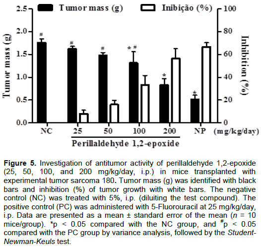
When comparing the PC group to the groups treated with test compound, statistical alterations were observed for the groups 25, 50 and 100 mg/kg/day (p < 0.05) and no statistically significant changes for the group 200 mg/kg/day (p > 0.05). These results indicate that dose of 100 and 200 mg/kg/day the perillaldehyde 1,2-epoxide has antitumor activity in vivo against the sarcoma 180 with more significant action at the highest dose.
Other monoterpenes p-menthane as perillaldehyde 8,9-epoxide and perillyl alcohol were also submitted to evaluation of antitumor activity in vivo using the same experimental tumor model. Both compounds were tested at doses of 100 and 200 mg/kg/day, demonstrating tumor mass growth inhibition rates of 38.4 and 58.7% for the perillaldehyde 8,9-epoxide and 35.3 and 45.4% for the perillyl alcohol, respectively (Andrade et al., 2016). Compared to the data obtained in this study with those obtained by Andrade and collaborators (Andrade et al., 2016)it was observed proximity between tumor mass growth inhibition rates and therefore a similar antitumor in vivo potency. The alcohol perillyl has been studied quite a long time to be able to inhibit the growth of tumor cells in cell culture and exert cancer preventive and therapeutic activity in a variety of animal tumor models.
Furthermore, the perillyl alcohol has been successfully used intranasally in the treatment of patients with malignant brain tumors (Chen et al., 2015). Thus, the results so far found with perillaldehyde 1,2-epoxide are promising for progression of studies.
Toxicological analyses
It is known that most of the currently used anticancer drugs are cytotoxic to tumor cells, but they also have non-specific action, because they affect healthy cells, leading to an undesirable side effect (Sun and Peng, 2008). Therefore we decided to evaluate toxicological characteristics of mice with tumor sarcoma 180 undergoing treatment with perillaldehyde 1,2-epoxide at doses of 100 and 200 mg/kg/day, to assess the cost and benefits of intervention. For this, some toxicological parameters were evaluated as variation in body mass, organ weights (liver, spleen, and kidney), liver (AST and ALT), renal (urea and creatinine), hematologic (total leukocytes) parameters and histopathological analyses (Dória et al., 2016).
There was no significant change (p > 0.05) in organ weights (liver, spleen and kidney), liver (AST and ALT) and renal (urea and creatinine) parameters in the groups treated with perillaldehyde 1,2-epoxide at doses of 100 and 200 mg/kg/day when compared with NC group (Table 2). These data are significant since many anticancer drugs cause changes in renal function, liver function and spleen organ volume as a result of the lack of specificity of antineoplastic, inducing severe adverse effects (Bezerra et al., 2008; Amaral et al., 2016).
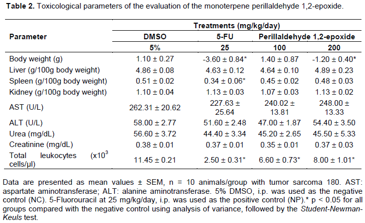
When analyzing the variation in body mass and total leukocyte count, a significant reduction in body mass (p < 0.05) and a decrease in total leukocytes were observed in the groups treated with perillaldehyde 1,2-epoxide at one dose of 200 mg/kg/day compared to NC group. The group administered with compost test at a dose of 100 mg/kg/day no significant change (p > 0.05) the mass variation but significant decrease in total leukocytes (p < 0.05). These data can be considered a limiting factor for the administration of higher doses of test compound but does not prevent the advancement of studies because adverse effects such as body weight loss and leukopenia are common in some marketed antineoplastics (Cao et al., 1998; Zamagni et al., 1998). An example of these adverse effects is observed in the use of 5-FU 25 mg/kg/day as a positive control in the present paper, where a significant reduction (p < 0.05) in the variation of body weight, spleen weight and some leukocytes was checked.
Histopathology is a test used to aid in data biochemical and microscopic analysis, detecting any morphological change or characteristic lesion in the evaluated tissues. Histopathological examination was performed on organs, liver, spleen, and kidney of animals with sarcoma 180 and all tissue structures are substantially preserved in treated groups with perillaldehyde 1,2-epoxide at a dose of 100 and 200 mg/kg/day (Data not presented).
CONCLUSION
Therefore, the present study indicates that the perillaldehyde 1,2-epoxide exhibits high cytotoxic activity against the human cancer cell lines HCT-116, OVCAR-8, SF-295 and HL-60 with the possible predominant process of cell death by apoptosis and in vivo antitumor effects without presenting substantial toxicity in experimental tumor model sarcoma 180. It is possible to continue the study for applicability of this compound in other tumor models or as prototypes for the development of antineoplastic agents.
CONFLICT OF INTERESTS
The authors have not declared any conflict of interests.
ACKNOWLEDGEMENTS
This research was supported by Conselho Nacional de Desenvolvimento Científico e Tecnológico (CNPq), Coordenação de Aperfeiçoamento de Pessoal de Nível Superior (CAPES), and, the Fundação de Apoio à Pesquisa e Inovação Tecnológica do Estado de Sergipe (FAPITEC/SE).
REFERENCES
|
Amaral RG, Andrade LN, Dória GAA, Barbosaâ€Filho JM, Sousa DP, Carvalho AA, Thomazzi SM (2016). Antitumour effects of the essential oil from Mentha x villosa combined with 5â€fluorouracil in mice. Flavour and Fragrance Journal 31(3):250-254. |
|
|
Amaral RG, Fonseca CS, Silva TKM, Andrade LN, França ME, Barbosaâ€Filho JM, Sousa DP, Moraes MO, Pessoa CÓ, Carvalho AA (2015). Evaluation of the cytotoxic and antitumour effects of the essential oil from Mentha x villosa and its main compound, rotundifolone. Journal of Pharmacy and Pharmacology 67(8):1100-1106. |
|
|
Andrade LN, Amaral RG, Dória GAA, Fonseca CS, Da Silva TKM, Albuquerque Júnior RLC, Thomazzi SM, Do Nascimento LG, Carvalho AA, De Sousa DP (2016). In vivo Anti-tumor activity and toxicological evaluations of perillaldehyde 8, 9-Epoxide, a derivative of perillyl alcohol. International Journal of Molecular Sciences 17(1):32. |
|
|
Andrade LN, Lima TC, Amaral RG, Pessoa CÓ, Soares BM, Nascimento LGD, Carvalho AA, De Sousa DP (2015). Evaluation of the cytotoxicity of structurally correlated p-menthane derivatives. Molecules 20(7):13264-13280. |
|
|
Aydın E, Türkez H, KeleÅŸ MS (2015). Potential anticancer activity of carvone in N2a neuroblastoma cell line. Toxicology and Industrial Health 31(8):764-772. |
|
|
Barros FW, Bezerra DP, Ferreira PM, Cavalcanti BC, Silva TG, Pitta MG, De Lima MDC, Galdino SL, Pitta IDR, Costa-Lotufo LV (2013). Inhibition of DNA topoisomerase I activity and induction of apoptosis by thiazacridine derivatives. Toxicology and Applied Pharmacology 268(1):37-46. |
|
|
Bezerra D, Castro F, Alves A, Pessoa C, Moraes M, Silveira E, Lima M, Elmiro F, Costa-Lotufo L (2006). In vivo growth-inhibition of Sarcoma 180 by piplartine and piperine, two alkaloid amides from Piper. Brazilian Journal of Medical and Biological Research 39(6):801-807. |
|
|
Bezerra DP, Castro FOD, Alves APN, Pessoa C, Moraes MOD, Silveira ER, Lima MAS, Elmiro FJM, Alencar N, Mesquita RO (2008). In vitro and in vivo antitumor effect of 5â€FU combined with piplartine and piperine. Journal of Applied Toxicology: An International Journal 28(2):156-163. |
|
|
Bezerra DP, Pessoa C, Moraes MOD, Silveira ER, Lima MAS, Martins Elmiro FJ, Costa-Lotufo LV (2005). Antiproliferative effects of two amides, piperine and piplartine, from Piper species. Zeitschrift für Naturforschung C 60:539-543. |
|
|
Bhanot A, Sharma R, Noolvi, MN (2011). Natural sources as potential anti-cancer agents: A review. International Journal of Phytomedicine 3:9. |
|
|
Britto AC, de Oliveira AC, Henriques RM, Cardoso GM, Bomfim DS, Carvalho AA, Moraes MO, Pessoa C, Pinheiro ML, Costa EV (2012). In vitro and in vivo antitumor effects of the essential oil from the leaves of Guatteria friesiana. Planta Medica 78:409-414. |
|
|
Butler MS (2008). Natural products to drugs: natural product-derived compounds in clinical trials. Natural Product Reports 25(3):475-516. |
|
|
Cao X, Cai R, Wen Ju D, Tao Q, Yu Y, Wang J (1998). Augmentation of hematopoiesis by fibroblast-mediated interleukin-6 gene therapy in mice with chemotherapy. Journal of Interferon & Cytokine Research 18(4):227-233. |
|
|
Carvalho AA, Andrade LN, De Sousa ÉBV, De Sousa, DP (2015). Antitumor phenylpropanoids found in essential oils. BioMed Research International 1-21. |
|
|
Chen TC, Da Fonseca CO, Schönthal AH (2015). Preclinical development and clinical use of perillyl alcohol for chemoprevention and cancer therapy. American Journal of Cancer Research 5(5):1580. |
|
|
Chinta G, Syed B, Coumar S, Periyasamy MS (2015). Piperine: A comprehensive review of pre-clinical and clinical investigations. Current Bioactive Compounds 11(3):156-169. |
|
|
Clark SS (2006). Perillyl alcohol induces c-Myc-dependent apoptosis in Bcr/Abl-transformed leukemia cells. Oncology 70(1):13-18. |
|
|
Costa-Lotufo L, Cunha G, Farias P, Viana G, Cunha K, Pessoa C, Moraes M, Silveira E, Gramosa N, Rao V (2002). The cytotoxic and embryotoxic effects of kaurenoic acid, a diterpene isolated from Copaifera langsdorffii oleo-resin. Toxicon 40(8):1231-1234. |
|
|
Dória GAA, Menezes PP, Lima BS, Vasconcelos BS, Silva FA, Henriques RM, Melo MG, Alves ÂV, Moraes MO, Pessoa CÓ (2016). In vivo antitumor effect, induction of apoptosis and safety of Remirea maritima Aubl.(Cyperaceae) extracts. Phytomedicine 23:914-922. |
|
|
Dos Santos Júnior HM, Oliveira DF, De Carvalho DA, Pinto JMA, Campos VAC, Mourão ARB, Pessoa C, De Moraes MO, Costa-Lotufo LV (2010). Evaluation of native and exotic Brazilian plants for anticancer activity. Journal of Natural Medicines 64(2):231-238. |
|
|
Garcia DG, De Castro-Faria-Neto HC, Da Silva CI, Gonçalves-de-Albuquerque CF, Silva AR, De Amorim LMDF, Freire AS, Santelli RE, Diniz LP, Gomes FCA (2015). Na/K-ATPase as a target for anticancer drugs: studies with perillyl alcohol. Molecular Cancer 14:1. |
|
|
Geng CX, Zeng ZC, Wang JY, Xuan S-Y, Lin CM (2005). Docetaxel shows radiosensitization in human hepatocellular carcinoma cells. World Journal of Gastroenterol 11(19):2990-2993. |
|
|
Jaafari A, Mous, H, M'Bark L, Tilaoui M, Elhansali M, Lepoivre M, Aboufatima R, Melhaoui A, Chait A, Zyad A (2009). Differential antitumor effect of essential oils and their major components of Thymus broussonettii: relationship to cell cycle and apoptosis induction. Herba Polonica 55:36-50. |
|
|
Jimenez PC, Fortier SC, Lotufo TM, Pessoa C, Moraes MEA, De Moraes MO, Costa-Lotufo LCV (2003). Biological activity in extracts of ascidians (Tunicata, Ascidiacea) from the northeastern Brazilian coast. Journal of Experimental Marine Biology and Ecology 287:93-101. |
|
|
Kang C, Munawir A, Cha M, Sohn ET, Lee H, Kim JS, Yoon WD, Lim D, Kim E (2009). Cytotoxicity and hemolytic activity of jellyfish Nemopilema nomurai (Scyphozoa: Rhizostomeae) venom. Comparative Biochemistry and Physiology Part C: Toxicology & Pharmacology 150:85-90. |
|
|
Mann J (2002). Natural products in cancer chemotherapy: past, present and future. Nature Reviews Cancer 2:143-148. |
|
|
Nikoletopoulou V, Markaki M, Palikaras K, Tavernarakis N (2013). Crosstalk between apoptosis, necrosis and autophagy. Biochimica et Biophysica Acta (BBA)-Molecular Cell Research 1833:3448-3459. |
|
|
Pita JCLR, Xavier AL, Sousa TKGD, Mangueira VM, Tavares JF, Júnior RJO Veras RC, Pessoa HLF, Silva MS, Morelli S (2012). In vitro and in vivo antitumor effect of trachylobane-360, a diterpene from Xylopia langsdorffiana. Molecules 17:9573-9589. |
|
|
Polu P, Nayanabhirama U, Khan S (2015). Herbal medicinal plants as an anticancer agents. Annals of Phytomedicine 4:37-45. |
|
|
Prakash O, Kumar A, Kumar P (2013). Anticancer potential of plants and natural products: A review. American Journal of Pharmacological Sciences 1:104-115. |
|
|
Qurishi Y, Hamid A, Majeed R, Hussain A, Qazi AK, Ahmed M, Zargar MA, Singh SK, Saxena AK (2011). Interaction of natural products with cell survival and signaling pathways in the biochemical elucidation of drug targets in cancer. Future Oncology 7(8):1007-1021. |
|
|
Ribeiro SS, De Jesus AM, Dos Anjos CS, Da Silva TB, Santos AD, De Jesus JR, Andrade MS, Sampaio TS, Gomes WF, Alves PB (2012). Evaluation of the cytotoxic activity of some Brazilian medicinal plants. Planta Medica 78(4):1601-1606. |
|
|
Sahin M, Perman S, Jenkins G, Clark S (1999). Perillyl alcohol selectively induces G0/G1 arrest and apoptosis in Bcr/Abl-transformed myeloid cell lines. Leukemia 13(10):1581. |
|
|
Su M, Mei Y, Sinha S (2013). Role of the crosstalk between autophagy and apoptosis in cancer. Journal of Oncology pp. 1-14. |
|
|
Suffness M, Pezzuto JM (1990). Assays related to cancer drug discovery. Methods in Plant Biochemistry: Assays for Bioactivity 6:71-133. |
|
|
Sun HX, Peng XY (2008). Protective effect of triterpenoid fractions from the rhizomes of Astilbe chinensis on cyclophosphamide-induced toxicity in tumor-bearing mice. Journal of Ethnopharmacology 119(2):312-317. |
|
|
Tantanak D, Vincent MA, Hillier IH (1998). Elucidation of the mechanism of alkene epoxidation by hydrogen peroxide catalysed by titanosilicates: a computational study. Chemical Communications 1031-1032. |
|
|
Topham CH, Taylor SS (2013). Mitosis and apoptosis: how is the balance set? Current Opinion in Cell Biology 25(6):780-785. |
|
|
Trevisi L, Pighin I, Bazzan S, Luciani S (2006). Inhibition of 3â€(4, 5â€dimethylthiazolâ€2â€yl)â€2, 5â€diphenyltetrazolium bromide (MTT) endocytosis by ouabain in human endothelial cells. FEBS Letters 580(11):2769-2773. |
|
|
Umadevi M, Kumar KS, Bhowmik D, Duraivel S (2013). Traditionally used anticancer herbs in India. Journal of Medicinal Plants Studies 1:56-74. |
|
|
Veras ML, Bezerra MZB, Braz-Filho R, Pessoa ODL, Montenegro RC, Do Ó Pessoa C, De Moraes MO, Costa-Lotufo LV (2004). Cytotoxic epimeric withaphysalins from leaves of Acnistus arborescens. Planta Medica 70:551-555. |
|
|
Yang W, Zhang H, Ji M, Pei F, Wang Y (2016). Antitumor effect of a polysaccharide isolated from Phellinus pullus as an immunostimulant. Biomedical Reports 4:361-364. |
|
|
Yeruva L, Pierre KJ, Elegbede A, Wang RC, Carper SW (2007). Perillyl alcohol and perillic acid induced cell cycle arrest and apoptosis in non small cell lung cancer cells. Cancer Letters 257:216-226. |
|
|
Zamagni C, Martoni A, Cacciari N, Gentile A, Pannuti F (1998). The combination of paclitaxel and carboplatin as first-line chemotherapy in patients with stage III and stage IV ovarian cancer: A phase I-II study. American Journal of Clinical Oncology 21(5):491-497. |
|
|
Zong WX, Thompson CB (2006). Necrotic death as a cell fate. Genes and Development 20:1-15. |
|
Copyright © 2024 Author(s) retain the copyright of this article.
This article is published under the terms of the Creative Commons Attribution License 4.0


