Full Length Research Paper
ABSTRACT
Anacardium occidentale nut shell liquid has been used for decades especially by natives of South America, Asia and Africa in the treatment of topical skin diseases, abrasion and cancerous ulcers amongst others. Chronic exposure to sunlight can lead to skin damages including erythema, edema, hyperplasia, formation of sunburn cells, photo-aging, suppression of the immune system and skin cancer. This study was aimed at evaluating the effect of methanol extract of A. occidentale nut shell liquid on UV radiation (UVR) induced skin damage and cancer initiation. Gas chromatography and mass spectroscopic (GC-MS) analysis was carried out on the extract. Skin damage was induced by exposing the hairless part of the experimental animals directly to UVR (180 mJ/cm2 daily) for 42 days alongside treatment with the extract. Percentage weight gain, relative organ weight, lipid peroxidation and histological studies were carried out. The methanol extract of CNS as determined by GC-MS analysis contains 1,2,3-Benzenetriol and benzofuran. A normal skin tissue and hepatocyte was observed in the normal control, severe wrinkling of skin epithelium with actinic keratosis of the dermal collagen and marked venous congestion in the liver of the negative control and complete recovery in the group treated with 300 mg/kg of the extract was observed. This study suggests that the administration of the extract exhibited a chemopreventive effect against skin damage which could lead to cancer initiation stage resulting from ultraviolet radiation by preventing its detrimental impact on the epidermis.
Key words: Anacardium occidentale (Cashew), chemo-preventive effect, bioactive compounds, skin damage, ultra-violet radiation.
INTRODUCTION
Chemotherapy has been one of the major treatment option for cancer but unfortunately there are side effects such as renal impairment, neurotoxicity and ototoxicity associated with chemotherapy (Ali et al., 2013) and this could at times be life threatening.
Cashew (Anacardium occidentale L.) is a widely distributed plant in South America, Africa and Asia mostly for nutritional and commercial purposes. Various parts of Anacardium occidentale have been reported to be traditionally used, across the world, in the treatment of various diseases (Iwu, 1993). Cashew nut shell liquid (CNSL), a reddish brown viscous liquid extracted from the pericarp of the cashew nut which is mostly a byproduct of industrial processing of cashew nut has various medicinal uses (Hamad and Mubofu, 2015). The major constituents of CNSL include anacardic acid, cardol, cardanol and methyl cardol. Based on the method of extraction, the ratio of these components varies. Cashew nut shell liquid is used in folk medicine by some natives in treating soles of feet abrasion, topical infections and also cancerous ulcers (Patel et al., 2012).
Chronic exposure to ultra-violet radiation (UVR) from sunlight can result to skin damages which may include erythema, edema, hyperplasia, formation of sunburn cells, photo-aging, suppression of the immune system and skin cancer. With these effects it could be said that UVR is doubly involved in the development of skin cancer through the induction of genetic alterations in keratinocytes leading to their neoplastic transformation and by the depression of the skin normal immune responses (Leffell and Brash, 1996).
Skin cancer is the most common form of cancer, accounting for nothing less than 40% of cases globally. The most common type is non-melanoma skin cancer, which affects about 2 to 3 million people per year (Dubas and Ingraffea, 2013; Harris and Randall, 2013).
There are primary and secondary methods involved in cancer prevention. The primary method is aimed at keeping the carcinogenic process from the beginning; the secondary prevention method is through the discovery of cancerous or precancerous conditions in their very early stage (Spratt, 1981).
This research was designed to evaluate the effect of methanol extract of Anacardium occidentale (cashew) nut shell on UV induced skin damage and cancer initiation.
MATERIALS AND METHODS
Collection of plant sample
Anacardium occidentale seeds were obtained from Salem University Lokoja, Kogi State, Nigeria and identified in the Department of Plant Science and Biotechnology of Kogi State University, Anyigba with voucher number (023). The nut shells of A. occidentale were washed in cold water, the nuts that floated were taken out and the rest were air dried. The kernels were shelled from the nut; the shells were then cut into smaller sizes and blended using an electric blender.
Soxhlet extraction
Extraction of A. occidentale shell was obtained using soxhlet extraction method, with methanol as solvent. The sample to be extracted was weighed into a thimble and loaded into the chamber of the soxhlet extractor; 150 ml of the solvent was poured into a different chamber attached to the soxhlet extractor, enough to siphon at least twice into the flask. The temperature of the experiment was set up to 750C and heated at reflux. This cycle was repeated over and over again over six hours. The extract was dried at room temperature and stored at 40C for further usage.
Experimental animals
Thirty (30) male albino rats with average weight of 196 g were obtained from the animal house section of Salem University Lokoja, Kogi State. The animals were acclimatized in the experimental room for one week. The experimental animals were allowed access to standard food and water at libitum and handled according to guidelines on the use of experimental animals by the Department of Biosciences, Salem University Lokoja.
Gas chromatography mass spectrophotometry (GC-MS)
GC-MS analysis of the methanol extract of A. occidentale nut shell was performed using a Perkin–Elmer GC Clarus 500 system comprising an AOC-20i auto-sampler and a Gas Chromatograph interfaced to a Mass Spectrometer (GC-MS) equipped with an Elite-5MS (5% diphenyl/95% dimethyl poly siloxane) fused a capillary column (30 × 0.25 μm ID × 0.25 μm df). For GC-MS detection, an electron ionization system was operated in electron impact mode with ionization energy of 70 eV. Helium gas (99.999%) was used as a carrier gas at a constant flow rate of 1 ml/min, and an injection volume of 2 μl was employed (a split ratio of 25:1). The injector temperature was maintained at 250°C, the ion-source temperature was 200°C, the oven temperature was programmed from 60°C (isothermal for 2 min), with an increase of 30°C/min to 120°C, ending with a 3-min isothermal at 290°C. Mass spectra were taken at 70 eV; a scan interval of 0.5 s and fragments from 45 to 700 Da. The solvent delay was 0 to 2 min, and the total GC/MS running time was 21 min.
Animal study
UV induced skin damage and cancer initiation
The experimental animals (n=30) were weighed, shaved of the fur on their back and divided into six (6) groups according to the experimental design below. The induction was done according to a modified method by Wang et al. (1992). The rats were exposed to UVR (180 mJ/cm2 daily) for 42 days.
Experimental design
Group 1- Normal Control
Group 2- Negative control (exposed but untreated)
Group 3- 100 mg/kg methanol extract
Group 4- 300 mg/kg methanol extract
Group 5- 500 mg/kg methanol extract
Group 6- normal + 500 mg/kg methanol extract
Treatment of experimental animals
The experimental animals were treated alongside with the induction process following the details of their grouping above. The treatment of the animals was done by applying the extract on their shaved skin 5 min after exposure to the UV radiation. At the end of the treatment, they were starved for 12 h, weighed and anesthetized and sacrificed.
Collection of organs
The vital organs (heart, spleen, liver, lungs and kidneys) of the animals were collected and weighed.
Preparation of serum sample
Blood was collected into sample bottles and was centrifuged at 3000 rpm (using a micro field centrifuge) for 15 min. The serum was decanted into a different sample bottle and was stored at 40C for further analysis.
Preparation of tissue homogenate
The organs collected from each animal were rinsed using normal saline and homogenized in ice cold buffer with pH 7.4 using a homogenizer. The homogenates were centrifuged at 3000 rpm for 15 min and the supernatant was collected.
Protein determination
Four hundred and fifty (450) micro liter of distilled water was pipetted into test tubes in duplicates and 50 μl of the sample was added after which 1.5 ml of biuret reagent was added. The absorbance was read at 540 nm using a spectrophotometer.
Lipid peroxidation
Two millimeter of thiobarbituric acid (TBA) and trichloro acetate (TCA) was added to 50 μl of the tissue homogenates, respectively. The mixture was incubated for 30 min at 80°C. The test tubes were allowed to cool under ice and centrifuged at 3000 rpm for 15 min. The supernatant was measured using a spectrophotometer at a wave length of 535 nm.
Histological studies
The skin and liver tissues of the experimental animals were stored in 10% formalin for 24 h. The formalin fixed specimens were embedded in paraffin, sectioned (3-5 μm) and stained with haematoxylin and eosin dye. The histological sections were evaluated by light microscopy.
Statistical analysis
Statistical analysis for this study was done using SPSS version 16 at significant level of p<0.05.
RESULTS
Gas chromatography mass spectroscopy (GC-MS)
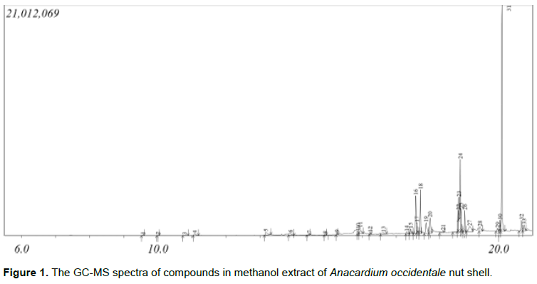
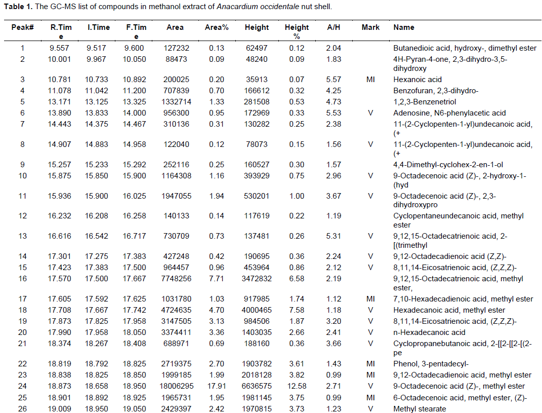
The gas chromatography mass spectroscopy (GC-MS) of the extract revealed (Figure 1 and Table 1) it contains bioactive compounds of pharmacological relevance.
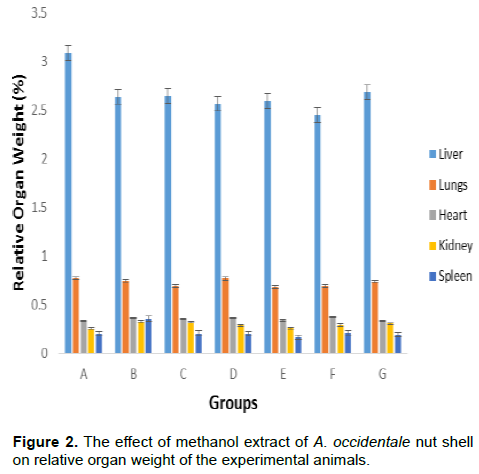
Relative organ weight
Figure 2 shows the effect of methanol extract of A. occidentale nut shell on relative organ weight of the experimental animals. There was no significant (p<0.05) decrease or increase in all the groups when compared to the normal and negative control.
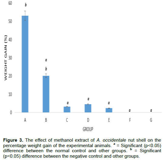
Percentage weight gain
The result given in Figure 3 represents the effect of methanol extract of A. occidentale nut shell on the percentage weight gain of the experimental animals.
There was a significant (p <0.05) weight decrease in all the groups when compared to the normal.
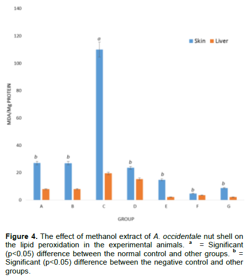
Lipid peroxidation
The result given in Figure 4 represents the effect of methanol extract of A. occidentale nut shell on the lipid peroxidation in the experimental animals. There was a significant (p<0.005) decrease in the malondialdehyde concentration between the negative control and all other groups except the group treated with the lowest dose (100 mg/kg) of the extract.
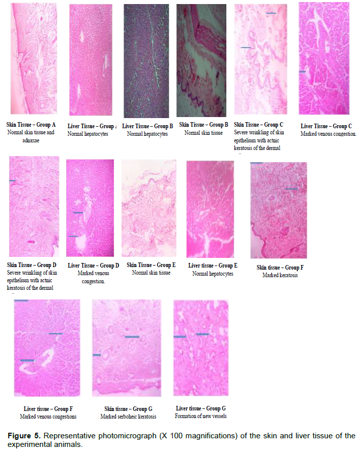
Histological studies
The result in Figure 5 is a representative photomicrograph (x100 Magnification) of the skin and liver tissue of the experimental animals. A normal skin tissue and hepatocyte was observed in the normal control, severe wrinkling of skin epithelium with actinic keratosis of the dermal collagen and marked venous congestion in the liver of the negative control was observed. Complete recovery was observed in the group treated with 300 mg/kg of the extract.
DISCUSSION
Plant-based and traditional medicine systems will continue to play essential roles in health care, with about 80% of the world’s inhabitants relying mainly on traditional medicines for their primary health care (Owolabi et al., 2007). Cashew nut shell liquid is used in folk medicine by some natives in treating soles of feet abrasion, topical infections and also cancerous ulcers (Patel et al., 2012).
The gas chromatography mass spectroscopy (GC-MS) of the extract revealed (Figure 1 and Table 1) it contains bioactive compounds of pharmacological relevance. Some of these compounds are believed to be responsible for the activity of the extract observed in this study. These bioactive compounds include 1,2,3-Benzenetriol also known as pyrogallol and benzofuran which according previous studies (Rida et al., 2006; Rice-Evans et al., 1996; Chen et al., 1998) exhibited anticancer, antioxidant and antibacterial activities, respectively.
There was no significant (p<0.05) change observed in the relative organ weight of the treated groups when compared with the normal and negative control (Figure 2). This indicates that the induction had little or no effect on the organ weight of the experimental animals.
A significant (p<0.005) decrease in malondialdehyde (MDA) concentration was observed in the skin of all the groups when compared to the negative control. There was also a significant (p<0.005) decrease in MDA concentration between the negative control and all other groups except the group treated with the lowest dose (100 mg/ml) of the extract, this effect was also observed in the liver of the treated group. This shows that the extract had effect against lipid peroxidation in the liver and skin of the treated animals.
The histological study revealed that the extract was able to bring the skin and liver tissues of the treated animals to a normal state especially at 300 mg/kg where a complete recovery was observed as shown in Figure 5. A normal skin tissue and hepatocyte was observed in the normal control, severe wrinkling of skin epithelium with actinic keratosis of the dermal collagen and marked venous congestion in the liver of the negative control was observed.
According to a UV radiation induced skin cancer model (Leffell and Brash, 1996), UV radiation penetration through the epidermis is the first step in the initiation of skin cancer and in this study the effect observed indicates that the extract exhibited a chemo-preventive effect against skin cancer initiation stage resulting from ultraviolet radiation, this effect is believed to be through the pharmacological action of the bioactive compounds identified in the extract amongst others.
CONCLUSION
This study suggests that the administration of methanol extract of A. occidentale nut shell exhibited a chemo-preventive effect against skin damage which could lead to skin cancer initiation stage resulting from ultraviolet radiation by preventing its detrimental impact on the epidermis which could in turn lead to DNA damage and subsequently skin cancer initiation.1,2,3-Benzenetriol and benzofuran, which according to previous studies, exhibits anticancer, antioxidant and antibacterial activities were present in the extract, these compounds amongst others are believed to be responsible for the pharmacological activities observed in the study. However, further study needs to be done to isolate and evaluate the effect of these bioactive compounds.
CONFLICT OF INTERESTS
The authors have not declared any conflict of interests.
REFERENCES
|
Ali I, Wani WA, Saleem K, Haque A (2013). Platinum compounds: A hope for future cancer chemotherapy. Anti-Cancer Agents in Medicinal Chemistry (Formerly Current Medicinal Chemistry-Anti-Cancer Agents) 13(2):296-306. |
|
|
Chen ZP, Schell JB, Ho CT, Chen KY (1998). Green tea epigallocatechin gallate shows a pronounced growth inhibitory effect on cancerous cells but not on their normal counterparts. Cancer Letters 129(2):173-179. |
|
|
Dubas LE, Ingraffea A (2013). Non-melanoma skin cancer. Journal of Facial Plastic Surgery Clinics of North America 21(1):43-53. |
|
|
Hamad F, Mubofu E (2015). Potential Biological Applications of Bio-Based Anacardic Acids and Their Derivatives. International Journal of Molecular Sciences16:(12)8569-8590. |
|
|
Harris RE, Randall E (2013). Epidemiology of Chronic Disease. Global Perspectives. Jones and Bartlett Learning. P. 271. |
|
|
Iwu MM (1993). Handbook of African medicinal plants. CRC Press. P 351. |
|
|
Leffell DJ, Brash DE (1996). Sunlight and skin cancer. Scientific American 275(1):52-59. |
|
|
Owolabi J, Omogbai EKI, Obasuyi O (2007). Antifungal and antibacterial activities of the ethanolic and aqueous extract of Kigella africana (Bignoniaceae) stem bark. African Journal of Biotechnology 6:882-885. |
|
|
Patel DK, Desai SN, Gandhi HP, Devkar RV, Ramachandran AV (2012). Cardio protective effect of Coriandrum sativum L. on isoproterenol induced myocardial necrosis in rats. Journal of Food and Chemical Toxicology 50(9):3120-3125. |
|
|
Rice-Evans CA, Miller NJ, Paganga G (1996). Structure-antioxidant activity relationships of flavonoids and phenolic acids. Free Radical Biology and Medicine 20(7):933-956. |
|
|
Rida SM, Hawash ESAM, Fahmyl HTY, Hazza AA, Meligy EMMM (2006). Synthesis and In Vitro Evaluation of Some Novel Benzofuran Derivatives as Potential Anti-HIV-1, Anticancer, and Antimicrobial Agents. Archives of Pharmaceutical Research 29(1):16-25. |
|
|
Spratt JS (1981). The primary and secondary prevention of cancer. Journal of Surgical Oncology18 (3):219-230. |
|
|
Wang ZY, Huang MT, Ho CT, Chang R, Ma W, Ferraro T, Reuhl KR, Yang CS, Conney AH (1992). Inhibitory effect of green tea on the growth of established skin papillomas in mice. Cancer Research 52(23):6657-6665. |
|
Copyright © 2024 Author(s) retain the copyright of this article.
This article is published under the terms of the Creative Commons Attribution License 4.0


