Full Length Research Paper
ABSTRACT
Albizia anthelmintica is a medicinal plant belonging to the Fabaceae family. It is widely used by smallholder farmers and pastoralists to treat internal parasites in their livestock. This study aimed to determine the antibacterial and antioxidant potential of A. anthelmintica on pathogenic veterinary isolates. 100% hexane (He100), 100% chloroform (Ch100), 100% ethanol (E100), and 70% ethanol (E70) extracts of the roots and barks of A. anthelmintica were tested against four bacterial strains (Escherichia coli, Clostridium perfringens, Salmonella enterica serovar Typhimurium and Proteus mirabilis). Thin layer chromatography- 2, 2-diphenyl-1- picryl hydrazyl (TLC-DPPH) assay was used to examine antioxidant potential of extracts. Antimicrobial activity was determined using the disc diffusion method and minimum inhibiting concentrations (MICs) values were determined using the micro-titre broth-dilution method. At a concentration of 500 µg/ml, E70 roots extract showed the highest % DPPH inhibition of 66.9%. Among the bark extracts, the highest free radical scavenging activity was observed in E70 extracts with 58.9% DPPH inhibition. Phytochemical analysis of the plant extracts revealed the presence of compounds which are known to exhibit medicinal properties such as tannins, terpenoids, quinones, saponins and fatty acids phenols. E100 bark extracts contained most of these compounds except flavonoids. Only alkaloids were not detected in any of the roots or bark extracts. Ch100 bark extracts showed the highest antimicrobial activity and all bacterial isolates were resistant to the E100 root extracts. Ch100 root extracts showed the lowest minimum inhibition concentration of 0.625 mg/ml against S. enterica serovar Typhimurium. Findings of this study show that some of the root and bark extracts of the A. anthelmintica plant have both antimicrobial and antioxidant properties. These findings can possibly be relevant in the development of novel medication against veterinary pathogens. Furthermore, this study will guide similar studies.
Key words: Antibacterial, antioxidant, Albizia anthelmintica, phytochemical, minimum inhibitory concentrations (MICs).
INTRODUCTION
Medicinal plants have been used for many decades around the world to prevent or treat infectious and non-infectious diseases. As a result of the resistance of disease causing bacteria to conventional therapy, the use of medicinal plants has been on an increase (Notka et al., 2004).
Around 80% of the world population relies on alternative medicines from plants with approximately 70,000 plants being utilised (Thomas, 2000). A variety of plant parts such as flowers, roots, stems and fruits have been found useful in drug development because of their medicinal properties (Prior, 2003).
Medicinal plants have great pharmacological importance because they possess bioactive molecules. These molecules are produced by metabolic pathways and genetic structures that are unique to each plant species. It has been proved that the environment can influence the amount of phytochemicals that can be found in each plant species (Thomas, 2000). Phyto-chemicals are therefore being used in the production of novel medicines as an alternative to synthetically produced drugs.
Pastoral farming remains very active in Botswana not only for the sustenance of families but also as a viable source of income through beef exports. Veterinary diseases remain a great threat to livestock production in Botswana. The diseases can adversely impact livestock health, lead to loss of income, lack of food security and cause productivity losses. There are different ways through which veterinary diseases can be managed. These include among others quarantining of diseased animals, breeding control, regulation of entry into farm lots and the development of better antibiotics, diagnostics tools, vaccines and vector control techniques (Sharma, 2016).
As Sharma (2016) explains, diseases cause poor livestock performance and medicines are too expensive and inaccessible to most smallholder farmers. Because of the cultural and ecological diversity of Botswana, there is substantial knowledge on the proper management and utilization of the different medicinal plants in the country to alleviate diseases that affect livestock. Therefore, without modern medicine, traditional medicinal plant extracts will continue to be an important component of smallholder farming in Botswana for the foreseeable future (Sharma, 2016).
Because of the great potential of medicinal plants in the manufacturing of antimicrobial drugs, this study screened the plant species Albizia anthelmintica for the antibacterial and antifungal activity of its extracts. A. anthelmintica belongs to the kingdom Plantae (Order Fabales; family Fabaceae; subfamily Mimosoideae) (Grade et al., 2008). Albizia species have high content of phenolic compounds such as triterpenoids and saponins.
In Africa, Albizia plant species are commonly used in the treatment of conditions such as diarrhea, coughs and rheumatism. Plants such as these have proved crucial to the identification of novel agents of therapy (Grade et al., 2008). The study aimed to determine the antibacterial potential of A. anthelmintica as a medicinal plant on pathogenic veterinary isolates. Additionally, its potential antioxidant properties were investigated.
MATERIALS AND METHODS
Sample collection and extract isolation
The sample site for this study was Sikwane village in Kgatleng district, Botswana. It is located at 55 km to the east of Gaborone. A. antihelmintica roots and bark samples were randomly collected and their identity confirmed at the University of Botswana Herbarium (Voucher No: 009). Four bacterial veterinary pathogenic strains identified as Escherichia coli, Clostridium perfringens, Salmonella enterica serovar Typhimurium (S. Typhimurium) and Proteus mirabilis were collected from the Department of Biological Sciences, University of Botswana, Gaborone. The bacterial strains were previously isolated from diseases cattle and goats in Botswana. Cultures of these bacteria were maintained in Mueller-Hinton nutrient agar slants at 28°C. Roots and bark samples were washed, dried and blender ground into powder. The powder was then soaked in 100% hexane, 100% chloroform, 100% ethanol and 70% ethanol. The solvents were then rota evaporated to retain crude extract.
Determination and measurement of antioxidant activity of extracts
Free radical scavenging activity of plant extracts was determined using the semi quantitative TLC- DPPH method. Briefly, 1 mg/1000 µl methanol solutions for each extract were prepared. To determine the scavenging capacity of extracts for the free radical 2, 2-diphenyl-1-picryl hydrazyl (DPPH), thin layer chromatography was used as described by Yeboah and Majinda (2004). Methanol was used as a control. The following equation was used to plot a bar graph of inhibition concentration at 500 µg/ml.

*Asample and Acontrol are absorbances of extract samples and the control, respectively.
Phytochemical screening and antimicrobial activity determination
Phytochemical screening was performed according to the methods described by Mazimba et al. (2006). Extracts were screened for the presence of the following: flavanoids, tannins, saponins, alkaloids,
terpenoids, quinones and fatty acids phenols. Antimicrobial activities of A. anthelmintica extracts (100% hexane, 100% chloroform 100% ethanol and 70% ethanol) were determined using a modified Kirby-Bauer disc diffusion method (Bauer et al., 1966). Antimicrobial assays were performed in duplicates and discs impregnated with 100 μl Trimethoprim and 100 μl of distilled water were used as positive and negative controls, respectively. Results were interpreted as follows: Sensitive (S), Intermediary Resist (IR) and Resistant (R) which is in accordance with the standard measurement of inhibitory zones in millimetre (mm).
Minimum inhibition concentrations (MIC) determination
MIC values were determined using the micro-titre broth-dilution method. Muller Hinton broth was used as the primary medium for the tube dilution to determine the MIC for each microorganism as described in Wikler (2008). MIC values of A. anthelmintica extracts were determined using two-fold broth micro-dilution to prepare extract concentrations of 80 and 40 mg/ml. Trimethoprim and Muller-Hinton broth containing bacterial isolates were used as positive and negative controls, respectively.
Statistical analysis
The data of various analyses were expressed as mean ± standard deviation. All tests were carried out in triplicate to improve accuracy. Data was analysed using one way analysis of variance (ANOVA) followed by Dunnet’s test. In the experiments, P<0.05 was taken to be significant.
RESULTS
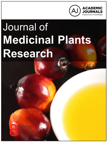
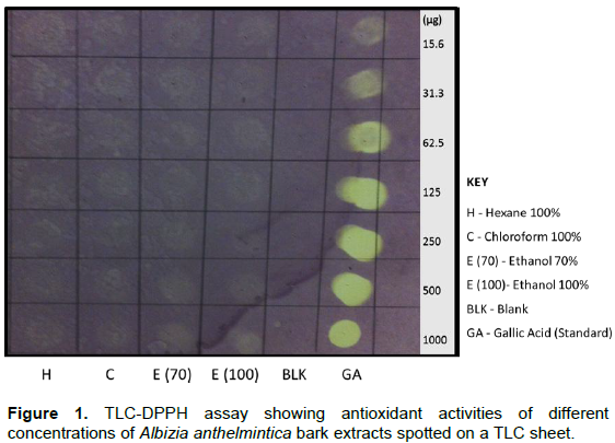
Plant extract concentrations (15.6 to 1000 µg/ml) in different solvents were observed on the TLC sheet (Figures 1 and 2). These concentrations showed moderate activities, indicated by the faint yellow colouration over the purple DPPH background in comparison with the bold colouration of the gallic acid standard. In the bark extracts, the highest activities were observed in the 100% chloroform extracts, 70% ethanol extracts and 100% ethanol extracts while the 100% hexane extracts showed little activity (Figure 1). Similar trends were observed in roots extracts (Figure 2).
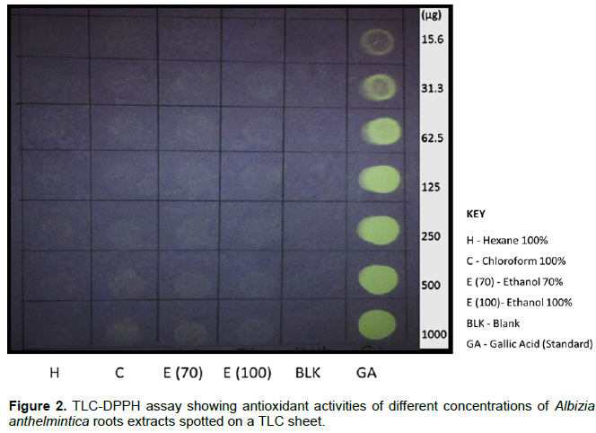
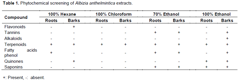
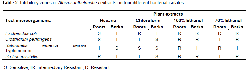
Percentage inhibitions of bark and roots extracts were determined at 500 µg/ml concentrations (Figure 3). From the highest inhibition to the lowest inhibition, the observed inhibition percentages for the different extracts were as follows: 70% ethanol roots (66.9%); 70% ethanol barks (58.9%); ethanol 100% roots (49.2%); chloroform barks (49%); hexane bark (25.5%); ethanol 100% barks (14.6%); chloroform roots (7.3%) and hexane roots (4.9%). The standard (gallic acid) displayed 86.7% DPPH inhibition at a dose of 500 µg/ml Phytochemical screening of all extracts was performed. Both 100% chloroform bark and roots extracts contained only one type of compound (terpenoids). Ethanol (100%) bark extracts contained all phenolic compounds except flavonoids (Table 1). Among all the compounds, terpenoids are the only ones present in all extracts. Chloroform barks showed the greatest antimicrobial activity of all the extracts. Barks of 100% ethanol extracts and 70% ethanol extracts showed the least antimicrobial activities (Table 2). All bacterial isolates were resistant to 100% ethanol roots extracts. Therefore, 100% ethanol roots displayed minimal antimicrobial activity.
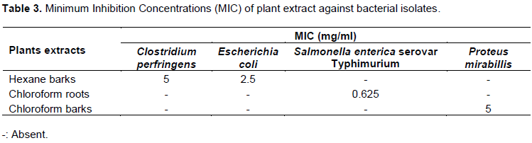
MIC values determination showed a 0.625 mg/ml for chloroform roots against S. Typhimurium. The highest recorded MIC value was 5 mg/ml for both chloroform barks and hexane barks against P. mirabilis and C. perfringens, respectively (Table 3). 100% hexane barks recorded an MIC value of 2.5 mg/ml against E. coli.
DISCUSSION
All the extracts of A. anthelmintica in this study showed antioxidant properties. Any substance that scavenges free radicals qualifies as an antioxidant. DPPH is a proton free radical. Scavenging of proton free radicals is a common mechanism of eliminating oxidants from biological systems (Krystyna and Anna, 2013). In this study, different A. anthelmintica extracts scavenged DPPH radicals to varying extents. Dasgupta and De (2007) has shown that plant extracts are mixtures of different scavenging compounds that operate synergistically to counter radical activity. The success of these extracts in suppressing DPPH radicals depends on their ability to donate a hydrogen or electron atom to react with DPPH radical. There were variations in the scavenging activities of the same extracts in different solvents. These variations could be due to the unequal distribution of the antioxidant molecules in the different parts of the plant (Krystyna and Anna, 2013).
Phytochemical analysis of the plant extracts showed that some of their constituents such as tannins, flavonoids, saponins and terpenoids may have medicinal potential. Audu et al. (2007) explain that phenolic compounds are amongst the largest and most abundant plant metabolites with a variety of biological properties such as antimicrobial, anti-apoptosis, anti-aging, anti-carcinogen and anti-inflammation. The study revealed the presence of fatty acids phenols in 4 of the 8 A. anthelmintica roots and bark extracts. Several studies, such as Himesh et al. (2011), have shown that most medicinal plants have phenolic compounds and natural antioxidants such as flavonoid, phenolic acids and tocopherols.
The present study revealed the presence of tannins in all ethanol roots extracts except 100% ethanol roots extracts. Zhao et al. (2010) has shown that tannins operate by binding to proline rich proteins to inhibit the synthesis of proteins. In this study, flavonoids were only detected in the 100% hexane bark extracts. Plants synthesize flavonoids to respond to infections by microbes and in vitro, they have been used effectively against different microorganisms (Parekh et al., 2006). Therefore, production of flavonoids is reactionary rather than routine which might explain their scarcity in most of the extract samples. The activity of flavonoids is dependent on their ability to form complexes with extracellular and soluble proteins as well as bacterial cell walls (Parekh et al., 2006).
Saponins were present in all ethanol extracts irrespective of the concentration of the solvent or the nature of the plant extract. They were however not present in any other solvent. Saponins do have the characteristic of foaming in aqueous solutions (Marin et al., 2001). In this study, terpenoids were the only compounds detected in all plant extracts. Croteau (1998) has shown that terpenoids have an important role as plant hormones and as a plant defence mechanism against microbial diseases and insect herbivores. This, therefore, means that they are indispensable to plant systems hence their ubiquitous presence across all extracts. Indeed, terpenoids have been attributed to a wide array of medicinal properties such as anti-carcinogenicity, anti-malaria, anti-ulcer, antimicrobial and diuretic activities (Aharoni et al., 2005). A. anthelmintica was previously associated with antibacterial activity because of monoterpenes that are present in essential oils made from this plant (Marin et al., 2001) which are effective against pathogenic veterinary isolates (Dasgupta and De, 2007).
In this study, 100% ethanol roots extract inhibited the growth of all bacterial isolates. C. perfringens is often involved in diseases in most domestic animals and some wildlife, including, poultry, sheep, goats, cattle, ostriches, dogs and cats (Niilo, 1993). Multidrug resistant S. Typhimurium and E. coli have been widely reported as causative agents of diarrhoea in cattle and small stock and still are a major cause of productivity and economic loss to cattle producers worldwide (Voetsch et al., 2004; Cho and Yoon, 2014).
P. mirabilis is one of the most common bacteria infecting the urinary tract in humans and dogs (Abe et al., 2017). S. Typhimurium showed intermediary inhibition and resistance to all ethanol extracts. MIC value of 0.625 mg/ml was observed on chloroform roots against S. Typhimurium. These MIC results were in agreement with the phytochemical screening results which indicated that chloroform roots contained only terpenoids.
In conclusion, findings of this study have shown that extracts of the A. anthelmintica plant species may be reliable sources of antioxidants and antimicrobials which can be used the development of novel drugs and the treatment of multi drug resistance veterinary pathogens.
Furthermore, this study provides valuable information that can not only guide future studies on medicinal plants but can also be an educational reference for students and scholars of Botswana.
CONFLICT OF INTERESTS
The authors have not declared any conflict of interests.
ACKNOWLEDGEMENTS
The authors are thankful to office of Research and Development, University of Botswana for funding this study.
REFERENCES
|
Abe T, Iizuka A, Kojima H, Kimura K, Shibahara T, Haritani M (2017). Necrotizing suppurative nephritis in a Japanese black feedlot steer due to Proteus mirabilis infection. The Journal of Veterinary Medical Science 79(4):709-713. |
|
|
Aharoni A, Jongsma MA, Bouwmeester HJ (2005). Volatile science? Metabolic engineering of terpenoids in plants. Trends in Plant Science 10(12):594-602. |
|
|
Audu SA, Mohammed I, Kaita HA (2007). Phytochemical screening of the leaves of Lophira lanceolata (Ochanaceae). Life Science Journal 4(4):75-79. |
|
|
Bauer A, Lewish W, Healter G (1996). Antibiotic susceptibility testing by a standard single dics method. Clinical pathology 45:493-496. |
|
|
Cho Y, Yoon KJ (2014). An overview of calf diarrhea - infectious etiology, diagnosis, and intervention. Journal of Veterinary Science 15(1):1-17. |
|
|
Wikler MA (2008). Performance standards for antimicrobial susceptibility testing: eighteenth informational supplement. Clinical and Laboratory Standards Institute (CLSI). |
|
|
Croteau RB (1998). The Discovery of Terpenes. Discoveries in Plant Biology Vol. I, eds. Shain-Dow Kung and Shang-Fa Yang. Singapore: World Scientific pp. 112-120. |
|
|
Dasgupta N, De B (2007). Antioxidant activity of some leafy vegetables of India: A comparative study. Food chemistry 101:471-474. |
|
|
Grade J, Arble B, Welad J, Van Damme (2008). Anthelmintic efficacy and dose determination of Albizia anthelmintica against gastrointestinal nematodes in naturally Infected Ugandan sheep. Veterinary Parasitology 157:267-274. |
|
|
Himesh S, Sarvesh S, Sharan PS, Mishra K (2011). Preliminary phytochemical screening and HPLC Analysis of Flavonoid from Methanolic Extract of Leaves of Annona squamosa. International Research Journal of Pharmacy 2(5):242-246. |
|
|
Krystyna P, Anna P (2013). Application of free radical diphenylpicrylhydrazyl (DPPH) to estimate the antioxidant capacity of food samples. Analytical Methods 5(17):4288-4295. |
|
|
Marin PD, Grayer RJ, Veitch NC, Kite GC, Harborne JB (2001). Acacetin glycosides as taxonomic markers in Calamintha and Micromeria. Phytochemistry 58(6):943-947. |
|
|
Mazimba A, Rajendra A, Punitha IS (2006). Antioxidant studies on the benzyl tetra isoquinoline alkaloid berberine. Biological and Pharmaceutical Bulletin 29(9):1906-1910. |
|
|
Niilo L (1993). Enterotoxemic Clostridium perfringens. Pathogenesis of bacterial infections in animals pp. 114-123. |
|
|
Notka F, Meier G, Wagner R (2004). Concerted inhibitory activities of Phyllanthus amarus on HIV replication in vitro and ex vivo. Antiviral Research 64(2):93-102. |
|
|
Parekh J, Karathia N, Chanda S (2006). Evaluation of antibacterial activity and phytochemical analysis of Bauhinia variegata L. bark. African Journal of Biomedical Research 9(1):53-56. |
|
|
Sharma SP (2016). Pathological findings in animals in Gaborone Area. Botswana Journal of Agriculture and Applied Sciences 10(1):24-29. |
|
|
Thomas L (2000). Root based responses account for A. anthelmintica survival at high nickel concentration. Journal of Plant Physiology 174:137-146. |
|
|
Voetsch AC, Van Gilder TJ, Angulo FJ, Farley MM, Shallow S, Marcus R, Cieslak PR, Deneen VC (2004). Tauxe RV Emerging Infections Program FoodNet Working Group. FoodNet estimate of the burden of illness caused by nontyphoidal Salmonella infections in the United States. Clinical Infectious Diseases 38(3):127–134. |
|
|
Yeboah E, Majinda RRT (2004). Erythrinaline alkaloids from the flowers and pods of Erythrina lysistemon and their DPPH radical scavenging properties. Phytochemistry 65(10):1397-1404. |
|
|
Zhao J, Zhang JS, Yang B (2010). Free radical scavenging activity and characterization of sesquiterpenoids in four species of curcuma using a TLC bioautography assay and GC-MS analysis. Molecules 11(15):7547-755. |
|
Copyright © 2024 Author(s) retain the copyright of this article.
This article is published under the terms of the Creative Commons Attribution License 4.0


