Full Length Research Paper
ABSTRACT
Phytochemicals possessing free radical scavenging and antiproliferative activities play an important role in cancer chemoprevention. In this study, the ethanolic extracts prepared from fresh and dried leaves, bark, and seed skin of Pra (Elateriospermum tapos Blume.) were tested for their antioxidant activity using a ferric reducing ability of plasma (FRAP) assay. The antiproliferative activities against four human cancer cell lines, breast (MCF-7), colon (HT-29, HCT116) and cervical (HeLa) cancer cells, were examined using the MTT assay. Fresh leaf extract exhibited the greatest antioxidant activity by giving the greatest FRAP value, followed by bark, seed skin and dried leaf extracts, respectively, which were strongly correlated to their flavonoid content. Dried leaf extract with the highest total phenolic content exhibited the greatest antiproliferative activity against all cancer cell lines tested. The growth of cervical cancer cell line (HeLa) was the most sensitive to all plant part extracts tested with IC50 values of 5.44 ± 0.57 µg/ml (fresh leaf), 5.41 ± 0.37 µg/ml (dried leaf), 6.39 ± 1.16 µg/ml (bark) and 7.03 ± 0.07 µg/ml (seed skin) at 72 h exposure. The non-cancer cell line (Vero) was more resistant to all plant part extracts when compared with the cancer cell lines. The ethanolic extracts of all E. tapos plant parts are promising for further purification and drug development.
Key words: Antioxidant, antiproliferation, chemoprevention, Elateriospermum tapos, ethanolic extract.
INTRODUCTION
The leakage of free radicals during respiration is an unavoidable consequence and may account for cell or tissue injury. The reactive oxygen species (ROS) are involved in both initiation and promotion stages of multistage carcinogenesis via damaging DNA, proteins and lipids (Klaunig and Wang, 2018; Perchellet, 1995). Antioxidants are generally known as the free radical scavengers, acting as the inhibitors at both initiation and promotion stages of carcinogenesis and protecting cells against oxidative damage. Chemoprevention involves the administration of chemical agents to block initiation and promotion processes of cancer development. The cancer prevention program of US National Cancer Institute using animal model or epidemiological studies includes (1) identification and characterization of chemopreventive agents, (2) establishment of efficacy and toxicity testing of candidate compounds in animal model systems, and (3) establishment of human intervention trials of promising chemopreventive agents (Boone et al., 1990; Golemis et al., 2018). The study at cellular level is definitely required prior to animal studies, although it has not literally mentioned.
Nowadays, cancer is the leading cause of the death of human especially breast, colon and cervical cancers (National Cancer Institute, 2009). In the United States, 1,735,350 new cancer cases and 609,640 cancer deaths are expected to occur in 2018 (Siegel et al., 2017). In Thailand, five types of cancers: breast, cervix, colorectal, liver and lung cancers are accounted for approximately 60% of the cancer burden (Virani et al., 2017). A tremendous number of cancer patients in the world face problem with high cost of chemotherapy especially in developing countries bringing about an inevitable option for development of herbal medicine. A number of research indicated that there are many types of plants and different parts of some edible plant extracts that contained high level of phenolic compounds and had significant anticancer activities (Ismail et al., 2012; Ramasamy et al., 2011; Sanseera et al., 2016; Senawong et al., 2014). Besides anticancer properties, these plants also possess antioxidant properties, preventing oxidative damage and tumorigenesis. Different prepared extracts of some plants and their parts have been reported to contain wide range of flavonoids, steroids, phenolics and saponins which may exert varied pharmacological activities like anticancer, antidiarrheal, antimicrobial and antioxidant (Gandhi and Mehta, 2013). Many research found different kinds of plants, vegetable, fruits that are able to cure cancer or prevent cancer, however the search for new anticancer plants is still needed in order to find more effective results and less toxic.
Elateriospermum tapos Blume, locally known in Thai as Pra, Kra, or Perah, is a plant in the family Euphorbiaceae, distributed across the national highland rain forest or the high humidity mountain located in the southern part of Thailand especially in the Bantad Mountain Range in Trang and Nakornsrithammarat provinces (Chayamarit and van Welzen, 2005). This plant is also abundant in lowland forests throughout Malaysia especially in northern parts of Peninsular Malaysia (Ling et al., 2006).
The well-known medicinal plant Suregada multiflora Baill. of the same family (Euphorbiaceae) has been recorded for its anticancer activity (Itharat et al., 2004) and has been used in Thai traditional medicine (Chayamarit and van Welzen, 2005), however, there is no recorded data for the use of E. tapos in cancer treatment. The white and sticky latex exuding from its bark, leaves and fruit stalks, is used for the treatment of cracked sole of the foot (Chai et al., 1989). Our recent data from a screen program searching for natural products with anticancer properties indicated that E. tapos plant parts exhibited a promising antiproliferative activity against several human cancer cell lines (Tisadoldilok and Senawong, 2017). In this study, the ethanolic extracts prepared from fresh and dried leaves, bark, and seed skin of Pra were further investigated for their antioxidant activity using ABTS and ferric reducing ability of plasma (FRAP) assays comparing with the previous results from (2,2-diphenyl-1-picryl-hydrazyl-hydrate) (DPPH) assay. The anticancer activities of the extracts characterized by the half maximal inhibitory concentration (IC50) values against four human cancer cell lines, breast (MCF-7), colon (HT29, HCT116) and cervical (HeLa) cancer cell lines, were evaluated using MTT assay.
MATERIALS AND METHODS
Plant materials
Fresh and dried leaves, bark, and seed skin of Pra (E. tapos) were collected from Nakornsrithamaraj Province, Thailand, during August - October, 2015. Taxonomic identification was approved by the Forest Herbarium, Department of National Parks, Wildlife and Plant Conservation, Ministry of Natural Resources and Environment, Bangkok, Thailand. A voucher specimen (voucher number ST15001) is deposited at Department of Biochemistry, Faculty of Science, Khon Kaen University, Khon Kaen, Thailand.
Preparation of plant extracts
The plant parts used in this study included fresh and dried leaves, seed skin and bark. The leaves, seed skin and bark of Pra (E. tapos) were cleaned with running tap water to remove any external material. The leaves of Pra were divided into fresh and dried samples. To prepare the dried samples, the plant parts were dried in a hot air oven at 40 to 50°C for 24 h. The fresh plant parts were cut into small pieces, whereas the dried plant samples were ground. Both fresh and dried samples of the plant parts (500 g) were soaked in 95% ethanol (2,000 ml) for seven days at room temperature. The supernatant was collected and filtered through a filter paper. The solid residue was repeatedly extracted 3 times with ethanol. The filtrates from each extraction were combined and the solvent was evaporated under reduced pressure using a rotary evaporator to yield the crude ethanolic extract of each plant part. All the crude extracts were weighed and kept in a refrigerator until they were used.
Determination of total flavonoid content
Total flavonoid content of the plant part ethanolic extracts was measured using a colorimetric method as previously described (Chuenchom et al., 2016). The flavonoid content was expressed as mg Rutin equivalent per gram of the extract.
FRAP assay
The antioxidant activities based on the ferric reducing ability of E. tapos ethanol crude extracts were analyzed according to the method of Benzie and Strain (1996) with some modifications. A reagent was prepared fresh by mixing 10 ml of 300 mM acetate buffer with 1 ml of 10 mM 2, 4, 6-Tris(2-pyridyl)-s-triazine (TPTZ) in 40 mM of hydrochloric acid (HCl) and 1 ml of 20 mM FeCl3.6H2O. The freshly prepared FRAP reagent was pre-warmed at 37°C for 5 min. Thereafter, a blank reading was taken at 595 nm using a plate reader. Subsequently, 3 µl of sample, standard or positive control (each dissolved in 10% DMSO) and 9 µl of water was added to 90 µl of the FRAP reagent. Absorbance readings were measured instantly upon addition of the FRAP reagent and again at 4 min after the start of the reaction. The change in absorbance in the 4 min reaction was calculated by comparison to the absorbance changes of FeSO4.7H2O against a standard curve (100-1,000 µM) tested in parallel. Results were expressed as millimoles (mmol) FeSO4 equivalents per gram of the extract. All experiments were carried out in triplicate.
Cell culture
Human cervical adenocarcinoma (HeLa) and colorectal adenocarcinoma (HT29) cell lines were obtained from Dr. P. Picha (National Cancer Institute, Bangkok, Thailand). Human colorectal carcinoma (HCT116) and breast adenocarcinoma (MCF-7) cell lines were kindly provided by Dr. O. Tetsu (University of California, San Francisco, U.S.A.). The non-cancer Vero cells were kindly provided by Dr. S. Barusrux (Khon Kaen University, Khon Kaen, Thailand). All cell lines were cultured in RPMI-1640 medium supplemented with 10% fetal bovine serum, penicillin (100 U/ml), and streptomycin (100 µg/ml) (Gibco-BRL) at 37°C in a humidified atmosphere with 5% CO2.
Antiproliferative activity assay
MTT (3-(4,5-dimethylthiazol-2-yl)-2,5-diphenyltetrazolium bromide) assay was used to assess antiproliferative activity of the plant extracts against human cancer cell lines. Briefly, cells were seeded in a 96-well plate (104 cells/well) and incubated for 24 h. Different concentrations (0.5-100 µg/ml) of the plant ethanolic extracts were added to the cells and incubated for 24, 48 and 72 h. Vehicle control groups were added with 0.5% dimethyl sulfoxide (DMSO). After exposure of the cells to the plant ethanolic extracts, the medium was removed, and the cells were incubated with MTT (Sigma Chemical Co., St Louis, MO) (0.5 mg/ml in PBS) for 2 h. DMSO was used to dissolve the formazan dye, which was detected by a microplate reader (Bio-Rad Laboratories, Hercules, CA, U.S.A.) at 550 nm. A reference wavelength used in this study was at 655 nm to subtract optical density caused by dead cells and cell debris. The absorbance of formazan dye was proportional to the number of viable cells. The percentage of viable cells was calculated using the following equation:
% Cell viability = [Sample (A550-A655)/Control (A550-A655)] × 100
The half maximal inhibitory concentration (IC50) values from each experiment were estimated by plotting x-y and fitting the data with a straight line (linear regression). The average of IC50 values was calculated from three independent experiments.
Statistical analysis
Data from three independent experiments were expressed as mean ± standard deviation (SD). Statistical analyses were carried out using the statistical program SPSS version 17.0 for windows (SPSS Corporation, Chicago, IL). The criterion for statistical significance was set at p<0.05.
RESULTS AND DISCUSSION
Antioxidant activities of plant part ethanolic extracts of Pra (E. tapos)
Our previous results indicated that the ethanolic extracts of E. tapos plant parts exhibited antioxidant activity by the DPPH radical scavenging assay (Tisadoldilok and Senawong, 2017). In this study, the antioxidant capacity of the plant part ethanolic extracts were determined by a FRAP assay, expressed as mg FeSO4/g sample, and compared the results with the previous DPPH results. According to DPPH and FRAP assays, fresh leaf extract, compared with those of other E. tapos plant part extracts, exhibited the greatest antioxidant capacity by giving the least IC50 value for DPPH assay and giving the greatest value for FRAP assay (Table 1). Fresh leaf and bark extracts appeared to exhibit antioxidant capacity more potent than dried leaf and seed skin extracts when compared the results from DPPH and ABTS assays. Fresh leaf extract showed approximately 31 times greater antioxidant activity than that of Trolox from DPPH assay.

Whereas in a FRAP assay, fresh leaf extract exhibited antioxidant activity values approximately 1.9 times less than those of Trolox. The FRAP assay was demonstrated to have similar predictive power as the DPPH assay on E. tapos plant part antioxidant activity. In addition, total flavonoids of the ethanolic plant part extracts correlated well with their antioxidant activities. The difference in antioxidant activities of the extracts from fresh and dried leaves might probably be due to the extraction procedure or drying process. Some active volatile compounds may have been destroyed or evaporated during the processing of samples so that the antioxidant activities of dried and fresh leaf extracts are different (Table 1). This observation is consistent with other studies (Alabri et al., 2014; Vaidya et al., 2014). The strong correlation between total flavonoids and the antioxidant capacity of E. tapos plant parts indicates that flavonoids of the plant parts may be responsible for antioxidant activity. Phenolic compounds including flavonoids may act as free radical scavengers by acting as hydrogen or electron donors, singlet oxygen quenchers and metal chelators (Riachi and De Maria, 2015).
Antiproliferative activities of ethanolic extracts of plant parts of Pra (E. tapos)
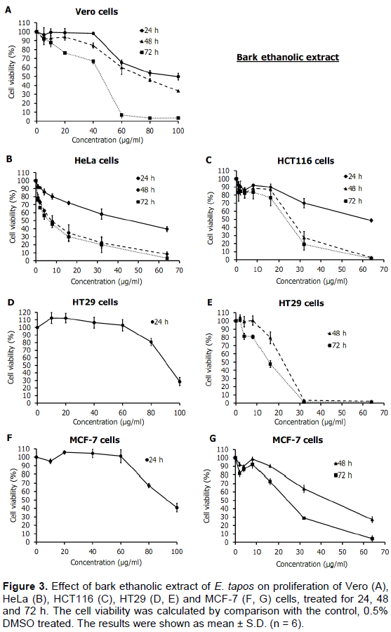
Our previous study demonstrated that ethanolic extracts of E. tapos plant parts exhibited antiproliferative activities against HeLa, HCT116, HT29 and MCF-7 cells at screening concentration of 100 µg/ml (Tisadoldilok and Senawong, 2017). In this study, antiproliferative activities of ethanolic extracts of the plant parts against four cancer cell lines (HeLa, HT29, HCT116 and MCF7) and a non-cancer cell line (Vero) were further investigated by MTT assay to obtain the IC50 values. According to dose-response curves (Figures 1 to 4), all plant part ethanolic extracts inhibited proliferation of all cancer cell lines tested in a dose- and time-dependent manner. Cellular sensitivities determined as the IC50 values against each cell line are summarized in Table 2 in accordance with the dose-response curves in Figures 1 to 4.
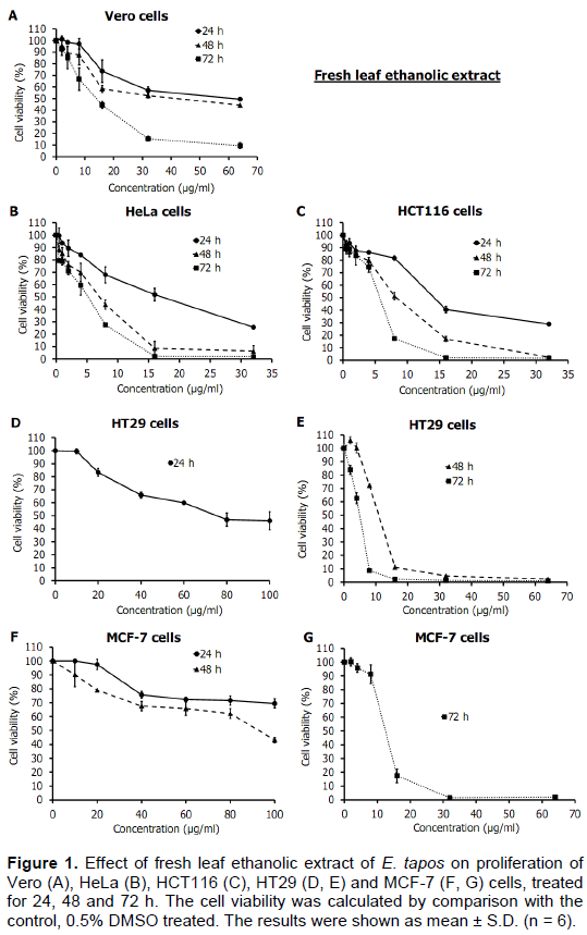
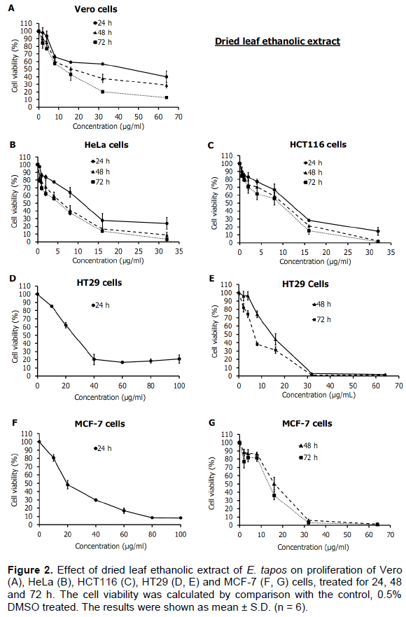
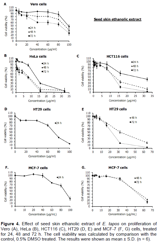
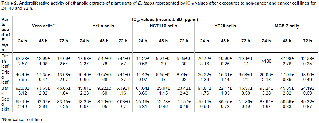
Cellular sensitivities differed depending on types of cells and extracts. In dried leaves, no further enzymatic or metabolic alteration of natural compounds would become possible further, whereas in fresh leaves there remains a possibility of formation of new compounds as secondary metabolites in responses to light and other factors. Although there is no significant difference in total phenolic contents between the extracts from dried and fresh leaves of moringa, the dried leaves significantly promoted the crude extract with higher total flavonoid content (Vongsak et al., 2013). In this study, fresh and dried leaf extracts of E. tapos were studied comparatively. Fresh leaf extract of E. tapos exhibited most effective growth inhibition against cervical cancer (HeLa) and colon cancer (HCT116) cell lines for 24 and 48 h exposures, and most effective inhibition against colon cancer (HT29) cell line for 72 h exposure (Figure 1 and Table 2). Dried leaf extract was more effective than fresh leaf extract on growth inhibition in all cancer cell lines tested for 24 h exposure (Figures 1, 2 and Table 2). However, no significant difference on growth inhibition in all cancer cell lines was observed between fresh and dried leaf extracts for 72 h exposure (Figures 1, 2 and Table 2).
Bark extract effectively inhibited the growth of HeLa, HCT116 and HT29 cells for 48 and 72 h exposures with less toxicity on the non-cancer Vero cells (Figure 3). The breast cancer MCF-7 cell line was more resistant than other cancer cell lines, but more sensitive to bark extract than the non-cancer cell line (Figure 3 and Table 2). Seed skin extract was more potent than Bark extract on growth inhibition of HeLa and HCT116 cells at exposure times of 24 and 48 h (Figures 3, 4 and Table 2). Among all plant part extracts used in this study, seed skin extract showed the least toxicity against the non-cancer Vero cells (Figure 4 and Table 2).
Based on the criteria of the American National Cancer Institute, the crude extract promising for further purification should have IC50 values lower than 30 µg/ml (Suffness and Pezzuto, 1990). In this study, IC50 values of ethanolic extracts from fresh leaves, dried leaves, and seed skin against HeLa and HCT116 cells at all exposure times (24, 24 and 72 h) are less than 30 µg/ml, indicating that ethanolic extracts from these plant parts are promising for further purification and drug development. The taraxerane triterpene, 2,3-seco-taraxer-14-ene-2,3,28-trioic acid 2,3-dimethyl ester, was previously isolated from the leaves of E. tapos and exhibited cytotoxic activity against small cell lung carcinoma cell line (NCI-H187) (Pattamadilok and Suttisri, 2008). Although antioxidant activity of the plant part extracts was strongly correlated with their flavonoid content, antiproliferative activity was not correlated with their flavonoid content (Tables 1 and 2), suggesting that type of flavonoids may have a greater impact on their antiproliferative activity. However, antiproliferative activity was correlated well with their total phenolic content.

CONCLUSION
Comparing to the antioxidant values of the other plant parts of E. tapos, fresh leaf showed the greatest antioxidant activity by giving the least IC50 value for DPPH assay and the greatest FRAP value. The growth of cervical cancer cell line (HeLa cells) was most sensitive to all plant part extracts in comparison to other cancer cell lines studied, especially to dried leaf extract. The viability of the non-cancer cell line (Vero cells) was also progressively decreased with increasing concentrations of the extracts, but more resistant to the extracts when compared with the cancer cell lines. The ethanolic extracts from these plant parts are promising for further purification and drug development as their IC50 values against cancer cell lines are less than 30 µg/ml. The data finding from this research will be used for further studies on development of the herbal cancer medicine in the future. In addition, the reported antineoplastic properties of these plant parts would promote the cultivation and preservation programs of this plant in local area.
CONFLICT OF INTERESTS
The authors have not declared any conflict of interests.
REFERENCES
|
Alabri THA, Musalami AHSA, Hossain MA, Weli AM, Al-Riyami Q (2014). Comparative study of phytochemical screening, antioxidant and antimicrobial capacities of fresh and dry leaves crude plant extracts of Datura metel L. Journal of King Saud University - Science 26(3):237-243. |
|
|
Benzie IFF, Strain JJ (1996). The ferric reducing ability of plasma (FRAP) as a measure of "antioxidant power": the FRAP assay. Analytical Biochemistry 239(1):70-76. |
|
|
Boone CW, Kelloff GJ, Malone WE (1990). Identification of candidate cancer chemopreventive agents and their evaluation in animal models and human clinical trials: a review. Cancer Research 50(1):2-9. |
|
|
Chai PPK, Lee BMH, Ismawi O (1989). Native Medicinal Plants of Sarawak. Forest Department. |
|
|
Chayamarit K, van Welzen PC (2005). Euphorbiaceae (Genera A-F). Euphorbiaceae (Genera AF). Forest Herbarium, National Part Wildlife and Plant Conservation Department. |
|
|
Chuenchom P, Swatsitang P, Senawong T, Jogloy S (2016). Antioxidant capacity and phenolic content evaluation on peanut skins from 3 peanut types. Chiang Mai Journal of Science 43(1):1177-1191. |
|
|
Golemis EA, Scheet P, Beck TN, Scolnick EM, Hunter DJ, Hawk E, Hopkins N (2018). Molecular mechanisms of the preventable causes of cancer in the United States. Genes and Development 32(13-14):868-902. |
|
|
Ismail M, Bagalkotkar G, Iqbal S, Adamu HA (2012). Anticancer properties and phenolic contents of sequentially prepared extracts from different parts of selected medicinal plants indigenous to Malaysia. Molecules 17(5):5745-5756. |
|
|
Itharat A, Houghton PJ, Eno-Amooquaye E, Burke PJ, Sampson JH, Raman A (2004). In vitro cytotoxic activity of Thai medicinal plants used traditionally to treat cancer. Journal of Ethnopharmacology 90(1): 33-38. |
|
|
Klaunig JE, Wang Z (2018). Oxidative stress in carcinogenesis. Current Opinion in Toxicology 7:116-121. |
|
|
Ling SK, Fukumori S, Tomii K, Tanaka T, Kouno I (2006). Isolation, purification and identification of chemical constituents from Elateriospermum tapos. Journal of Tropical Forest Science 18(1):81-85. |
|
|
Pattamadilok D, Suttisri R (2008). Seco-terpenoids and other constituents from Elateriospermum tapos. Journal of Natural Products 71(2):292-294. |
|
|
Perchellet (1995). Oxidative stress and multistage skin carcinogenesis. Skin cancer: mechanisms and human relevance pp. 145-180. |
|
|
Ramasamy S, Wahub NA, Abidin NZ, Manickam S (2011). Cytotoxicity evaluation of five selected Malaysian Phyllanthaceae species on various human cancer cell lines. Journal of Medicinal Plants Research 5(11):2267-2273. |
|
|
Riachi LG, De Maria CAB (2015). Peppermint antioxidants revisited. Food Chemistry 176:72-81. |
|
|
Sanseera D, Liawruangrath B, Pyne SG, Liawruangrath S (2016). Determination of antioxidant and anticancer activities together with total phenol and flavonoid contents of Cleidion javanicum Bl. and Bridelia retusa (L.) A. Juss. Chiang Mai Journal of Science 43: 534-545. |
|
|
Senawong T, Khaopha S, Misuna S, Komaikul J, Senawong G, Wongphakham P, Yunchalard S (2014). Phenolic acid composition and anticancer activity against human cancer cell lines of the commercially available fermentation products of Houttuynia cordata Thunb. Science Asia 40:420-427. |
|
|
Siegel RL, Miller KD, Jemal A (2017). Cancer Statistics. CA: A Cancer Journal for Clinicians 68:7-30 |
|
|
Suffness M, Pezzuto JM (1990). Assays related to cancer drug discovery. Methods in plant biochemistry: assays for bioactivity 6:71-133. |
|
|
Tisadondilok S, Senawong T (2017). Antioxidant activities and anticancer screening of ethanolic extracts from Baccaurea macrophylla Muell and Elateriospermum tapos Blume. Journal of Thai Interdisciplinary Research 12(5):11-18. |
|
|
Vaidya BN, Brearley TA, Joshee N (2014). Antioxidant capacity of fresh and dry leaf extracts of sixteen Scutellaria species. Journal of Medicinally Active Plants 2(3):42-49. |
|
|
Virani S, Bilheem S, Chansaard W, Chitapanarux I, Daoprasert K, Khuanchana S, Leklob A, Pongnikorn D, Laura S. Rozek LS, Siriarechakul S, Suwanrungruang K, Tassanasunthornwong S, Vatanasapt P, Sriplung H (2017). National and Subnational Population-Based Incidence of Cancer in Thailand: Assessing Cancers with the Highest Burdens. Cancers 9(8):108. |
|
|
Vongsak B, Sithisarn P, Mangmool S, Thongpraditchote S, Wongkrajang Y, Gritsanapan W (2013). Maximizing total phenolics, total flavonoids contents and antioxidant activity of Moringa oleifera extract by the appropriate extraction method. Industrial Crops and Products 44:566-571. |
|
Copyright © 2024 Author(s) retain the copyright of this article.
This article is published under the terms of the Creative Commons Attribution License 4.0


