Full Length Research Paper
ABSTRACT
Caryocar brasiliense (pequi) is an exotic fruit, high in monounsaturated fat acids (MUFA) and bioactive compounds, which have beneficial effects on cardiometabolic risk factors. However, this fruit is poorly studied in this context. In this study, the effects of pequi pulp intake on cardiometabolic risk factors of rats were evaluated. Therefore, 16 male weaned rats were divided into two groups: Control group and Pequi group. Control group was feed a standard diet and pequi group, the same diet added pequi pulp (3.26 g.100-1) for 15 weeks. At the end, plasma lipids, glucose, insulin, Homeostasis Model Assessment of Insulin Resistance index (HOMA-IR), blood pressure, heart rate, hepatic and fecal lipids and intestinal histomorphometric parameters were accessed. Liver and heart samples were harvested for redox status assays. There were no differences between experimental groups for blood pressure, heart rate, glucose, insulin, HOMA-IR, triglycerides, cholesterol, HDL-cholesterol, and liver and heart redox status (p<0.05). Pequi group had lowered lipid hepatic deposition and increased fecal output (p<0.05), increased intestinal villus height and crypt depth. Thus, pequi pulp intake minimized liver fat deposition by increasing its intestinal output and improved intestinal structure of rats, which can contribute for reducing cardiometabolic risk factors. MUFA, carotenoids and fibres can be associated, at last in part, with these effects.
Key words: Caryocar brasiliense, pequi, cardiometabolic risk, lipid metabolism, redox status.
INTRODUCTION
Non communicable diseases (NCD), such as type 2 diabetes and cardiovascular diseases, are major causes of mortality worldwide (up to 38 million by year), and are responsible for 80% of deaths occurring in developing countries (WHO, 2014). Increasing the intake of fruits and vegetables are one of the main recommendations for reducing NCD risk. From this perspective, consumer interest in foods with functional properties is increasing (Bumgarner et al., 2012; Jackix et al., 2013; Schreckinger et al., 2010).
In this context, pequi (Caryocar brasiliense) is an exotic fruit from a Brazilian savannah-like biome. Its pulp is sweet, yellowish and has a high energy density and lipid content (~33%), being monounsaturated fatty acids (MUFAs) (especially oleic, ~54%) its major constituent (Lima et al., 2007). It also has substantial amounts of fibres (18%) (Teixeira et al., 2013) and carotenoids (~42 mg.100-1g), especially violaxanthin, lutein, zeaxanthin, β-cryptoxanthin, neoxanthin and β-carotene (Azevedo-Meleiro and Rodriguez-Amaya, 2004).
From pequi pulp chemical constituents, MUFAs have been associated with improvements in lipid profile, reduced platelet aggregation, favourable modulation of blood pressure, insulin sensitivity and glycemic control (Hammad et al., 2016). Fibres are related to the regulation of intestinal function, control of body weight and lipid metabolism by decreasing the absorption and increasing excretion of cholesterol and triglycerides, and they have indirect effects on the blood pressure and serum glucose control (Lattimer and Haub, 2010). Carotenoids are powerful antioxidants, and some, such as β-carotene, have vitamin A activity. They are associated with reduced risk for cancer and cardio-vascular diseases, and protection from cell oxidative damage (Gülçin, 2012; Ried and Fakler, 2011).
Thus, the composition of nutrients and bioactive compounds of pequi pulp suggests that it could be a food supplement and it can exert effects on metabolism, cardiovascular function and cell redox status as a functional food. However, this fruit has been poorly studied. To the author’s knowledge, there are only some research showing healing (Quirino et al., 2009; Bezerra et al., 2015), chemopreventive (Palmeira et al., 2016; Colombo et al, 2015) and anti-inflammatory (Miranda-Vilela et al., 2009) properties of pequi oil. Studies regarding functional properties from pequi pulp intake are scarce (Teixeira et al., 2013).
Therefore, the aim of this study was to evaluate the effects of pequi pulp intake on cardiometabolic risk markers of rats. The in vitro antioxidant activity and the chemical composition of pequi pulp, were determined previously because some compounds could be related to its potential health benefits.
MATERIALS AND METHODS
Pequi pulp samples
Ripe pequi fruits were acquired from the local market of Diamantina city, Minas Gerais State, Brazil. They were washed with tap water and subsequently with distilled water. After drying at room temperature, each fruit was cut in half and the pulp was separated from the almond manually. Afterwards, the pulps were placed on trays and dried at 65°C for 48 h (Teixeira et al., 2013). After drying, the material was grounded, wrapped in a plastic bag, labeled and stored at -18±2°C until the analysis.
Chemical composition and in vitro antioxidant activity of pequi pulp
Protein, total lipids, dietary fibres (enzymatic–gravimetric method) and total carotenoids were determined as described by The Association of Official Analytical Chemists - AOAC methods (AOAC, 1995). Carbohydrates were calculated by difference, and the total energy value (TEV) was estimated using the Atwater factors (Buchholz & Schoeller 2004). Fatty acids were analyzed by gas chromatography (CGC Agilent 6850 Series GC System) according to The American Oil Chemist´s Society – AOCS (AOCS, 2009).
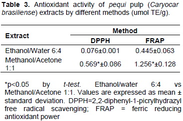
The in vitro pequi pulp antioxidant activity was performed in both 6:4 ethanol: water and 1:1 methanol: acetone extracts. Briefly, dehydrated pequi pulp samples were extracted with 40 mL of 1:1 methanol/water solution for 1 h, at room temperature. Afterwards, the mixture was centrifuged (Biosystems, Modelo 80-2B, Curitiba-PR) at 3.000 rpm for 15 min. The supernatant was harvested and the step was repeated, using a 7:3 acetone/water solution (Larrauri et al., 1997). After the solvents evaporation, the mixtures were diluted in 6:4 ethanol : water and 1:1 methanol : acetone solutions. The 2,2-diphenyl-1-picrylhydrazyl free radical scavenging (DPPH) and ferric reducing antioxidant power (FRAP) methods were used according to Rufino et al. (2010).
Rat study
Experimental protocols were performed in accordance with the EU Directive 2010/63/EU for animal experiments. They were approved by the Ethics Committee on Animal Use/Federal University of Vales do Jequitinhonha e Mucuri, Diamantina, MG, Brazil (Protocol 010/2012).
Sixteen male Wistar rats, four weeks aged, were housed in individual stainless steel cages and maintained in a room with controlled temperature (22±2°C) and a 12 h light/dark cycle, with free access to food and water during the experimental period.
A commercial chow (RhosterLab®) was used as a standard diet, and its energy density was 3.28 kcal.g-1 (13.77 kJ g-1). Based on the lipid composition of the pequi pulp, the standard diet was added, pequi pulp at 3.26.100 g-1, which resulted in a 50% increase in total lipid content, so its energy density turned into 3.39 kcal g-1 (or 14.2 kJ.g-1, a 3.35% increase). The pequi pulp supplementation was also added, 1.19 g.100 g-1 of oleic acid; 0.63 g.100 g-1 of fibres and 0.14 mg.100 g-1 of carotenoids to the standard diet.
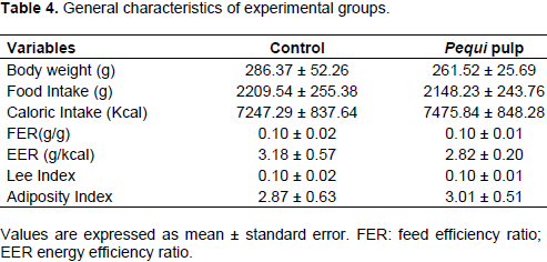
All 16 animals were randomly assigned to two treatment groups (n=8): Control group - animals fed the standard diet and Pequi pulp group - animals fed the standard diet added pequi pulp. The study lasted for 15 weeks. During this period, body weight and food intake were monitored for energy efficiency ratio (EER = weight gain/kcal) and feed efficiency ratio (FER = weight gain/g of diet) calculations. Faeces were collected in the last 72 h of the experiment, dried and kept at -80°C until analysis. The body weight and length (nose–anus length) were measured in all anaesthetized rats (quetamin + xilazin/50 mg/kg + 10 mg/kg) in the previous day to the euthanasia for the Lee index calculation (weight body.g 0.33 /nose–anus length).
On the last day, overnight fasted animals were anesthetized (quetamin + xilazin/50 mg/kg+10 mg/kg), euthanized by decapitation for blood and tissues (adipose tissues, liver, heart, duodenum) harvesting. Retroperitoneal and epididymal fat pads were used for adiposity index ([retroperitoneal + epididymal pads/body weight – (retroperitoneal + epididymal pads)]*100)) calculation. Blood was centrifuged in heparinized tubes to obtain plasma, and aliquots were transferred to Eppendorf tubes and kept at -80°C until analysis. Liver and heart tissues were processed for redox status analysis. Duodenum fragments of 5 cm were harvested and stored in a 10% formaldehyde solution for complementary histological analyses.
Cardiometabolic risk factors
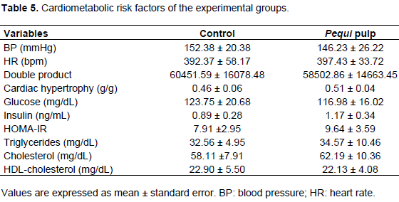
Tail blood pressure (BP) and heart rate (HR) were measured in the last week of the experimental protocol by the non-invasive tail plethysmography method. The animals were heated to cause vasodilation of the caudal artery. The pulses were recorded by system (AD Instruments Ltd, UK). The BP and HR values were used for the double product calculation (BP x HR). The heart weight and body weight were used for cardiac hypertrophy evaluation, by heart weight/body weight calculation.
Fasted plasma glucose levels (GLU) were measured by means a commercial kit according the procedures recommended by the manufacturer and using a semi-automatic biochemical analyzer (PIOWAY-3000). Fasted plasma insulin (INS) was determined using a commercially available Enzyme-Linked Immunosorbent Assay kit (Linco Research Inc., St. Louis, MO, USA) and a micro-plate reader (Spectra MAX 190, Molecular Devices, USA). Insulin resistance was accessed by the homeostasis model assessment of insulin resistance (HOMA-IR index), from fasted glucose and insulin levels according to Matthews et al. (1985).
Total plasma cholesterol (CHOL), high-density lipoprotein cholesterol (HDL-C) and triglyceride (TG) levels were determined using a semi-automatic biochemical analyzer (PIOWAY-3000) and commercial kits according the procedures recommended by the manufacturer. Liver and faeces samples were oven-dried (60°C ± 2°C for 72 h) after harvesting, grounded, and their lipids were extracted according to Folch et al. (1957). CHOL and TG levels were determined using commercial kits according the procedures recommended by the manufacturer and using a semi-automatic biochemical analyzer (PIOWAY-3000).
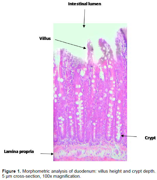
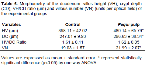
Considering the possible influences of some chemicals from pequi pulp in the intestinal morphology, which could affect nutrient digestion and absorption, we also preceded histomorphometric assays. For that, fragments of duodenum were removed and fixed in a 4% buffered formaldehyde solution. After dehydration and fixation in paraffin, two 5 µm cross-sections which were stained with haematoxylin/eosin was performed. Results were obtained by means of a digital camera coupled to a microscope. All images were analysed using the Axion Vision software. The villus height (VH) and the crypt depth (CD) were expressed as the arithmetic mean determined from 20 measurements of each sample. The villus height/crypt depth ratio (VH/CD) was also calculated. The villi density per optical field (920764.14 μm²) was taken from five photos from each animal. All measurements were made in µm, at 100x magnification (Figure 1).
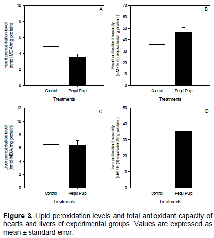
For the redox status analysis, liver and heart samples were homogenized in phosphate-buffered saline (PBS) (T 20 basic ULTRA-TURRAX; IKA Labortechnik, China), pH 7.2, and centrifuged for 10 min at 10,000x and 4°C (Jouan BR4i, Thermo Fischer Scientiï¬c, USA). The supernatant was harvested and used for the protein determination (Bradford, 1976) and the biochemical assays described below. The total antioxidant capacity was measured using the ferric reducing antioxidant power (FRAP) assay, according to the method of Benzie and Strain (1996). The formation of thiobarbituric acid-reactive substances (TBARS) during a hot acid reaction was used as an index of lipid peroxidation, according to Ohkawa et al. (1979).
Statistical analysis
Results from chemical composition and the in vitro antioxidant activity assays were expressed in mean ± standard deviation. Results from rat study were expressed in mean ± standard error. The experiment was performed in a completely randomized design with two treatments (experimental groups) and eight repetitions. Data were analyzed by one way ANOVA at p<0.05, using the Statistica 10.0 software. Figures were drawn by means of the SigmaPlot 11.0 software.
RESULTS
Chemical composition of pequi pulp and in vitro antioxidant assays
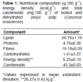
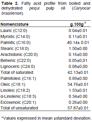
Pequi pulp had expressive amounts of total lipids (Table 1). The main fatty acids from pequi lipids were oleic followed by palmitic (Table 2). Pequi pulp also had high amounts of fibres, being majorly insoluble, and total carotenoids (Table 1). For the in vitro antioxidant activity assays, pequi pulp methanol/acetone extract had higher antioxidant capacity, by both FRAP and DPPH methods (p<0.05) (Table 3).
Rat study
Similar body weights were found at the beginning and end of the experiment. The food and caloric intake, FER, EER, Lee index and adiposity index did not differ between groups (Table 4). There were no differences for BP, HR, double product, hypertrophy index, plasma markers of glucose and lipids metabolism markers (glucose, insulin, HOMA-IR, triglycerides, cholesterol and HDL-C) (Table 5).
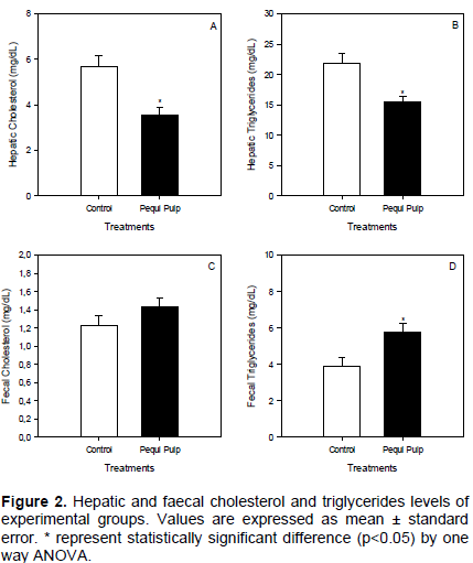
Regarding hepatic and faecal lipids, pequi pulp animals had lower hepatic levels of CHOL and TAG when compared with controls (p<0.05) (Figure 2A and B). Faecal CHOL did not differ between groups. Pequi group had higher faecal TAG levels when compared with C (p<0.05) (Figure 2D).
From the duodenum histomorphometric assays, an increase in the villus height (VH), Crypt depth (DC) and number of villous (NV) per optical field for pequi pulp group (p<0.05) was observed. However, differences were not observed between treatments for VH/DC ratio (Table 6). There were no differences between groups for both liver and heart lipid peroxidation levels and antioxidant capacity (Figure 3). However, for hearts, pequi group had a 23% increase in the antioxidant capacity and a 28% decrease in the peroxidation levels as compared to the control.
DISCUSSION
Pequi is an exotic fruit with a heavy potential to be a functional food, since it has a peculiar nutritional composition and is high in several bioactive compounds. In this study, the amount of lipids found in the pequi pulp samples was in accordance with previous data from the laboratory (Teixeira et al., 2013) and higher than that found by Cardoso et al. (2013) and Lima et al. (2007). Also, pequi pulp has a paradoxal composition in fatty acids, since oleic acid, a MUFA, is its major constituent and related to cardiometabolic risk reduction. Otherwise its second higher constituent is palmitic acid, a saturated fatty acid with several cytotoxic effects described (Akazawa et al., 2010; Eitel et al., 2002).
Its amount of fibres and carotenoids were comparable to other commonly consumed foods that are high in those compounds (Rodriguez-Amaya et al., 2008). Otherwise, carotenoids were lower than other samples analysed elsewhere (Cardoso et al., 2013; Lima et al., 2007; Teixeira et al., 2013). These differences may be due the different processing forms of pequi pulp samples in those studies. In these samples, boiling and dehydrating may have lowered carotenoid content and also, increased other nutrient concentrations, such as lipids and fibres. The higher antioxidant activity of methanol/acetone pequi pulp extract indicated that lipophilic compounds account significantly for the pulp antioxidant power.
Based on these findings, it is clear that this fruit has expressive amounts of certain nutrients and bioactive compounds that have been associated with protection in many biochemical processes that underly the development of cardiometabolic diseases. Therefore, study of some biological effects of these compounds together in a single food which is still poorly studied, is proposed.
Adding pequi pulp did not influence body weight gain, food intake, glucose and plasma lipids. In addition, this increase also did not affect BP and HR and it did not cause cardiac overload, since no changes in the double product and cardiac hypertrophy index was observed. There was 50% increase in total lipid content of standard diet by adding pequi pulp. Although, there was increase in calories and lipids, the diet did not turned into a high fat, which could cause metabolic disturbance. According to Buettner et al. (2006) and Hariri and Thibault (2010), to have high fat, a diet must have at least 30% of its energy from lipids. The pequi group diet had 18.51%.
Although, the main chemical constituents from pequi pulp (mainly MUFAs and fibre) are related to CHOL, TG and LDL-lowering effects (Ried and Fakler, 2011) as well as having antioxidant properties (carotenoids) (Gülçin, 2012), we did not observe these effects on plasma lipids. Otherwise, there were pronounced effects from pequi pulp supplementation in the hepatic and faecal lipids. The lower hepatic levels of TRI and CHOL and higher TRI faecal output in the pequi group may be associated with the higher pequi pulp fibre content.
Fibres, especially soluble, can increase the lumen viscosity, bind to cholesterol, triglycerides, bile acids and other lipids, impairing their digestion/absorption and increasing their faecal output (Lattimer and Haub, 2010). They can also be fermented by microflora and generate products, as acetate, propionate and butyrate, which affect the endogenous synthesis of theses lipids (Ngoc et al., 2012). Insoluble fibres, in turn, regulate intestinal transit-time, contributing to the lower absorption of those nutrients (Lattimer and Haub, 2010). Furthermore, insoluble fibres are related to a higher expression of hepatic genes that increase fatty acid oxidation (Isken et al., 2010).
Therefore, the pequi pulp could have modulated the function of the gastrointestinal tract to increase the lipids excretion. Histological data corroborated these findings. Pequi group showed higher villous height and crypt depth, implying this food exerted a positive effect in the mucosa integrity. Conversely, the DC increase in this group indicated a high rate of cell differentiation in crypts. In addition, the increase in VH indicated cell migration and renovation to the villus (Rosa et al., 2010).
Some compounds of pequi pulp can be related to that. Fibres were associated with VH increase (Ashraf et al., 2013) and oleic acid was associated with a better gut development and a DC increase (Rosa et al., 2010). In addition, carotenoids, as vitamin A precursors can act on intestinal cell growth and differentiation (Allen et al., 2002). As antioxidants, they can decrease damage caused by oxidative agents and therefore, contribute to cell preservation (Turan et al., 2009). Then, it can be inferred that pequi pulp may have increased the duodenal absorption surface area and cell renovation, helping the maintenance of the mucosa integrity.
Regarding redox status, statistical differences were not observed between groups for livers and hearts. According to several authors (Feillet-Coudray et al., 2009; Sour et al., 2015), changes on redox status parameters are easily detectable when dietary lipid and caloric overload occurs, or when there are some physiological disturbance, such as inflammation, obesity and dyslipidaemia. Adding pequi pulp did not increase significantly lipid content of the diet. In addition, the lipid lower liver accumulation and increased faecal output upon pequi pulp intake may also be related to these results, since it can have contributed to a less generation of reactive oxygen and nitrogen species with subsequent lower peroxidation of membrane lipids.
However, in the heart, it is important to consider that pequi pulp led to a trend in increasing antioxidant capacity and decreasing lipid peroxidation levels. It seems that the heart was the more sensitive organ upon pequi pulp intake. Carotenoids are natural antioxidants, have lipophilic characteristic and may be incorporated into mitochondrial membranes, which are the main site for free radical production during the electron flow (Vega et al., 2009). Furthermore, this in vitro assay showed that methanol and ethanol extracts of pequi pulp had a high antioxidant capacity.
CONCLUSION
Taken together, the results indicate that pequi pulp intake minimized liver fat deposition by increasing its faecal output and improved intestinal structure, which could account for reduction of cardiometabolic risk in rats. Fibres, MUFA and carotenoids from this fruit may be responsible, at last in part, for these effects.
CONFLICTS OF INTERESTS
The authors have not declared any conflict of interest.
ACKNOWLEDGEMENTS
This work was supported by the Brazilian agencies Fundação de Amparo à Pesquisa do Estado de Minas Gerais (FAPEMIG) and Conselho Nacional de Desenvolvimento Científico e Tecnológico (CNPq). The authors gratefully acknowledge the Instituto de Pesquisas e Estudos de Lassance (IPEL) for the contribution of the biochemical kits.
REFERENCES
|
American Oil Chemists' Society (AOCS) (2009). Official methods and recommended practices of the AOCS Urbana: AOCS. |
|
|
Akazawa Y, Cazanave S, Mott JL, Elmi N, Bronk SF, Kohno S, Charlton MR, Gores GJ (2010). Palmitoleate attenuates palmitate-induced Bim and PUMA up-regulation and hepatocyte lipoapoptosis. J. Hepatol. 52:586-593. |
|
|
Allen S, Maden M, Price J (2002). A role for retinoic acid in regulating the regeneration of deer antlers. Dev. Biol. 251:409-423. |
|
|
Association of Official Analytical Chemists (AOAC) (1995). Ofï¬cial Methods of Analysis of AOAC International Washington DC: AOAC International. |
|
|
Ashraf S, Zaneb H, Yousaf M, Ijaz A, Sohail M, Muti S, Usman M, Ijaz S, Rehman H (2013). Effect of dietary supplementation of prebiotics and probiotics on intestinal microarchitecture in broilers reared under cyclic heat stress. J. Anim. Physiol. Anim. Nutr. (Berl). 97:68-73. |
|
|
Azevedo-Meleiro CH, Rodriguez-Amaya DB (2004). Confirmation of the identity of the carotenoids of tropical fruits by HPLC-DAD and HPLC-MS. J. Food Compost. Anal. 17:385-396. |
|
|
Benzie IF, Strain J (1996). The ferric reducing ability of plasma (FRAP) as a measure of "antioxidant power": the FRAP assay. Anal. Biochem. 239:70-76. |
|
|
Bezerra NKMS; Barros TL, Coelho NPMF. A ação do óleo de pequi (Caryocar brasiliense) no processo cicatricial de lesões cutâneas em ratos (2015). Rev. Bras. Plant Med. 17:875-880. |
|
|
Bradford MM (1976). A rapid and sensitive method for the quantitation of microgram quantities of protein utilizing the principle of protein-dye binding. Anal. Biochem. 72:248-254. |
|
|
Buchholz AC, Schoeller DA (2004). Is a calorie a calorie? Am. J. Clin. Nutr. 79(5):899S-906S. |
|
|
Buettner R, Parhofer K, Woenckhaus M, Wrede C, Kunz-Schughart L, Schölmerich J, Bollheimer L (2006). Defining high-fat-diet rat models: metabolic and molecular effects of different fat types. J. Mol. Endocrinol. 36:485-501. |
|
|
Bumgarner NR, Scheerens JC, Kleinhenz MD (2012). Nutritional Yield: A proposed index for fresh food improvement illustrated with leafy vegetable data. Plant Foods Hum. Nutr. 67:215-222. |
|
|
Cardoso LDM, Reis BDL, Hamacek FR, Sant'ana HMP (2013). Chemical characteristics and bioactive compounds of cooked pequi fruits (Caryocar brasiliense Camb.) from the Brazilian savannah. Fruits 68:3-14. |
|
|
Colombo NBR, Rangel MP, Martins V, Hage M, Gelain DP, Barbeiro DF, Capelozzi VL (2015). Caryocar brasiliense camb protects against genomic and oxidative damage in urethane-induced lung carcinogenesis. Braz. J. Med. Biol. Res. 48:852-862. |
|
|
Eitel K, Staiger H, Brendel MD, Brandhorst D, Bretzel RG, Häring H-U, Kellerer M (2002). Different role of saturated and unsaturated fatty acids in β-cell apoptosis. Biochem. Biophys. Res. Commun. 299:853-856. |
|
|
Feillet-Coudray C, Sutra T, Fouret G, Ramos J, Wrutniak-Cabello C, Cabello G, Cristol J, Coudray C (2009). Oxidative stress in rats fed a high-fat high-sucrose diet and preventive effect of polyphenols: Involvement of mitochondrial and NAD (P) H oxidase systems. Free Radic. Biol. Med. 46:624-632. |
|
|
Folch J, Lees M, Sloane-Stanley G (1957). A simple method for the isolation and purification of total lipids from animal tissues. J. Biol. Chem. 226:497-509. |
|
|
Gülçin I (2012). Antioxidant activity of food constituents: an overview. Arch. Toxicol. 86:345-391. |
|
|
Hammad S, Shuaihua P, Jones PJ (2016). Current evidence supporting the link between dietary fatty acids and cardiovascular disease. Lipids 51(5):507-17. |
|
|
Hariri N, Thibault L (2010). High-fat diet-induced obesity in animal models. Nutr. Res. Rev. 23:270-299. |
|
|
Isken F, Klaus S, Osterhoff M, Pfeiffer AFH, Weickert MO (2010). Effects of long-term soluble vs. insoluble dietary fiber intake on high-fat diet-induced obesity in C57BL/6J mice. J. Nutr. Biochem. 21:278-284. |
|
|
Jackix AE, Monteiro EB, Raposo HF, Amaya-Farfán J (2013). Cholesterol reducing and bile-acid binding properties of taioba (Xanthosoma sagittifolium) leaf in rats fed a high-fat diet. Food Res. Int. 51:886-891. |
|
|
Lattimer JM, Haub MD (2010). Effects of dietary fiber and its components on metabolic health. Nutrients 2:1266-1289. |
|
|
Larrauri JA, Rupérez P, Sauracalixto F (1997). Effect of drying temperature on the stabilitity of polyphenols and antioxidant activity of red grape pomace peels. J. Agric. Food Chem. 45:1390-1393. |
|
|
Lima AD, Silva AMdOe, Trindade RA, Torres RP, Mancini-Filho J (2007). Composição química e compostos bioativos presentes na polpa e na amêndoa do pequi (Caryocar brasiliense Camb.). Rev. Bras. Frutic. 29:695-698. |
|
|
Matthews D, Hosker J, Rudenski A, Naylor B, Treacher D, Turner R (1985). Homeostasis model assessment: insulin resistance and β-cell function from fasting plasma glucose and insulin concentrations in man. Diabetologia 28:412-419. |
|
|
Miranda-Vilela AL, Pereira LCS, Gonçalves CA, Grisolia CK (2009). Pequi fruit (Caryocar brasiliense Camb.) pulp oil reduces exercise-induced inflammatory markers and blood pressure of male and female runners. Nutr. Res. 29:850-858. |
|
|
Ngoc T, Hong T, Len N, Lindberg J (2012). Effect of fibre level and fibre source on gut morphology and micro-environment in local (Mong Cai) and exotic (Landrace Yorkshire) pigs. Asian-Australas J. Anim. Sci. 25:1726-1733. |
|
|
Ohkawa H, Ohishi N, Yagi K (1979). Assay for lipid peroxides in animal tissues by thiobarbituric acid reaction. Anal. Biochem. 95:351-358. |
|
|
Palmeira SM, Silva PR, Ferrão JS, Ladd AA, Dagli ML, Grisolia CK, Hernandez-Blazquez FJ (2016). Chemopreventive effects of pequi oil (Caryocar brasiliense Camb.) on preneoplastic lesions in a mouse model of hepatocarcinogenesis. Eur. J. Cancer Prev. 25(4):299-305. |
|
|
Quirino GS, Leite GO, Rebelo LM, Tomé AR, Costa JGM, Cardoso AH, Campos AR (2009). Healing potential of Pequi (Caryocar coriaceum Wittm.) fruit pulp oil. Phytochem. Lett. 2:179-183. |
|
|
Ried K, Fakler P (2011). Protective effect of lycopene on serum cholesterol and blood pressure: Meta-analyses of intervention trials. Maturitas 68:299-310. |
|
|
Rodriguez-Amaya DB, Kimura M, Amaya-Farfan J (2008). Fontes brasileiras de carotenoides: Tabela brasileira de composição de carotenoides em alimentos Brasília: Ministério do Meio Ambiente. |
|
|
Rosa DD, Sales RLd, Moraes LFdS, Lourenço FC, Neves CA, Sabarense CM, Ribeiro SMR, Peluzio MdCG (2010). Flaxseed, olive and fish oil influence plasmatic lipids, lymphocyte migration and morphometry of the intestinal of Wistar rats. Acta Cir Bras. 25:275-280. |
|
|
Rufino MSM, Alves RE, de Brito ES, Pérez-Jiménez J, Saura-Calixto F, Mancini-Filho J (2010). Bioactive compounds and antioxidant capacities of 18 non-traditional tropical fruits from Brazil. Food Chem. 121:996-1002. |
|
|
Schreckinger ME, Lotton J, Lila MA, de Mejia EG (2010). Berries from South America: a comprehensive review on chemistry, health potential, and commercialization. J. Med. Food 13:233-246. |
|
|
Sour S, Belarbi M, Sari N, Benammar C, Baghdad C, Visioli F (2015). Argan oil reduces, in rats, the high fat diet-induced metabolic effects of obesity. Nutr. Metab. Cardiovasc. Dis. 25:382-387. |
|
|
Teixeira TN, Esteves EA, Oliveira LG, Oliveira MLP, Santana RC, Rodrigues AP (2013). Caryocar brasiliense pulp increases serum HDL and reduces hepatic lipid accumulation in rats fed a high fat diet. J. Med. Plant Res. 7:963-969. |
|
|
Turan A, Gill R, Dudeja PK, Mohan H, Mahmood A (2009). Effect of fat feeding on pro-oxidant and anti-oxidant enzyme systems in rat intestine: possible role in the turnover of enterocytes. Dig. Dis. Sci. 54:1229-1236. |
|
|
Vega VA, Anzulovich AC, Varas SM, Bonomi MR, Giménez MS, Oliveros LB (2009). Effect of nutritional vitamin A deficiency on lipid metabolism in the rat heart: Its relation to PPAR gene expression. Nutrition 25:828-838. |
|
|
World Health Organization (WHO) (2014). Global status report on noncommunicable diseases 2014. |
|
Copyright © 2024 Author(s) retain the copyright of this article.
This article is published under the terms of the Creative Commons Attribution License 4.0


