Full Length Research Paper
ABSTRACT
Hyraceum (HM) used in traditional medicine in Southern Africa is produced by the herbivore Procavia capensis. It is fossilized excreta derived from urine, faecal matter and plant material. In this study a qualitative phytochemical screening, determination of the in vitro antioxidant activity using the 1, 1-Diphenyl-2-picrylhydrazyl (DPPH) and hydrogen peroxide scavenging methods, and determination of the total phenolic content in the crude methanolic (95%) extract were done. Phytochemical screening detected the major phytochemical classes in the hyraceum extract as terpenoids, saponins, polyphenols, quinones, phlobatannins and coumarins with the minor components as flavonoids, alkaloids, tannins, simple phenols, anthocyanins, anthraquinones and amino acids. Total phenolics content was 37.339 mg gallic acid equivalents per gram dry weight (mgGAE/g DW). Effective concentration at 50% (EC50) for HM and L-ascorbic (AA) in DPPH assay was 5.983 and 0.429 µg/ml respectively while in H2O2 scavenging assay EC50 was 5.059 and 1.666 µg/ml, respectively. The antioxidant activity of HM could have been due to the various phenolic and terpenoid antioxidants in the HM. The findings implied that HM was slightly stronger at scavenging H2O2 than at scavenging DPPH. Bioactive compounds in HM could potentially be exploited in further studies as potential antioxidants of therapeutic value.
Key words: Phytochemicals, 1, 1-Diphenyl-2-picrylhydrazyl (DPPH), hydrogen peroxide.
INTRODUCTION
The production of reactive oxygen species (ROS) and reactive nitrogen species (RNS) is the trigger of non-communicable environmental disease (Zeliger, 2015). The ROS and RNS damage biological molecules, in vivo via oxidative stress. Elevated levels of ROS, acting via molecular level toxic effects, are now thought to be responsible for a wide spectrum of diseases, including lipid peroxidation of cellular membranes which causes decrease in membrane fluidity, DNA attack, adduction, enzyme inhibition, oxidative attack on the central nervous system and cell signaling; all of which have been linked to non-communicable diseases (NCDs) including neurodegenerative diseases (such as Alzheimer's disease, Parkinson's disease, amyotrophic lateral sclerosis), cancer, cardiovascular disease, diabetes, and others (Basu et al., 1999; Ames et al., 1993; Kovacic and Somanathan, 2012).
Reactive oxygen species (ROS) are continuously generated inside the human body while the generated ROS are detoxified by antioxidants present in cells. However, over production of ROS or inadequate antioxidant defences can lead to oxidative damage of various biomolecules including proteins, lipids, lipoproteins and DNA. Free radicals are the major cause of chronic and degenerative diseases such as coronary heart diseases, inflammatory stroke, diabetes and cancer (Scalbert et al., 2005).
An antioxidant is generally defined as any substance that effectively prevents or delays the adverse effects caused by free radicals and the amount of the antioxidant is less than that of the substance to be oxidized (Halliwell and Gutteridge, 1999). An antioxidant significantly delays or prevents oxidation of cell components (susceptible to oxidation) such as proteins, lipids, carbohydrates and deoxyribonucleic acid (DNA). Due to their redox properties, antioxidants act as reducing agents, hydrogen donors, singlet oxygen quenchers and chelating metal (Tung et al., 2009; Lauro and Francis, 2000). Although several synthetic antioxidants such as butylated hydroxyanisole (BHA) and butylated hydroxytoluene (BHT) are commercially available, their toxicity has always been a concern and strong restrictions have been placed on their application in pharmaceuticals. There is therefore a need for more effective, less toxic and cost effective antioxidants. Recently, there has been an upsurge of interest in the therapeutic potential of plant-derived antioxidants in reducing free radical-induced tissue injury and the current trend is to substitute synthetic with naturally occurring antioxidants (Barlow, 1990). Several biologically active compounds of plant origin (phytochemicals) have been found to possess antioxidant, free radical scavenging activity and many are being applied therapeutically for free radical associated disorders (Lee et al., 2000).
The concoctions used in traditional medicine contain many secondary metabolites (SMs) of plant origin such as polyphenolic compounds and polysaccharides which interact synergistically (Eid et al., 2013, 2012; Mulyaningsih et al., 2010; Wink, 2015). The therapeutic benefit of plant-based traditional medicine is often attributed to the antioxidant properties and potential of the constituent phytochemicals (Hertog et al., 1993; Zhang et al., 2001).
In Lesotho, as in many other countries in the world, a system of traditional medicine based on the use of plants, birds, animals, their products and their combinations to treat a broad spectrum of communicable and non-communicable diseases is still being practiced (Padmanabhan and Sujana, 2008).
One of such products used in Lesotho from animals is the fossilized excreta of Procavia capensis (rock hyrax) called hyraceum or “moroto oa pela” in Sesotho. The Basotho use hyraceum to treat respiratory infections, urinary tract or bladder infections, measles and non-communicable diseases such as diabetes mellitus.
The hyraceum is also used in combination with other medicinal plant species to enhance its efficacy (Seleteng-Kose et al., 2015). The strongly aromatic hyraceum is also a well-known Khoikhoi medicine, often used as a post-natal medicine for mothers and babies and a remedy for hysteria and epilepsy (van Wyk, 2008). The fossil is formed from the faeces and urine of P. capensis as the major components which accrete to form dark brown, resin-like masses to which plant material, pollen grains and other digestive remains are trapped. Fossilization occurs with time in arid regions. P. capensis inhabits shelters in rocky outcrops in a variety of biomes and feeds on a variety of grasses, shrubs, tree leaves, fruits and berries including the bark of the tree (Olsen et al., 2008).
The only literature found on the biological activity of hyracei was that on its effect on the GABA-benziodiazepin receptor, which indicated that since the hyracei exhibited a high affinity for GABA-benziodiazepin they could be used to treat epilepsy, a non-communicable disease (Olsen et al., 2008). Other than that, no records were found on the scientific validation of traditional medicinal uses of hyraceum (van Wyk, 2008).
In view of complex nature of hyraceum, composed of animal metabolic waste and plant materials and its use to treat both communicable and non-communicable diseases, the present study was performed and involved the analyses of the crude methanolic (95%) extract of hyraceum for the content of phytochemical and antioxidant activity and the relationship between the total phenolic content and antioxidant activity.
MATERIALS AND METHODS
Hyraceum material
A sample of hyraceum was purchased at the main Maseru open air market. Maseru is the capital of Lesotho with the geographical coordinates of; Latitude: -29° 19' 0.01" S, Longitude: 27° 28' 59.99" E. Part of the sample was fragmented and dried in a fanned oven (Labcon) at 35°C to a constant weight and brittle. The dried hyraceum was ground to a fine powder using an electrical pulverizer (Kenwood) and assessed for antioxidant activity.
Preparation of extract
The crude methanolic (95%) extract of hyraceum was prepared according to the method of Adedapo et al. (2009) with slight modifications. Ground material was soaked with 95% methanol (v/v) in distilled water at 2 ml/g and shaken on an orbital shaker for 24 h. The extract was filtered through No. 1 Whatman filter paper using suction. The filtrate was concentrated using a Gallenkamp rotary evaporator and oven dried at 35ºC overnight to constant weight and stored in glass Petri dish at 4°C until used.
Chemicals
All solvents and reagents were of analytical grade. L-ascorbic (AA), gallic acid (GA), Folin-Ciocalteau’s phenol reagent and 1, 1- Diphenyl-2-picrylhydrazyl (DPPH) were acquired from Sigma-Aldrich, St Louis, MO, USA. Methanol (absolute) was a product of Associated Chemical Enterprises (Pty) Ltd (Johannesburg, South Africa); Hydrochloric acid, Glacial acetic acid and Hydrogen peroxide were products of UNILAB (Krugersdorp, South Africa). All spectrophotometric measurements were done on the Shimadzu 1201 UV-VIS Spectrophotometer (Shimadzu, Kyoto, Japan).
Qualitative phytochemical screening
The hyraceum extract was subjected to a qualitative screening for the presence of major phytochemical classes using standard phytochemical methods and the appropriate reagents and chemicals according to the modified methods of Trease and Evans (1984; Trease and Evans, 2002) by Nwaoguikpe et al. (2014), Soni and Sheetal (2013) and Uddin et al. (2014). The reaction mixture was visually assessed as in Lu et al. (2014), for precipitation, foam formation, colour change and colour intensity according to the following key: (+), Low presence; (++), Moderate presence; (+++), High presence and (-), Absence. The colour intensity of the solutions of hyraceum and (or) the appearance of solids or precipitates enabled a semi-quantitative evaluation for the presence of antioxidants in extract solutions (Chukwudi and Yusha’u, 2016).
Determination of total phenolic content (TPP)
Based on the results of the colorimetric phytochemical profile of hyraceum extract, which showed that most of the compounds present in the hyraceum were phenolic, only the total phenolic content was determined. The total phenolic content was determined by the methods of McDonald et al. (2001) and Sharma and Joshi (2011) using the Folin-Ciocalteu reagent and expressed in milligrams of gallic acid equivalents per gram dry weight (mgGAE/gram DW) of extract. A 1.0 mL volume of HM extract solution (5.0 g/L in ethanol) was mixed thoroughly with Folin-Ciocalteau reagent (1.0 mL) in a volumetric flask. After 3 min, 4.0 mL of sodium carbonate (Na2CO3) solution (0.7M) was added, and then the mixture was allowed to stand for 2 h at room temperature in the dark with intermittent shaking. The absorbance of the mixture was measured at 760 nm in a Shimadzu 1201 UV-VIS spectrophotometer. The absorbance of the solutions was measured at 760 nm. All determinations were done in triplicates. A standard calibration curve was obtained by mixing 1ml ethanolic solutions of gallic acid of various concentrations (0.025-0.200 mg/ml) with 1ml Folin-Ciocalteau’s reagent (ten-fold dilution) and 4.0 ml sodium carbonate (0.7M) and treated in the same manner as the hyraceum extract. All tests were done in triplicate.
The total phenolic content of the extract was determined in milligrams of gallic acid equivalents per gram dry weight (mgGAE/g DW) of extract using the gallic acid standard curve, calculated from the following formula:
C = c×V/m (1)
Where; C = Total phenolic content in mgGAE/g DW of hyraceum extract, c = Concentration of the gallic acid established from the gallic acid standard curve (mg/ml), V = Volume of the extract used in the assay (ml) and m = mass of the hyraceum extract used in the assay (g) (Genwali et al., 2013).
Antioxidant activities
The 2-diphenyl-2-picrylhydrazyl (DPPH) free radical and Hydrogen peroxide (H2O2) scavenging activity of hyraceum extract were used to determine antioxidant activity of the hyraceum determination of milligram gallic acid equivalents per gram of dry weight (mgGAE/g DW) of extract and the concentration of extract needed to inhibit oxidation by 50% (EC50).
1, 1-Diphenyl-2-picrylhydrazyl (DPPH) free radical scavenging activity of hyraceum extract
The free radical scavenging activity of the extract was evaluated using the in vitro DPPH free radical scavenging assay (Nwaoguikpe et al., 2014). Crude hyraceum extract solutions were prepared by dissolving 0.03 g powder of the extract in 10 ml of 50% methanol (v/v in water) to obtain a stock solution of 3000 µg/ml from which serial dilutions of concentrations (2000, 1500, 1000, 500, 200, 100 and 0 µg/ml) were obtained. An aliquot of 0.1 ml of each of the sample dilutions was placed into reaction tubes, followed by the addition of 1.0 ml of 0.1 mM DPPH solution dissolved in absolute methanol and 0.45 ml of 50 mM Tris-HCl buffer (pH 7.40). The contents were mixed and incubated at room temperature for 30 min.
The absorbance of the mixture was measured at 517 nm against the corresponding blank solution (50% v/v methanol in distilled water) in a Shimadzu 1201 UV-VIS spectrophotometer. The assay was performed in triplicates. L-ascorbic acid was used as positive control (Magama et al., 2013).
DPPH free radical scavenging was calculated as percentage inhibition of DPPH free radical based on the control reading, which contained DPPH and distilled water only using the formula:
DPPH scavenged (%) = [(Acont-Atest)/Acont] x 100, where Acont and Atest are absorbance of control and test respectively.
The assay was performed in triplicates. The antioxidant activity of the hyraceum extract was expressed as EC50; the EC50 value being defined as the concentration (in µg/ml) of the extract that was required to scavenge 50% of the initial concentration of the DPPH free radical and was calculated from the equation of the linear regression curve of the graph of concentration versus percent scavenging of DPPH free radical using the means of the three determinations (Moyo et al., 2013; Do et al., 2014; Elfalleh et al., 2018).
Hydrogen peroxide scavenging activity
The hydrogen peroxide scavenging activity of the hyraceum was estimated by replacement titration (Nwaoguikpe et al., 2014) using 100 µl of the solutions (3000, 2000, 1000, 500 and 0 µg/ml). All tests were done in triplicate. The percentage of hydrogen peroxide scavenged was calculated as follows (Vinodhini and Lokeswar, 2014; Abayomi et al., 2014):
Hydrogen peroxide scavenged (%)=[(Vcont-Vtest)/Vcont] x 100, (2)
Where Vcont and Vtest was the volume of sodium thiosulphate used to titrate control sample in the presence of hydrogen peroxide (without extract) and volume of sodium thiosulphate solution used in the presence of the extract, respectively.
The EC50 value in the hydrogen peroxide scavenging assay was defined as the concentration (in µg/ml) of the extract that reduced the initial hydrogen peroxide concentration by 50% (Moyo et al., 2013) and was calculated from the equation of the linear regression curve of the graph of concentration versus percent inhibition of H2O2 using the mean values obtained from three determinations (Do et al., 2014). L-ascorbic was used as a standard.
Data analysis
Data was expressed as means ± standard deviations of three replicate determinations using Microsoft excel 2013. Differences between controls and treatment groups were determined using Student’s t-test. P-values of less than 0.05 were considered statistically significant using the
RESULTS
Qualitative phytochemical profile of the hyraceum extract
The yield of the hyraceum crude methanol extract from the whole mass of hyraceum was 6.17%. In Table 1 is presented the qualitative biochemical profile of hyraceum extract obtained from the various tests. Colour intensities of the hyraceum solutions (ranging from absence of antioxidant (-) to high presence (+++) for the most intense colour reaction, gave a semi-quantitative indication for the presence of different classes and relative the amounts of phytochemicals in the crude extract (Lu et al., 2014; Chukwudi and Yusha’u, 2016). Most of the classes of compounds identified in the hyraceum were phenolic in nature (flavonoids, alkaloids, tannins, terpenoids, saponins, phenols, polyphenols, anthocyanins, quinones, phlobatannins, anthraquinones and coumarins) and amino acids. No sterols, cardiac glycosides, reducing sugars, fatty acids and proteins were detected in the hyraceum extract. The highest colour intensities (+++) indicating a high presence were exhibited by terpenoids, saponins, polyphenols, quinones, phlobatannins and coumarins followed by anthocyanins (++). The lowest colour intensities, indicating a low presence (+), were given by flavonoids, alkaloids, tannins, phenols, anthraquinones, and amino acids.

Quantitative determination of total phenolics content (TPP)
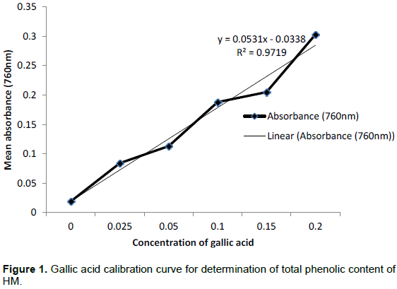
In Figure 1 is presented the gallic acid calibration curve for determination of total phenolics of the hyraceum extract. The regression equation obtained was y = 0.053x - 0.033, R2=0.971; where y was the mean absorbance of the sample at 760 nm and x, the concentration established from the gallic acid calibration curve. The total phenolic content of the hyraceum extract was found to be 37.339 mg GAE/g dry weight (37.339 mg GAE/g DW).
The results of the DPPH assay are presented in Table 2. A dose-dependent decrease in absorbance was observed which also reflected a dose-dependent increase in the percentage scavenging of the DPPH free radical for both the hyraceum and L-ascorbic acid. The highest concentration (3000 µg/ml) of the hyraceum crude extract and the reference compound L-ascorbic acid gave the lowest absorbance values at 517 nm while the lowest concentration (0 µg/ml), being the negative control, gave the highest absorbance as shown.
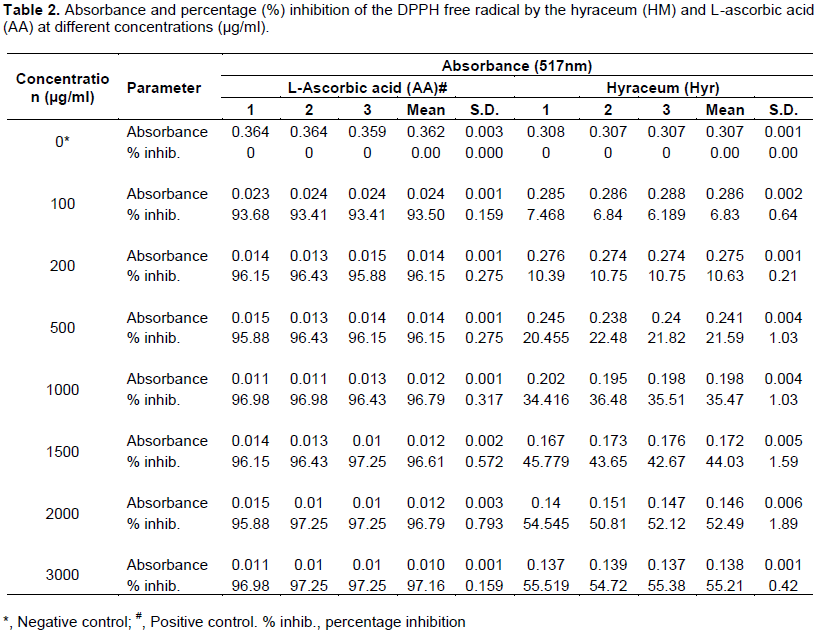
As shown in Table 2, the highest percentage (%) inhibition of the DPPH free radical by the hyraceum extract (55.21±0.42%) was observed at the maximum concentration of 3000 µg/ml used in the study, while at the same concentration, L-ascorbic acid scavenged 97.16 ±0.16% of the DPPH free radical. The lowest value for the DPPH free radical scavenging activity by the hyraceum extract (6.83±0.64%) was observed at100 µg/ml while at that same concentration L-ascorbic acid scavenged 93.50±0.16% of the free radical. The EC50 for hyraceum (HM) extract was 5.983 µg/ml calculated from the linear regression equation y = 8.651x-10.7, R2=0.981 while the EC50 for L-ascorbic acid (AA) was 0.4293 µg/ ml, calculated from the equation y = 8.292x + 46.44, R2 = 0.359 shown in Figure 2.

The crude hyraceum extract showed significant DPPH free radical scavenging activity, of 55.21±0.42% at the maximum concentration of 3000 µg/ml. At the highest concentration used in the study, there was a significant (p<0.05) difference in DPPH free radical scavenging activity between the hyraceum extract and L-ascorbic acid.It was observed that antioxidant values of the hyraceum extract were lower than those of L-ascorbic acid and with a significant difference from those of the standard L-ascorbic acid (p≤0.05).
In Table 3 is presented the results of the hydrogen peroxide scavenging assay. From 0 to 500 µg/ml the hydrogen peroxide scavenging effect of the hyraceum was insignificant, being similar to that of the negative control (0%), while at 5 µg/ml L-ascorbic acid scavenged 77.78±4.81% of the hydrogen peroxide. L-ascorbic acid in the concentration range from 500 to 3000 µg/ml was very potent; the hydrogen peroxide scavenging reaction being instantaneous following the addition of one drop of thiosulphate and was therefore recorded as being greater than 77.78±4.81%.

The EC50 for hydrogen peroxide scavenging by L- ascorbic acid (AA) was calculated from the equation y = 16.17x+23.06, R2=0.571 while that for the hyraceum (HM) was calculated from the regression equation y = 16.05x -31.2, R2=0.844 (Figure 3). The EC50 obtained for AA (1.666 µg/ml) was lower than that for HM (5.059 µg/ml) indicating a higher antioxidant activity of the AA in this assay.

DISCUSSION
The qualitative phytochemical screening of HM revealed the presence of different classes of phytochemicals in the crude methanolic (95%) extract of the hyraceum. There was a high presence (+++) of terpenoids, saponins, polyphenols, quinones, phlobatannins and coumarins (Table 1) in the solution of the hyraceum based on the intensity of the colour in the colorimetric tests, which indicated that these phytochemicals were most abundant. Other phytochemicals that were detected at smaller quantities were anthocyanins with a moderate presence (++), while flavonoids, alkaloids, tannins, simple phenols, anthraquinones and amino acids exhibited a low presence (+). Sterols, cardiac glycosides and cardenolides, reducing sugars, proteins and fatty acids were not detected and hence assumed to be absent or below detection limit of the methods used. With the exception of terpenoids, all the phytochemicals detected in the hyraceum extract were phenolic in nature even though they vary in structure. Terpenoids are highly conjugated molecules also with antioxidant properties just like phenolic compounds. According to Bang et al. (2015) phytochemicals such as carotenoids, terpenoids, ascorbates, reducing carbohydrates and tocopherols contribute to the antioxidant activity of natural products. The antioxidant activity of the crude methanolic (95%) extract of HM could therefore be attributed to the antioxidants detected in the hyraceum solution (Table 1). Phenolic compounds have antioxidant activity because they can donate hydrogen atoms to neutralize free radicals and the phenolic compounds form will be stabilized by resonance. Tannins act as antioxidants because of their ability to stabilize lipids and inhibition of lipoxygenase. Hydrolysable tannins have anti-ischemic activity. Alkaloids, particularly indole, are efficient at inhibiting the chain reaction of free radicals. All types of phenolic compounds are reported to exhibit antioxidant activity but to different degrees (Ahmad et al., 2014) and flavonoids are reported to exhibit antitumour, anti-inflammatory. The antioxidant activity of phenolic compounds is mainly due to their redox properties which make them act as reducing agents, hydrogen donors, singlet oxygen quenchers and also may have a metallic chelating potential (Rice-Evans et al., 1996). Synergism among the antioxidant compounds in the crude extract makes the antioxidant activity is not only dependent on the concentration but also on the structure and interaction between the antioxidant compounds (Djeridane et al., 2006).
The phytochemicals identified in the hyraceum listed in Table 1 were also identified in the beaver castoreum, another complex product of animal metabolic waste and plant materials which exudes from the castor sacs of the mature North American beaver (Castor canadensis) and the European beaver (Castor fiber) (Müller-Schwarze, 2003).
Pyrolysis gas chromatography mass spectrometry analysis of hyraceum identified nitrogen-containing aromatic compounds, notably benzamide (Carr et al., 2010). The solvent-extractable lipids of hyraceum comprised homologous suites of long-chain n-alkanes (C24–C34) and n-alkanols (C16–C26) characteristic of higher plant leaf waxes, along with an abundance of animal-derived sterols, higher plant sterols and terpenoids as well as benzamide (Carr et al., 2010). As early as 1879, Green and Parker (1879) analyzed hyraceum and found that about 56% of it was soluble in water, the remaining insoluble material was organic and composed of woody fibre, sand and other inorganic substances as mixtures of various salts such as soda and lime (having the highest proportion among the salts). The organic matter contained traces of urea together with uric, hippuric, and benzoic acids due to the fact that hyraceum is partly derived from urine and faecal matter (Green and Parker, 1879). No study was found on the phytochemical composition of the hyraceum with which to compare the results.
Only the total phenolics content of hyraceum crude extract was determined in this study. The determined value of total phenolics (Figure 1) of the hyraceum extract of 37.339 mg GAE/g dry sample of the hyraceum was much higher than the highest value of 1.699 mg GAE/g observed with cow dung, another complex product of animal metabolic waste and plant materials by Jirankalgikar et al. (2014) most probably due to the fossilized nature of the hyraceum.
The hyraceum extract was tested for antioxidant activity using the DPPH free radical and the hydrogen peroxide (H2O2) scavenging assays.
As shown in Table 2, at 3000 µg/ml the highest percentage inhibition of the DPPH free radical and the EC50 for the hyraceum (HM) extract were 55.21±0.42% and 5.983 µg/ml respectively and for L-ascorbic acid (AA) were 97.16±0.16% and 0.4293 µg/ml respectively. The EC50 is the concentration of extract that causes a 50% decrease in the initial concentration of the DPPH free radical (Do et al., 2014). A lower IC50 represents higher antioxidant activity (Do et al., 2014; Proestos et al., 2013). Jarald et al. (2008) observed an EC50 of 5.10 µg/ml in studies with cow urine while Jirankalgikar et al. (2014) observed an EC50 range of between 12.810 and 41.554 mg/ml with cow dung samples collected at different times of the day in a six day period of study. In the same study by Jirankalgikar et al. (2014), the EC50 of L-ascorbic acid was 20.13 µg/ml. The EC50 of the hyraceum extract of 5.983 µg/ml obtained in the current study is similar to that obtained by Jarald et al. (2008) of 5.10 µg/ml in studies with cow urine but lower than the values of between 12.810 and 41.554 mg/ml obtained in studies with cow dung by Jirankalgikar et al. (2014).
In this study, hydrogen peroxide scavenging activity was estimated by the replacement titration method. As shown in Table 3, at 3000 µg/ml the highest percentage scavenging of H2O2 and the EC50 for the hyraceum (HM) extract were 73.61±8.56% and 5.059 µg/ml, respectively and for L-ascorbic acid (AA) were greater than 77.78±4.81% and 1.666 µg/ml, respectively. No literature on hydrogen peroxide scavenging activities by mammalian metabolic waste products was found for comparison with the results of this study. However, in a study by Vinodhini and Lokeswari (2014) the methanolic leaf extract of Toona ciliate scavenged a maximum of 55.69 ± 1.04% of hydrogen peroxide.
CONCLUSION
The colorimetric tests in the present study detected the major phytochemical components of the hyraceum extract as terpenoids, saponins, polyphenols, quinones, phlobatannins and coumarins and the minor phytochemicals as flavonoids, alkaloids, tannins, simple phenols, anthocyanins, anthraquinones and amino acids. Sterols, cardiac glycosides and cardenolides, reducing sugars, proteins and fatty acids were not detected. The total phenolic content of the hyraceum extract was 37.339 mg GAE/gram dry weight. The hyraceum extract
was more effective at scavenging H2O2 (73.61±8.56%) than the DPPH free radical (55.21±0.42%). Bioactive compounds in HM could potentially be exploited in further studies as potential antioxidants of therapeutic value in preference to synthetic antioxidants as they have negative side-effects in the body.
CONFLICT OF INTERESTS
The authors have not declared any conflict of interests.
ACKNOWLEDGMENTS
This work was supported by a Research grant provided by the Research and conference committee, National University of Lesotho, 2014.
REFERENCES
|
Abayomi M, Adebayo AS, Bennett D, Porter R, Campbell JS (2014). In vitro antioxidant activity of Bixa orellana (Annatto) Seed Extract. British Journal of Pharmaceutical Research 4(11):1387. |
|
|
Adedapo AA, Jimoh FO, Afolayan AJ, Masika PJ (2009). Antioxidant properties of methanol extracts of the leaves of Celtis africana. Records of Natural Products 3:23-31. |
|
|
Ahmad AR, Wisdawati S, Asrifa WO (2014). Study of Antioxidant activity and determination of Phenol and Flavonoid content of Pepino's Leaf extract (Solanum muricatum Aiton). |
|
|
Ames BN, Shigenaga MK, Hagen TM (1993). Oxidants, Antioxidants and the Degenerative Disease of Aging. Pearsoneducation limited. London. Hal. pp. 791-798. |
|
|
Bang RP, Rosario RMD, Palmes ND (2015). Phytochemical profiles and antioxidant activity of selected indigenous vegetables in Northern Mindanao, Philippines. |
|
|
Barlow SM. 1990. Toxicolgical aspect of antioxidants used as food additives: In Hudson BJF (ed.) Food antioxidants. London, UK: Elsevier. Pp.253-307. |
|
|
Basu TK, Temple NJ, Garg MI (1999). Antioxidants in human health and disease. Cabi publishing,UK. |
|
|
Carr AS, Boom A, Chase BM (2010). The potential of plant biomarker evidence derived from rock hyrax middens as an indicator of palaeo environmental change, Palaeogeography, Palaeoclimatology, Palaeoecology 285(3-4):321-330. |
|
|
Chukwudi IE, Yusha'u M (2016). Phytochemical screening and brine shrimp lethality assay of the leaf extracts of Cucurbita maxima, Euphorbia hirta, Leptadenia hastata and Mitracarpus scaber. |
|
|
Djeridane A, Yousfi M, Nadjemi B, Boutassouna D, Stocker P, Vidal N (2006). Antioxidant activity of some Algerian medicinal plants extracts containing phenolic compounds. Food Chemisty 97(4):654-660. |
|
|
Do QD, Angkawijaya AE, Tran-Nguyen PL, Huynh LH, Soetaredjo FE, Ismadji S, Ju YH (2014). Effect of extraction solvent on total phenol content, total flavonoid content, and antioxidant activity of Limnophila aromatica. Journal of Food and Drug Analysis 22:296-302. |
|
|
Eid SY, El-Readi MZ, Eldin EEMN, Fatani SH, Wink M (2013). Influence of combinations of digitonin with selected phenolics, terpenoids, and alkaloids on the expression and activity of P-glycoprotein in leukemia and colon cancer cells. Phytomedicine 21(1):47-61. |
|
|
Eid SY, El-Readi MZ, Wink M (2012). Synergism of three-drug combinations of sanguinarine and other plant secondary metabolites with digitonin and doxorubicin in multi-drug resistant cancer cells. Phytomedicine 19(14):1288-1297. |
|
|
Elfalleh W, Hannachi H, Tlili N, Yahia Y, Nasri N, Ferchichi A (2012). Total phenolic contents and antioxidant activities of pomegranate peel, seed, leaf and flower. Journal of Medicinal Plants Research 6(22):4724-4730. |
|
|
Genwali GR, Acharya PP, Rajbhandari M (2013). Isolation of gallic acid and estimation of total phenolic content in some medicinal plants and their antioxidant activity. Available at: |
|
|
Greene WH, Parker AJ (1879). Note on hyraceum. American Journal of Pharmacy (1835-1907):363. |
|
|
Halliwell B, Gutteridge JMC (1999). Free Radicals in Biology and Medicine. 3rd ed. New York:Oxford University Press, pp.1-35. |
|
|
Hertog MG, Feskens EJ, Hollman PC, Katan MB, D Kromhout (1993). Dietary antioxidant flavonoids and risk of coronary heart disease: the Zutphen elderly study. The lancet 342(8878):1007-1011. |
|
|
Jarald E, Edwin S, Tiwari V, Garg R, Toppo E (2008). Antioxidant and antimicrobial activities of cow urine. Global Journal of Pharmacology 2(2):20-22. |
|
|
Jirankalgikar N, Nariya P, De S (2014). In vitro antioxidant activity evaluation and HPTLC profile of Cow dung. International Journal of Green Pharmacy (IJGP) 8(3). |
|
|
Kovacic P, Somanathan R (2012). Redox processes in neurodegenerative disease involving reactive oxygen species. Current Neuropharmacology 10(4):289-302. |
|
|
Lauro GJ, Franscis FJ (2000). Natural Food Colurants. New York, USA: Marcel Dekker. pp. 1-10. |
|
|
Lee YM, Kim H, Hong EK, Kang BH, Kim SJ (2000). Water extract of 1:1 mixture of phellodendron cortex and Aralia cortex has inhibitory effects on oxidative stress in kidney of diabetic rats. Journal of Ethnopharmacology 73: 429-43. |
|
|
Lu Y, Knoo T, Wiart C (2014). Phytochemical analysis and antioxidant activity determination on crude extracts of Melodinus eugeniifolus barks and leaves from Malaysia. Pharmacology & Pharmacy 5:773-780. |
|
|
McDonald S, Prenzler PD, Autolovich M, Robards K (2001). Phenolic content and antioxidant activity of olive extracts. Food Chemisty 73:73-84. |
|
|
Moyo M, Amoo SO, Ncube B, Ndhlala AR, Finnie JF, Van Staden J (2013). Phytochemical and antioxidant properties of unconventional leafy vegetables consumed in southern Africa. South African Journal of Botany 84:65-71. |
|
|
Müller-Schwarze D (2003). The Beaver: Its Life and Impact. Page 43. |
|
|
Mulyaningsih S, Sporer F, Zimmermann S, Reichling J, Wink M (2010). Synergistic properties of the terpenoids aromadendrene and 1,8-cineole from the essential oil of Eucalyptus globulus against antibiotic-susceptible and antibiotic resistant pathogens. Phytomedicine 17(13):1061-1066. Nwaoguikpe RN, Ujowundu CO, Emejulu AA (2014). The antioxidant and free radical scavenging effects of extracts of seeds of some neglected legumes of south-east Nigeria. Scholars Academic Journal of Biosciences 2:51-59. |
|
|
Olsen A, Prinsloo LC, Scott L, Jager AK (2008). Hyraceum, the follized metabolic product of rock hyraxes (Procavia capensis), shows GABA-benzodiazepine receptor affinity. South African Journal of Science 103:437-438. |
|
|
Oyono VA, Fokunang C, Assam JPA, Voundi S, Tsafack P, Mouafo ET, Ngandjui BT, Beng VP (2014). Acute toxicity studies, antioxidant and in vitro antibacterial activities of extract from the barks of Ricinodendron heudoletti (Euphorbiaceae). Journal of Pharmacognosy and Phytotherapy 6(4):47-53. |
|
|
Padmanabhan P, Sujana KA (2008). Animal products in traditional medicine from Attappady hills of western Ghats. |
|
|
Proestos C, Lytoudi K, Mavromelanidou OK, Zoumpoulakis P, Sinanoglou VJ (2013). Antioxidant capacity of selected plant extracts and their essential oils. Antioxidants 2:11-22. |
|
|
Rice-Evans C, Miller NJ, Paganga G (1996). Stucture–antioxidant activity: activity relationships of flavonoids and phenolic acids. Free Radical Biology and Medicine. 20(7):933-956. |
|
|
Scalbert A, Manach C, Remecsy C, Morand, C (2005). Dietary polyphenols and the prevention of diseases Critical Reviews in food Science and Nutrition 45:287-306. |
|
|
Seleteng-Kose L, Moteetee A, van Vuuren S (2015). Ethnobotanical survey of medicinal plants used in the Maseru district of Lesotho. Journal of Ethnopharmacology 170:184-200 |
|
|
Sharma M, Joshi SJ (2011). Comparison of antioxidant activity of Andrographis paniculata and Tinospora cordifolia leaves. |
|
|
Soni A, Sheetal S (2013). Phytochemical analysis and free radical scavenging potential of herbal and medicinal plant extracts. Journal of Pharmacognosy and Phytochemistry 2:22-29. |
|
|
Trease GE, Evans CW (1984). Pharmacognos, 12th ed. Balliere Tindall, London, UK, London. |
|
|
Trease GE, Evans WC (2002). Textbook of Pharmacognosy. 15th Ed. Saunders Publishers, London. |
|
|
Tung YT, Wub JH, Huang CY, Chang ST (2009). Antioxidant activities and phytochemical characteristic of extract of Acacia confuse bark. Bioresource Technology 100:509-514. |
|
|
Uddin G, Feroz S, Ali J, Rauf A (2014). Antioxidant, antimicrobial activity and phytochemical investigation of Pterospermum acerifolium (Leaf petiole). Wudpecker Journal Agriculture Resource 3:058-062. |
|
|
van Wyk BE (2008). A review of Khoi-San and Cape Dutch Medical Ethnobotany. Journal of Ethnopharmacology 119(3):331-341. |
|
|
Vinodhini V, Lokeswari TS (2014). Antioxidant activity of the isolated compounds, methanolic and hexane extracts of Toona ciliata leaves. International Journal of Engineering and Technology 4(3). |
|
|
Wink M (2015). Modes of Action of Herbal Medicines and Plant Secondary Metabolites. Medicines 2(3):251-286. |
|
|
Zeliger HI (2015). Causes, Mechanisms and Prevention of Environmental Diseases. |
|
|
Zhang Z, Chang Q, Zhu M, Huang Y, Ho WKK, ZY Chen (2001). Characterization of antioxidants present in hawthorn fruits. The Journal of Nutritional Biochemistry 12:144-152. |
|
Copyright © 2024 Author(s) retain the copyright of this article.
This article is published under the terms of the Creative Commons Attribution License 4.0


