Full Length Research Paper
ABSTRACT
This study evaluated if the oral administration of a bottled medicine containing Echinacea, Graviola, Purple Ipe, Sucupira and Cat’s claw could cause changes in biochemical, coagulation, hematological and histopathological parameters in rats. This study arised after a report to Ceatox of a patient who used a bottled medicine and developed hematological, renal and histopathological changes. The bottled medicine possesses 4.32 mg alcohol.mL-1 and by the phytochemical screening and UV/Scan tests showed some classes of secondary metabolites. In experiments using Wistar rats, two treatments were performed, single dose and 30 days, administering water, alcohol and bottled medicine by gavage. In the single dose treatment, alterations of uric acid, cholesterol, creatinine, alkaline phosphatase, iron and glucose (p<0.05) were found. Already in the 30 days treatment were alterations of uric acid, total and indirect bilirubins, creatinine, alkaline phosphatase, glucose, triglycerides, erythrocytes and leukocytes (p<0.05), and hepatic sinusoidal dilatation. The data showed that the alcohol present in the bottled medicine can alter biochemical, hematological parameters and cause liver damage in rats. The bottled medicine, although not showing alterations in important biochemical and hematological parameters, had more pronounced liver histopathological alterations.
Key words: Bottled medicine, biochemical, coagulation and hematological parameters.
INTRODUCTION
The popular culture of the use of medicinal plants in preparations as bottled medicine are common practices being used in the prophylaxis and treatment of many diseases (Silva and Freire, 2010).
The bottled medicine is a popular formulation prepared with components of vegetable, mineral, animal, spice and alcohol as vehicle. They are products sold in free markets, distributed by health centers, without any type of registration or quality control and who promise to cure various types of diseases (Camargo, 2011). The common mistake that natural products are non-toxic and have no adverse effects leads people to misuse without guidance, resulting in health problems, especially in cases of overuse or chronic use (UNESCO, 2013).
Studies in the scientific literature show that the administration of infusions of plant extracts in experiments with rats, both acute and chronic, causes alterations in biochemical parameters, especially hepatic enzymes, alanine aminotransferase (ALT) and aspartate aminotransferase (AST), urea, creatinine, uric acid, glucose and hematological parameters (Da Paz et al., 2005; Campelo et al., 2013).
In popular medicine, many plant extracts are used for the treatment of various types of diseases, among them Echinacea (Echinacea purpurea), Graviola (Annona muricata), Purple Ipe (Tabebuia avellanedae), Sucupira (Pterodon emarginatus) and Cat’s claw (Uncaria tomentosa), which are commonly found in bottled medicine of popular use (Lorenzi and Matos, 2008).
Studies have shown that E. purpurea has an in vitro effect on hematopoiesis, with stimulation of blood proliferative activity, induces mononuclear leukocyte production, stimulates granulocyte activity and increases the proportion of CD4/CD8 markers (BiaÅ‚as-Chromiec et al., 2003). In the liver, pyrrolising plant alkaloids lead to inhibition of CYP3A4 (Schrøder-Aasen, 2012).
Arthur et al. (2011) showed that the aqueous extract of A. muricata, when administered to rats, lowers glucose and low-density lipoprotein (LDL-cholesterol) levels and increases high-density lipoprotein (HDL-cholesterol) levels. They also showed that in prolonged exposure, damage to renal cells occurs, leading to decreased renal function. In this study, the increase in serum creatinine and damage to tubular cells was proportional to the increase in dose employed. The same was demonstrated by Dayeef et al. (2013) in histological sections, where renal cell damage proved renal failure. In another study, also with animals, Barata et al. (2013) found renal damage with decreased urinary volume and clearance of creatinine. In addition, they observed a large increase in blood lymphocyte count.
Studies with a napachtoquinone lapachol present in the plant T. avellanedae leads to harmful effects such as anemia, increased clotting time and gastrointestinal problems (Silva et al., 2003). Its structural similarity to vitamin K promotes inhibition of vitamin K epoxide reductase and vitamin K quinone reductase enzymes, leading to coagulation disorders (Hussain et al., 2007). An infusion of Purple Ipe bark causes vascular and hepatic congestion, focal inflammation between hepatocytes and portal space (Junior et al., 2006).
In a report of P. emarginatus poisoning in cattle that ingested leaves and fruits of the plant, serum AST, ALT, gamma glutamyltransferase (GGT) and bilirubin levels were high. Microscopic changes showed hepatocellular degeneration and necrosis, biliary hyperplasia and vacuolar degeneration in the contorted renal tubules (De Sant'Ana et al., 2012) and hypoglycemic activity (Macedo and Ferreira, 2004).
A detailed toxicity study with U. tomentosa capsules was performed in rats, and biochemical, hematological and histopathological changes were found. In acute toxicity, there was increased liver and kidney weight, reduced white blood cells and increased platelet count. In subacute toxicity, there was increased liver and body weight, increased AST and ALT levels, reduced white blood cells, mild hepatic and pulmonary congestion. In chronic toxicity, there was increased weight of the liver, heart, spleen and body aas well as reduced weight of the lungs. There was an increase in AST, ALT and platelet count (Ibrahim et al., 2009).
This study is based on a report in the Ceatox (Toxicology Assistance Center) of the University Hospital of Western Parana, of a case registered in the Datatox (Brazilian Poisoning Data System) about a patient who used an industrialized bottled medicine containing Echinacea, Graviola, Purple Ipe, Sucupira and Cat's claw and evolved to multiple changes, hematological, hepatic and renal.
The aim of the present study was to verify which alterations the industrialized medicinal bottle can cause in coagulation, biochemical, hematological and histopathological parameters in rats.
MATERIALS AND METHODS
Acquisition and identification of the sample
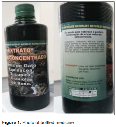
The bottled medicine, Naturlife®, Figure 1, from CSJ Mendes ME Industry and Natural Products Trade, was acquired in the city of Toledo, Parana, Brazil, in August 2015.
The dosage indication is 2 tablespoons 3 times a day for adults and 1 tablespoon 3 times a day for kids. The sample was analyzed as its physicochemical characteristics, such as color, odor, density (in refractometer), cold (25ºC) and hot (40ºC) pH (Alpax® pH meter), solubility, macroscopic and microscopic characteristics, alcohol, phytochemical screening and UV-scan spectrum.
Alcohol assay
Distillation technique was used to separate the alcohol from the vegetal matrix, obtaining the distillate, which was cold oxidized by nitrochromic solution. The amount of alcohol was calculated by the consumption of nitrochromic solution used in the oxidation of alcohol (Heck, 2007). The alcohol content was expressed as mg alcohol.mL-1 of bottled medicine.
Phytochemical screening
The phytochemical characterization was made through reactions with color development or precipitation, which involves searching for saponins (foam test), steroids and triterpenoids (Liebermann-Burchard reaction), alkaloids (Dragendorff Mayer reaction), tannins (reaction with ferric chloride), coumarins (reaction with KOH and UV) and flavonoids (Matos, 2009).
Uv/scan absorption spectrophotometry
For determination of maximum wavelength (λmax.) and maximum absorbance (Amax.), an aqueous solution at the concentration of 300 mg.L-1 was prepared. Scanning was performed between wavelengths of 200 to 400 nm, with resolution of 2 nm. Distilled water was used for the baseline and spectrophotometer UV/Visible, model DR5000, HACH®.
Animals
A total of 30 animals, Rattus novergicus Wistar, male, adult and healthy, weighing an average of 250 g, were collected from the State University of Western Parana Central Laboratory. The animals were housed in polypropylene boxes, air-conditioned (22°C ± 2°C), light-dark cycle of 12 h, constant exhaust system, water and feed ad libitum. After 1 week of adaptation, the animals were randomly divided into 6 experimental groups with 5 animals in each group (Figure 2). All experimental protocols were approved by the Ethics Committee of the State University of Western Parana (Cascavel, Brazil) for the care and use of laboratory animals.
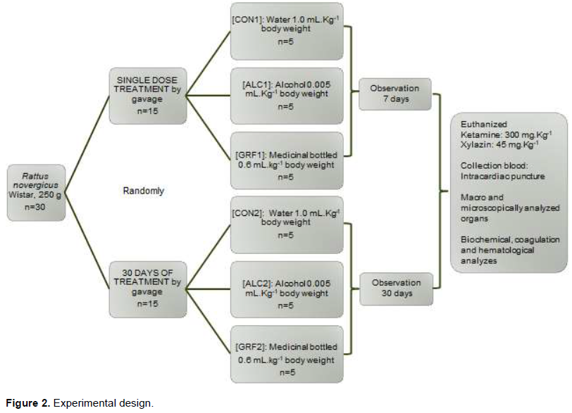
Single dose treatment
This experiment was based and adapted on qualitative and semi-quantitative toxicological analysis methodology (Jesus et al., 2012). After overnight fasting, the animals were treated by gavage, once only, according to:
Control Group [CON1]: received water (1.0 mL.kg-1 body weight); Alcohol Group [ALC1]: received alcohol (0.005 mL.kg-1 body weight);
Bottled medicine Group [GRF1]: received bottled medicine (0.6 mL.kg-1 body weight).
After treatment the animals received water and food ad libitum. All animals were observed for signals and symptons at times 0, 5, 10, 15, 30 and 60 min; 2, 4, 6 and 24 h and finally, 2, 4 and 7 days. Thereafter, activity and motor coordination, body tremors, fasciculations, ptosis eyelid, tearing, salivation, bleaching and hyperemia of the ears, paralysis of the legs, piloerection, analgesia, anesthesia, corneal and auricular reflex, temperature, diarrhea and other signs and symptoms were observed (Jesus et al., 2012). The body weight of the animals was monitored daily. After the seventh day of the experiment, all animals were fasted overnight. After overnight fasting, the animals were euthanized with an overdose of ketamine (300 mg.kg-1) and xylazine (45 mg.kg-1) for collection of blood by intracardiac puncture. Organs (adipose tissue, brain, heart, kidneys, liver, lungs, small intestine, spleen, stomach and testicles) were removed, weighed, macroscopically and histologically observed. Biochemical, coagulation and hematological parameters were also evaluated.
30 days of treatment
This experiment was based on toxicological analysis methodology after 30 days of treatment (Silva et al., 2015). After overnight fasting, the animals were treated by gavage, once daily, for 30 days, according to:
Control Group [CON2]: received water (1.0 mL.kg-1 body weight);
Alcohol Group [ALC2]: received alcohol (0.005 mL kg-1 body weight);
Bottled Medicine Group [GRF2]: received bottled medicine (0.6 mL.kg-1 of body weight).
The general behavior was observed, during the 30 days of experiment, in times of 5 and 30 min; 1, 2, 4 and 6 h after gavage.
Respiratory changes, locomotion alteration, tremor, convulsions, excitability, reduction of activity, ataxia, piloerection, drowsiness, ptosis eyelid, abdominal constriction, salivation and diarrhea were observed. The body weight of the animals was monitored daily. After the thirtieth day of the experiment, all animals were fasted overnight.
After overnight fasting, the animals were euthanized with an overdose of ketamine (300 mg.kg-1) and xylazine (45 mg.kg-1) for collection of blood by intracardiac puncture. Organs (adipose tissue, brain, heart, kidneys, liver, lungs, small intestine, spleen, stomach and testicles) were removed, weighed, macroscopically and histologically observed. Biochemical, coagulation and hematological parameters were also evaluated.
The doses of 0.6 mL.kg-1 of the bottled medicine per body weight, usual adult dose on the bottled medicine label and 0.005 mL.kg-1 of alcohol per body weight, referred to the alcohol content found in the bottled medicine. Control groups received water.
Data analysis
The data obtained were tabulated and statistically evaluated in the statistical software Statistica7®. The results obtained with the different groups were evaluated by the One-way ANOVA method followed by Fisher's post-hoc test, and the Kruskal-Wallis test was used to analyze non-parametric variables. The results were expressed as mean ± standard error (SEM) of n which reflects the number of animals. It was accepted as statistically significant p<0.05.
Blood collection
The collected blood was distributed in tubes containing the 3.2% sodium citrate anticoagulant (1.8 mL) for coagulation determinations and in tubes with sodium heparin anticoagulant at 151.58 IU (10 mL) for biochemical and hematological dosages. A drop of blood from the animals without anticoagulant was used for the blood smear on slide. Tubes for biochemical and coagulation parameters were centrifuged for 10 min at 2,500 xg for plasma separation.
Biochemical analyzes
The animals were dosed uric acid, ALT, AST, total bilirubin (BT), direct (BD) and indirect bilirubin (BI) cholesterol, creatinine, glucose, GGT, iron, alkaline phosphatase (AP), triglycerides and urea. Biochemical parameters were measured using an automated AU680 analyzer (Beckman Coulter®). The analytes were uric acid, ALT, AST, bilirubin, cholesterol, AP, glucose, GGT, triglycerides and urea by enzymatic method, iron by colorimetric method, creatinine by kinetic method and PCR by nephelometric method (Beckman Coulter®).
Coagulation analyzes
Prothrombin activation time (PT), International normalized ratio (INR), partial thromboplastin activation time (APTT) and ratio were determined. The PT and INR determinations were performed by automated coagulation equipment, by the clot detection method, CA500 (Siemens®). APTT and ratio measurements were performed by manual clot detection using reagent composed of rabbit brain cephalin, ellagic acid and calcium chloride (0.025 mol.L-1).
Hematological analyzes
Erythrocytes, hemoglobin (HB), hematocrit (HTC), mean corpuscular volume (MCV), mean corpuscular hemoglobin (HCM), mean corpuscular hemoglobin concentration (CHCM), erythrocyte volume variation (RDW), total leukocytes, relative values of bands, neutrophils, lymphocytes, monocytes and eosinophils, platelets, mean platelet volume (MPV) and reticulocytes were evaluated. The hematological parameters were analyzed using automated counter LH700 (Beckman Coulter®) by resistivity, impedance and colorimetric method. The slides were evaluated by optical microscopy after staining of May Grünwald Giemsa performed with commercial kit (NewProv®). All assays were performed in accordance with the manufacturers’ instructions and protocols.
Organ weights and histopathological analyzes
Adipose tissue, brain, heart, kidneys, liver, lungs, small intestine, spleen, stomach and testicles were isolatedly heavy. All organs of the animals were examined macroscopically. Organs with macroscopic alterations were fixed in 10% formaldehyde and organs of the control group were also analyzed histologically. Histological sections were stained with hematoxylin and eosin (HE) for microscopic examination.
RESULTS AND DISCUSSION
The bottled medicine is an industrialized liquid product of dark coloration, low viscous, soluble in water, with odor characteristic of propolis, refractive index of 20.0°Bx. Microscopically, it presents with few vegetal artifacts. In its formulation are described the plants Echinacea, Graviola, Purple Ipe Sucupira and Cat’s claw. In the label, it is said to be free of registration in ANVISA (National Agency of Sanitary Surveillance), does not contain alcohol and non-toxic product. Other than the one advertised on your label, the alcohol assay showed a content of 4.32 mg.mL-1. The presence of alcohol is worrying, although in its label there was indication and dosage for kids. The pH of the bottled medicine is very acidic 3.12 (25ºC) and 3.09 (40ºC).
Phytochemical screening showed the presence of some classes of secondary metabolites, such as saponins, coumarins and flavonoids. By absorption spectrophotometry UV, it was possible to detect λmax. at 278 nm. The probable secondary metabolites found by the UV/Scan for the sample studied were the alkaloids in the region between 254-365 nm (Wagner, 1984), saponins at 284 nm (Vigo et al., 2003) and tannins at 280 nm (Pinelo et al., 2006).
In relation to the treatments of single dose and 30 days treatment, there was a decrease in motor activity in three (3) animals (n=5) in the [ALC1] group and in 2 animals (n=5) in the [ALC2] group, after administration of alcohol at a concentration of 0.005 mL.kg-1. This effect was reversible and occurred within 4 to 6 h after administration. Other signs and symptoms were not observed during the treatments. The depressant effect of alcohol on the central nervous system is already well characterized, mainly by increasing the inhibitory response of the gamma-aminobutyric acid neurotransmitter (GABA) and by inhibiting the response of the excitatory neurotransmitter glutamate to N-methyl-D-Aspartate receptors (NMDA) (Imam, 2010).
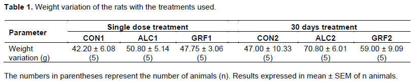
Table 1 shows the mean body weight variation of the animals during the treatments. Despite the weight variations observed between the groups, the body weight gain of the rats in this study was not significant. Another author showed significant changes in weight, being that the decreases and increases in weight occurred according to the amount of alcohol exposed (Silva et al., 2015).
The Table 2 presents the results of the biochemical analyzes with the treatments used. The rats treated with alcohol and bottled medicine showed a high and significant level of uric acid. This increase occurred with the [ALC1], [GRF1] and [ALC2] groups. The uric acid increased by 104% in the [ALC1] group (p<0.005) and by 84% (p<0.05) in the [GRF1] group compared to the control group. In the [ALC2] group, the increase of uric acid was even more expressive, being that values of 0.45 ± 0.11 mg.dL-1 of uric acid in the control group increased to 3.38 ± 0.17 mg.dL-1 of uric acid (p<0.05). For the [GRF2] group, this analyte was not significant, but remained in low concentrations.
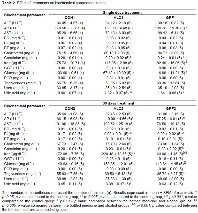
It has been demonstrated that alcohol use induces hyperuricemia (Kang et al., 2002). The increase in uric acid in these groups is probably due to the presence of alcohol in the bottled medicine. This increase is relevant because with only one dosage there was a significant change in this parameter. With the 30 days of treatment, some substances present in the bottled medicine attenuated the hyperuricemic effect of alcohol on rats [GRF2] by some unknown mechanism. The level of uric acid is controlled by the rate of degradation of endogenous and exogenous purines in uric acid and also by the rate of excretion of this analyte (Choi and Curhan, 2004). Any factor that changes hepatic or renal function may influence serum uric acid levels (Bugdayci et al., 2008).
We can verify that the creatinine levels of the [GRF1] and [ALC2] groups were significantly lower in the control groups (p<0.05). What caused the decrease in the serum creatinine level is not clear, since the renal function of the rats may be impaired by the considerable increase of uric acid found.
The total and indirect bilirubin levels of the [ALC2] and [GRF2] groups had their levels decreased significantly compared with the control group. It is known that the decrease in bilirubin levels shows an improvement in hepatic function. Early changes in bilirubin in the blood are an important prognostic factor in patients with alcoholic hepatitis (Mathurin et al., 2003). We found in our study decreased bilirubin levels for both the bottled medicine group and the alcohol group. This finding is different from what was expected, since alcohol consumption leads to an increase in serum bilirubin because it inhibits its conjugation (O’Malley et al., 2015).
We observed that the AP units decreased significantly from the [GRF1] group in relation to control (p<0.05). Already in the 30 days of treatment, the AP units of the [ALC2] group increased by 28% relative to the control (p<0.05). The AP units of the [GRF2] group remained low, but only significant in relation to the [ALC2] group (p<0.005). It is well known that increases in serum alkaline phosphatase levels are observed in response to chronic exposure to ethanol (Ravel, 2009) and also in non-alcoholics hepatics diseases (Pantsari and Harrison, 2006). In the liver, the AP is formed by hepatocytes and by cells of the mucosa of the biliary tract. Although the AP of hepatic origin may be increased in serum due to any type of active hepatic disease, the serum level is especially sensitive to obstruction of the biliary tract, be it intrahepatic or extrahepatic, mild or intense degree, localized in a small area of ​​the liver or in a more intense region (Ravel, 2009).
Thus, as described in the literature, we also found an increase of this serum enzyme in rats after 30 days of treatment with alcohol. Substances presents in the bottled medicine may have attenuated the action of alcohol, since in the [GRF2] group the AP units remained significantly lower. We have already demonstrated hepatic and renal protective effects of airborne portions of E. purpurea extract on diethylnitrosamine-induced injury in rats (Rezaie et al., 2013) and also anti-inflammatory effects on arsenic-induced hepatic toxicity (Heidari et al., 2012).
On the determination of serum iron, there was an increase in levels in the [GRF1] group, but were not significant in relation to the control (p>0.05). With the 30 days of treatment, there was a significant increase in levels in the [ALC2] group in relation to the control (p<0.005), and this increase was 33%. The regular consumption of alcohol is responsible for disrupting the normal metabolism of iron, resulting in excess and deposition of this element in the liver. It is likely that the two main proteins of its metabolism, ferritin and transferrin are involved in this process. Ferritin is the form of iron storage, while transferrin is the iron carrier protein. Alcohol affects the structure of the transferrin molecule causing it to become deficient carbohydrate, resulting in an increase in plasma ferritin concentration (Fletcher et al., 1999).
Harmful effects of alcohol and its derivatives on hepatic cell function are relevant. Exposure to alcohol causes steatosis, dysfunction of mitochondrial and cellular membranes, hypoxia and oxidative stress. At millimolar concentrations, alcohol affects directly the microtubular and mitochondrial function and membrane fluidity. The hepatic toxicity' pathogenesis by alcohol is related to acetaldehyde (the main alcohol intermediate metabolite), inducing peroxidation and formation of the acetaldehyde-protein adduct, disrupting the cytoskeleton and the membranes’ function. The metabolism by cytochrome P-450 produces reactive oxygen species (ROS), which react with cellular proteins, damage membranes and alter hepatic cell function. In addition, impairment of hepatic metabolism of alcohol-induced methionine causes a decrease in intrahepatic levels of glutathione, thereby sensitizing the liver to oxidative damage. The induction of CYP2E1 and other cytochrome P450 enzymes in the liver by alcohol increases alcohol catabolism in the endoplasmic reticulum and enhances conversion to toxic metabolites (Mitchell et al., 2012).
Glucose dosages of the [ALC1] group were lower compared to the [CON1] (p<0.05) and that of the [GRF1] group were superiors in relation to the [ALC1] group (p<0.05). The alcohol also demonstrates a direct relationship with glucose metabolism (Ting and Lautt, 2006). Low doses of alcohol have been associated with increased insulin sensitivity, which is a beneficial effect. Furthermore, the alcohol influences on glucose metabolism requires that some factors like time of the treatment and the ingested concentration of alcohol are considered (Furuya et al., 2005). This also influences on the glucose metabolism inhibiting both gluconeogenesis and glycogenolysis. The acute ingestion of alcohol in fasting may cause hypoglycemia, especially in cases of depleted glycogen stores. The ethanol causes hypoglycemia by interfering with a gluconeogenesis as well as glycogen synthesis (Cryer et al., 2009), justifying the low concentrations of glucose found in the [ALC1] group.
Acute alcohol intoxication may trigger a moderate and transient hyperglycemia due to the effect of oxidative stress resulting from the production of acetaldehyde. Already in a second phase, the chronic alcohol consumption can trigger the decrease of glycemic levels, and such a situation occurs due to blocking of the Krebs’ cycle (Mincis et al., 1995).
Already in the 30 days of treatment, glucose levels of [GRF2] decreased relative to the [ALC2] group (p<0.005). As in the alcohol treated group, the same result was not obtained, probably the hypoglycemic effect is the result of compounds of the vegetal species present in the bottled medicine. The aqueous extract of A. muricata, for example, has antidiabetic activity with antioxidant and protective action on pancreatic β cells, which improve glucose metabolism (Florence et al., 2014) and also with the species P. emarginatus (Macedo and Ferreira, 2004), that has been described as having hypoglycemic activity in a study on traditional plant use.
The effects on cholesterol and triglycerides levels were different in both treatments. The cholesterol determinations were significant for the [ALC1] group compared to the control (p<0.05), increasing by 19%. Already, the triglycerides levels were significant for the [GRF2] group, decreasing by 36% in relation to the control (p<0.05) and increasing levels in [ALC2] compared to [GRF2] (p<0.05). The increase in cholesterol levels at 13.98 mg.dL-1 in the [ALC1] group may reflect the increased absorption of dietary cholesterol or the induction of cholesterol absorption by alcohol (Latour et al., 1999). These findings were consistent with previous studies indicating that alcohol affected lipid metabolism (Toffolo et al., 2012) with increased cholesterol levels in rats treated with alcohol (Silva et al., 2015).
Triglycerides levels decreased significantly in the bottled medicine group (20.13 mg.dL-1) and increased in the alcohol group (24.83 mg.dL-1) compared to the bottled medicine. The decrease in triglycerides levels in the [GRF2] group may be related to an effect found in T. avellanedae extract. In this study, ovarectomized rats fed with Purple Ipe extract presented weight loss, decreased body fat and triglycerides levels. This anti-obesity effect of the extract was due to the ability to prevent the accumulation of triglycerides in adipocytes shown in the study (Iwamoto et al., 2016). Understandably, alcohol causes accumulation of hepatic triglycerides, and can lead to steatosis and alcohol-dependent hypertriglyceridemia. This accumulation of triglycerides is due to an increase in the hepatic synthesis of very low-density lipoproteins (VLDL) directly linked to the increase in the NADH/NAD ratio, which is responsible for the blocking of fatty acid oxidation and the Krebs’ cycle. Excess fatty acids are used for the synthesis of triglycerides (Mincis et al., 1995).
The accumulation of fat and cholesterol deposits (Thoolen et al., 2010) also reduces hepatic function. Some authors consider that this deterioration is progressive, beginning with some alterations of the lipid profile, followed by a potentially compensatory phase and, finally, resulting in hepatic insufficiency. In addition, this progressive deterioration of the liver may be responsible for the disappearance of the hyperlipidemia found in some cases of chronic alcohol consumption (Sozio and Crabb, 2008) which would explain our cholesterol findings in animals treated for 30 days.
Other results showed that biochemical parameters, AST, ALT, BD, GGT, PCR and urea, of the single dose and 30 days treatments were not significant between the groups (p>0.05).
Coagulation determinations PT, INR, APTT and ratio also did not show significant differences (p>0.05) in the treatments used. There is strong evidence in the literature that alcohol impairs the function of coagulation factors.
Acetaldehyde, product of alcohol metabolism, mediates these effects by the inactivation of thrombin, factor Xa, fibrinogen, II, VII and X, inhibits factor XIIIa transglutaminase activity and forms complexes with glycosaminoglycans to synergistically inhibit factors IX, IXa, X and Xa (Suchocki and Brecher, 2007) with consequent prolongation of coagulation times. Besides that, a compound present in T. avellanedae, lapachol, also showed changes in hemostasis, because it has a structure similar to vitamin K, though it is a reversible inhibitor of this vitamin (Hussain et al., 2007).
Table 3 shows the results of the different hematological parameters measured. Only the number of erythrocytes (p<0.05) and leukocytes (p<0.05) of the [ALC2] group were significant in relation to the control.
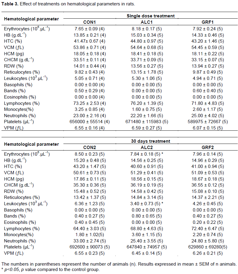
Erythrocytes and leukocytes decreased in the group exposed to alcohol, and this reduction was 8 and 47% for the respective parameters. These hematological parameters were also reduced in the bottled medicine group, but not significant. Compounds present in the bottled medicine have attenuated this effect of alcohol, although there are studies with Purple Ipe, for example, showing important hematological changes such as anemia (Silva et al., 2003). The consequences of alcohol on the hematological system include its toxic effects on bone marrow, on cellular precursors, on the maturation of erythrocytes, on kinetics and on the function of leukocytes and platelets. Alcohol causes suppression in the production of red blood cells, as it interferes with the metabolism of folic acid, iron, phosphate and vitamin B12, which have an essential role in the development of hematopoietic cells (Ballard, 1997), thus causing anemia, leukopenia, thrombocytopenia and increased VCM on blood peripheral (Costa et al., 2007), which would explain the decreased values of erythrocytes and leukocytes found in our study.
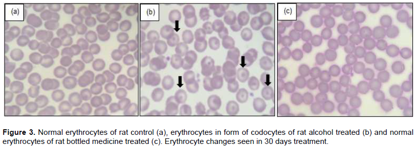
Some poikilocytosis are usually expected in peripheral blood from exposure to alcohol. Erythrocytes in the form of stomatocytes, which have a membrane defect by an unknown mechanism, but alcoholic abstinence make them disappear from the blood, and acanthocytes, by incorporating excess cholesterol into the cell membrane (Ballard, 1997). Other than that, we found erythrocyte changes in the form of codocytes (indicated by arrows) on the microscopy of the 5 slides of the [ALC2] group (n=5), Figure 3. In groups [CON2] (a) and [GRF2] (c), these alterations were not visualized.
Codocytes or Target cells are erythrocytes that have central halo, a hemoglobin concentration in the rounded form, which gives it the appearance of a target. These cells are characteristic of iron deficiency anemia, hemoglobinopathies and thalassemias (Silva et al., 2009). However, they also occur when there are alterations in plasma lipid composition, whose cholesterol and lecithin molecules are in continuous exchange with the erythrocyte membrane, since this phenomenon is responsible for the codocytosis of obstructive jaundice (Failace, 2015). Alterations in lipid composition could explain these findings in rats treated with alcohol, since changes in the triglycerides levels in this group were observed.
Other hematological parameters, such as HB, HTC, VCM, HCM, CHCM, RDW, reticulocytes, bands, basophils, eosinophils, lymphocytes, monocytes, neutrophils, platelets and VPM were not statistically significant (p>0.05).
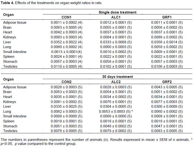
Table 4 shows the organ weight ratios, and no changes were observed in the macroscopic and histopathological analyzes in the single dose experiment. In the 30 days experiment, there was a significant reduction in lung weight of the [ALC2] group in relation to the control, but macroscopic and histopathological alterations of the lungs were found in both the alcohol and in the control group, invalidating it.
Although hepatic aminotransferases were not altered, macroscopic and histopathological alterations of the livers of the [ALC2] and [GRF2] groups were observed in the treatment of 30 days, in relation to control, which can be seen in Figure 4.
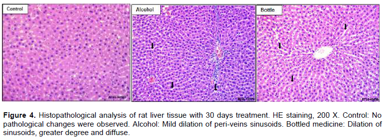
In this histopathological image of the liver of the alcohol group, we can notice the portal space and hepatocytes trabeculaes separated by intratrabeculaes spaces, that is, dilated peri-veins (indicated by arrows).
The sinusoidal dilation is mild. And in the histopathological image of the liver of the bottled medicine group, we noted hepatocytes trabeculaes separated by sinusoid dilatation, but of a greater degree and more diffused (indicated by arrows) than the alcohol group.
Sinusoidal dilatation occurs when there is an increase in hepatic vein pressure, hepatocytes atrophy or interruption of sinusoidal reticulin fibers (MacSween, 1994). Sinusoids can show a spectrum of pathological changes, from dilation and congestion to lesions affecting the subendothelial (Space of Disse). The width of sinusoids in hepatic biopsy specimens is variable, and is related to the amount of connective tissue in the sinusoidal wall. Among the various causes that lead to the increase of sinusoids are alcoholic hepatic disease and other toxic products (Lefkowitch, 2016).
Chronic use of alcohol causes hepatic disease, which progresses from simple steatosis to stages of steatohepatitis, fibrosis, cirrhosis and hepatic failure (Setshedi et al., 2010). Alcohol hepatotoxicity causes portal hypertension, with increased liver fat and inflammation. This will increase the intrahepatic volume and the degree of compression of the hepatic venules, sinusoids and intersinusoidal communications (Mincis and Mincis, 2011), and may explain the histopathological findings of the hepatic tissues of the rats.
As hepatic histopathological findings were found in both the [ALC2] group and the [GRF2] group, we can claim that these microscopic changes are related to alcohol. However, the microscopic changes found in the bottled medicine group were more prominent, that is, besides alcohol, there are other compounds present in the bottled medicine that have further compromised the hepatic tissue.
CONCLUSION
The data presented showed that the alcohol present in a bottled medicine can alter lipid profile, glucose, kidney function, some blood cells and cause damage to the liver of rats, but does not change the coagulation parameters. Although the bottled medicine did not show changes in biochemical, coagulation and hematological parameters, it presented more pronounced liver histopathological changes than alcohol in rat liver. That is, in addition to alcohol, there are other compounds present in the bottled medicine that further compromised the liver tissue.
This study demonstrates the importance of considering possible actions of alcohol present in herbal medicinal preparations. It is noteworthy that even at low doses, continuous exposure to alcohol of 0.005 mL alcohol.kg-1 body weight can cause significant changes in some biochemical, hematological and histopathological parameters of rats.
These data have considerable repercussions because natural products are mistakenly considered non-toxic and are used without professional guidance. Thus, toxicity studies should evaluate and contribute to show the safety of the active principles and their formulations.
Regarding the notification of toxicity, we cannot say that the hematological, renal and histopathological changes that the patient had were actually caused by the bottled medicine. However, these changes may have been exacerbated by alcohol or active plant compounds present in its formulation.
CONFLICT OF INTERESTS
The authors have not declared any conflict of interests.
REFERENCES
|
Arthur FKN, Woode E, Terlabi EO, Larbie C (2011). Evaluation of acute and subchronic toxicity of Annona muricata (Linn.) aqueous extract in animals. European Journal of Experimental Biology 1(4):115-124. |
|
|
Ballard HS (1997). The hematological complications of alcoholism. Alcohol Health and Research World 21(1):42-52. |
|
|
Barata LES, Alencar AAJ, Tascone M, Tamashiro J (2013). Plantas medicinais brasileiras. IV. Annona muricata L. (Graviola). Revista Fitos 4(1):132-138. |
|
|
BiaÅ‚as-Chromiec B, KapaÅ‚ka B, Radomska-LeÅ›niewska D, SkopiÅ„ska- Różewska E, Sommer E, Sokolnicka I, Filewska M, Demkow U (2003). The in vivo effect of Echinacea purpurea succus on various functions of human blood leukocytes. Central European Journal of Immunology 28(3):126-130. |
|
|
Bugdayci G, Balaban Y, Sahin O (2008). Causes of hypouricemia among outpatients. Laboratory Medicine 39(9):550-552. |
|
|
Campelo LML, Sá CG, Feitosa CM, Sousa GF, Freitas RM (2013). Chemical constituents and toxicological studies of the essential oil extracted from Citrus limon Burn (Rutaceae). Revista Brasileira de Plantas Medicinais 15(4):708-716. |
|
|
Choi HK, Curhan G (2004). Beer, liquor, and wine consumption and serum uric acid level: the Third National Health and Nutrition Examination survey. Arthritis Care and Research 51(6):1023-1029. |
|
|
Costa AC, Ribeiro B, Costa E (2007). Índices plaquetários em indivíduos com doença hepática alcoólica crônica. Arquivos de Gastroenterologia 44(3):201-204. |
|
|
Cryer PE, Axelrod L, Grossman AB, Heller SR, Montori VM, Seaquist ER, Service FJ (2009). Evaluation and management of adult hypoglycemic disorders: an Endocrine Society Clinical Practice Guideline. The Journal of Clinical Endocrinology and Metabolism 94(3):709-728. |
|
|
Da Paz J, Baldochi MR, Contrera MGD, Ribeiro AF, Regalo SCH, Da Paz K, Lopes RA, Sala MA (2005). Hepatotoxicidade de plantas medicinais. XXXII. Ação da infusão de Achyrocline satureioides (Lam.) DC no rato. Investigação - Revista Científica da Universidade de Franca 5(1):154-159. |
|
|
Dayeef AYM, Karyono S, Sujuti H (2013). The influence of Annona muricata leaves extract in damaging kidney cell and inducing caspase-9 activity. Journal of Pharmacy and Biological Sciences 8(5):48-52. |
|
|
Camargo MTLA (2011). A garrafada na medicina popular: uma revisão historiográfica. Dominguezia 27(1):41-49. |
|
|
De Sant'Ana FJ, Perin JN, Bilego UO (2012). Spontaneous poisoning of cattle by Pterodon emarginatus (Fabaceae) in Goiás, Brazil. Pesquisa Veterinária Brasileira 32(6):485-489. |
|
|
Failace RR (2015). Hemograma: manual de interpretação. Porto Alegre, RS: Artmed P 102. |
|
|
Fletcher L, Halliday J, Powell L (1999). Interrelationships of alcohol and iron in liver disease with particular reference to the ironâ€binding proteins, ferritin and transferrin. Journal of Gastroenterology and Hepatology 14(3):202-214. |
|
|
Florence NT, Benoit MZ, Jonas K, Alexandra T, Désiré DDP, Pierre K, Théophile D (2014). Antidiabetic and antioxidant effects of Annona muricata (Annonaceae), aqueous extract on streptozotocin-induced diabetic rats. Journal of Ethnopharmacology 151(2):784-790. |
|
|
Furuya DT, Binsack R, Onishi ME, Seraphim PM, Machado UF (2005). Low ethanol consumption induces enhancement of insulin sensitivity in liver of normal rats. Life Sciences 77(15):1813-1824. |
|
|
Hussain H, Krohn K, Ahmad VU, Miana GA, Green IR (2007). Lapachol: an overview. Arkivoc 2:145-171. |
|
|
Heck A (2007). Modelling intake and clearance of alcohol in humans. Electronic Journal of Mathematics and Technology 1(3): 232-244. |
|
|
Heidari M, Rezaie A, Broojeni MP, Najafzadeh H, Mohammadian B (2012). Histopathologic effects of Echinacea purpurea extract on sodium arsenite-induced hepatic disorders. Comparative Clinical Pathology 21(6):1629-1632. |
|
|
Ibrahim KE, Al-Ashban RM, El-Sammani SA (2009). A study of the toxicity of Cat's claw herbal medicine. Research Journal of Pharmacology 3(3):52-57. |
|
|
Imam I (2010). Alcohol and the central nervous system. British Journal of Hospital Medicine 71(11):635-639. |
|
|
Iwamoto K, Fukuda Y, Tokikura C, Noda M, Yamamoto A, Yamamoto M, Yamashita M, Zaima N, Iida A, Moriyama T (2016). The anti-obesity effect of Taheebo (Tabebuia avellanedae Lorentz ex Griseb) extract in ovariectomized mice and the identification of a potential anti-obesity compound. Biochemical and Biophysical Research Communications 478(3):1136-1140. |
|
|
Jesus NZ, Silva Júnior IF, Lima J, Colodel EM, Martins DT (2012). Hippocratic screening and subchronic oral toxicity assessments of the methanol extract of Vatairea macrocarpa heartwood in rodents. Revista Brasileira de Farmacognosia 22(6):1308-1314. |
|
|
Junior CGE, Carvalho D, De Souza MA, Kasai A, Lopes RA, Lopes PEVP, Sala MA, Regalo SCH, Petenusci SO (2006). Hepatotoxicidade de plantas medicinais. LIII. Ação da infusão de Tabebuia avellanedae lor. ex griseb. no rato. Investigação - Revista Científica da Universidade de Franca 6(1):13-16. |
|
|
Kang DH, Nakagawa T, Feng L, Watanabe S, Han L, Mazzali M, Truong L, Harris R, Johnson RJ (2002). A role for uric acid in the progression of renal disease. Journal of the American Society of Nephrology 13(12):2888-2897. |
|
|
Latour MA, Patterson BW, Kitchens RT, Ostlund RE, Hopkins D, Schonfeld G (1999). Effects of alcohol and cholesterol feeding on lipoprotein metabolism and cholesterol absorption in rabbits. Arteriosclerosis, Thrombosis and Vascular Biology 19(3):598-604. |
|
|
Lefkowitch JH (2016). Liver biopsy interpretation. New York, EUA: Elsevier Health Sciences pp. 256-257. |
|
|
Lorenzi H, Matos FJA (2008). Plantas Medicinais no Brasil: nativas e exóticas. Nova Odessa, SP: Instituto Plantarum pp. 67-288. |
|
|
Macedo M, Ferreira AR (2004). Plantas hipoglicemiantes utilizadas por comunidades tradicionais na Bacia do Alto Paraguai e Vale do Guapore, Mato Grosso - Brasil. Revista Brasileira de Farmacognosia 14(1):45-47. |
|
|
MacSween RNM, Anthony PP, Scheuer PJ, Burt AD, Portmann BC (1994). Pathology of the liver. London, UK: Churchill Livingstone pp. 243-267. |
|
|
Matos FJA (2009). Introdução à fitoquímica experimental. Fortaleza, CE: Edições UFC P 150. |
|
|
Mathurin P, Abdelnour M, Ramond MJ, Carbonell N, Fartoux L, Serfaty L, Valla D, Poupon R, Chaput JC, Naveau S (2003). Early change in bilirubin levels is an important prognostic factor in severe alcoholic hepatitis treated with prednisolone. Hepatology 38(6):1363-1369. |
|
|
Mincis M, Mincis R (2011). Álcool e o fígado. GED-Gastroenterologia Endoscopia Digestiva 30(4):152-162. |
|
|
Mincis M, Chebli JM, Khouri ST, Mincis R (1995). Ethanol and the gastrointestinal tract. Arquivos de Gastroenterologia 32(3):131-139. |
|
|
Mitchell RN, Kumar V, Abbas AK, Fausto N, Aster JC (2012). Robbins & Cotran: fundamentos de patologia. Rio de Janeiro, RJ: Elsevier P 867. |
|
|
O'Malley SS, Gueorguieva R, Wu R, Jatlow PI (2015). Acute alcohol consumption elevates serum bilirubin: An endogenous antioxidant. Drug and Alcohol Dependence 149:87-92. |
|
|
Pantsari MW, Harrison SA (2006). Nonalcoholic fatty liver disease presenting with an isolated elevated alkaline phosphatase. Journal of Clinical Gastroenterology 40(7):633-635. |
|
|
Pinelo M, Rubilar M, Sineiro J, Nuñez MJ (2006). Effect of bubbling nitrogen and pulsed flow on the antiradical activity of grape residues. Journal of Food Engineering 73(3):269-275. |
|
|
Ravel R (2009). Laboratório clínico: aplicações clínicas dos dados laboratoriais. Rio de Janeiro, RJ: Guanabara Koogan P 275. |
|
|
Rezaie A, Fazlara A, Karamolah MH, Shahriari A, Zadeh HN, Pashmforosh M (2013). Effects of Echinacea purpurea on hepatic and renal toxicity induced by diethylnitrosamine in rats. Jundishapur Journal of Natural Pharmaceutical Products 8(2):60. |
|
|
Setshedi M, Wands JR, De La Monte SM (2010). Acetaldehyde adducts in alcoholic liver disease. Oxidative Medicine and Cellular Longevity 3(3):178-185. |
|
|
Silva FC, De Souza JGDL, Reichert AM, Antonangelo RP, Suzuki R, Itinose AM, Marek CB (2015). Influence of the alcohol present in a phytotherapic tincture on male rat lipid profiles and renal function. Evidence-Based Complementary and Alternative Medicine, 2015. |
|
|
Silva PH, Hashimoto Y, Alves HB (2009). Hematologia Laboratorial. Rio de Janeiro, RJ: Revinter P 84. |
|
|
Silva MN, Ferreira FF, Souza MCBV (2003). An overview of the chemistry and pharmacology of naphthoquinones with emphasis on β-lapachone and derivatives. Química Nova 26(3):407-416. |
|
|
Silva TS, Freire EMX (2010). Abordagem etnobotânica sobre plantas medicinais citadas por populações do entorno de uma unidade de conservação da caatinga do Rio Grande do Norte, Brasil. Revista Brasileira de Plantas Medicinais 12(4):427-435. |
|
|
Schrøder-Aasen T (2012). Effects of Purple Coneflower (Echinacea purpurea) on CYP3A4 Metabolism and P-glycoprotein Mediated Transport in vitro. Thesis of Philosophiae Doctor, Norwegian University of Science and Technology, Trondheim. |
|
|
Sozio M, Crabb DW (2008). Alcohol and lipid metabolism. American Journal of Physiology-Endocrinology and Metabolism 295(1):10-16. |
|
|
Suchocki EA, Brecher AS (2007). The effect of acetaldehyde on human plasma factor XIII function. Digestive Diseases and Sciences 52(12):3488-3492. |
|
|
Thoolen B, Maronpot RR, Harada T, Nyska A, Rousseaux C, Nolte T, Malarkey DE, Kaufmann W, Küttler K, Deschl U, Nakae D, Gregson R, Vinlove MP, Brix AE, Singh B, Belpoggi F, Ward JM (2010). Proliferative and nonproliferative lesions of the rat and mouse hepatobiliary system. Toxicologic Pathology 38(7):5-81. |
|
|
Ting JW, Lautt WW (2006). The effect of acute, chronic, and prenatal ethanol exposure on insulin sensitivity. Pharmacology and Therapeutics 111(2):346-373. |
|
|
Toffolo MCF, De Aguiar-Nemer AS, Da Silva-Fonseca VA (2012). Alcohol: effects on nutritional status, lipid profile and blood pressure. Journal of Endocrinology and Metabolism 2(6):205-211. |
|
|
UNESCO (2013). Report of the International Bioethics Committee on Traditional Medicine Systems and their ethical implications. |
|
|
Vigo CLS, Narita E, Marques LC (2003). Validação da metodologia de quantificação espectrofotométrica das saponinas de Pfaffia glomerata (Spreng.) Pedersen-Amaranthaceae. Revista Brasileira de Farmacognosia 13(2):46-49. |
|
|
Wagner H, Bland S, Zgainski EM (1984). Plant Drug Analysis. Berlin, DE: Springer-Verlag P 320. |
|
Copyright © 2024 Author(s) retain the copyright of this article.
This article is published under the terms of the Creative Commons Attribution License 4.0


