ABSTRACT
The aim of this study was to evaluate acetophenones isolated from Croton spp. for antifungal activity in vitro against Candida albicans, Microsporum canis and Trichophyton rubrum, and to evaluate the toxicity of these substances in vivo. The minimum inhibitory concentration (MIC) and minimum fungicidal concentration (MFC) were determined using the broth microdilution method. The combined effect of the 2-hydroxy-3,4,6-trimethoxyacetophenone with ketoconazole was evaluated using the checkerboard technique. The toxicity was determined using Artemia salina L. The compounds were active against T. rubrum, with MICs ranging from 1.25 to 2.5 mg/mL. Similar results were obtained for M. canis, with MICs ranging from 5.0 to 10.0 mg/mL. In the toxicity tests, 2-hydroxy-4,6-dimethoxyacetophenone and 2-hydroxy-3,4,6-trimethoxyacetophenone presented LC50 of 46.22 and 75.55 µg/mL, respectively. 2-hydroxy-3,4,6-trimethoxyacetophenone exhibited a synergistic effect with ketoconazole.
Key words: Checkerboard, Croton spp., fungal infections, Microsporum canis, Trichophyton rubrum.
Plants have been in use for therapeutic purposes in many nations and are still a widely acceptable alternative treatment method not only in urban centers, but also in small rural communities. Among plant families comprising the world’s flora.
Euphorbiaceae is one of the largest groups of dicotyledons, containing about 300 genera and 5,000 species. In Brazil, there are 72 genera and around 1,300 species, with Croton being the second largest genus, comprising approximately 700 species (Salatino et al., 2007).
Phytochemical studies of Croton species have identified the presence of terpenoids (Barreto et al., 2013; Sousa et al., 2015), alkaloids (Risco et al., 2003; Araújo-Junior et al., 2004), flavonoids (Zou et al., 2010; Barreto et al., 2013), triterpenes and steroids (Catalán et al., 2003; Maciel et al., 2006). These secondary metabolites are responsible for a wide variety of pharmacological activities against fungi and bacteria (Salatino et al., 2007; Carneiro et al., 2011). These pharmacological activities are of great importance because the usual antimicrobial agents are becoming less effective, especially due to the fungi and bacterial resistance caused by the widespread use of these drugs (Sá et al., 2012). Research into new substances with antimicrobial activity can make a major contribution to human health worldwide, by finding a more efficient and less toxic antimicrobial substances in the fight against pathogenic microorganisms’ resistance (Saúde-Guimarães et al., 2007).
Cutaneous mycoses are among the most common fungal infections in humans and are mostly caused by dermatophytes which are keratinophilic filamentous fungi belonging to the genera Trichophyton, Microsporum and Epidermophyton (Havlickova et al., 2008; Guo et al., 2012). These fungi are cosmopolitan and according to the World Health Organization (WHO), they affect 25% of the world population. Till date, about 30 species have been identified as pathogenic to humans. It is estimated that about 30 to 70% of adults are asymptomatic host of these pathogens and that the incidence of mycoses is influenced by the individual’s age (Peres et al., 2008; White et al., 2008; Seebacher et al., 2010).
In recent years, the prevalence of infections caused by Candida species (candidiasis) has been on the increase. In fact, these species are now ranked as the fourth leading cause of infections. This increase in infections caused by Candida is generally explained by the growing use of medical devices such as implants, prostheses and catheters, excessive use of antimicrobial agents, general aging of the population and the rising number of immunocompromised patients (Negri et al., 2012).
There are many literature reports on the isolation of fungal strains that are resistant to the antifungal drugs available in the market (Posteraro et al., 2006; Brito et al., 2007; Yenisehirli et al., 2007; Eksi et al., 2013). This fact together with the high cost of most antifungals has prompted the search for effective alternative therapies to increase the number of treatment options and to find a more effective and less expensive compounds. In this context, there is the need to find natural products with antimicrobial properties and phytotherapeutic potential (Mehraboni et al., 2013).
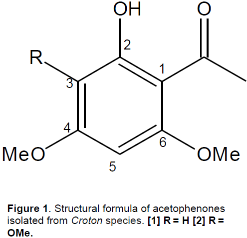
The phytochemical study of Croton anisodontus and Croton nepetaefolius led to the isolation of two compounds; 2-hydroxy-3,4,6-trimethoxyacetophenone and 2-hydroxy-4,6-dimethoxyacetophenone, respectively (Figure 1). These acetophenones are phenolic compounds with biological effects such as anti-inflammatory (Sala et al., 2001; Malaviya et al., 2010), antispasmodic (Cechinel Filho et al., 1995; Niero et al., 1996), antiproliferative (Pisco et al., 2006) and antimicrobial (Bonifait et al., 2012).
The aim of this study was to assess the modulatory and antifungal activities of acetophenones isolated from Croton spp. against strains of dermatophytes and yeasts.
Plant material
The plant material (stems) of C. anisodontus Müll.Arg. and C. nepetaefolius Baill. were collected in the city of Itapiuna, Ceará State, Brazil, in March 2011, at coordinates 04° 33' 50" S and 38° 55' 19" W, and in the city of Caucaia, Ceará, Brazil, respectively. The botanical identification was performed by Professor Edson Nunes of Ceará Federal University (UFC). Voucher specimens of the species were deposited in the Prisco Bezerra Herbarium of UFC under numbers 48.964 and 33.582, respectively.
Experimental
Adsorption column chromatography was carried out with silica gel 60 (63 to 200 mm, 70 to 230 mesh, Vetec). Column length and diameter were varied according to the sample amount. TLC used aluminum chromatoplates (20 x 20 cm) with silica gel 60 GF254 (Merck). Substances on the plates were revealed by exposure to a vanillin solution followed by heating. Infrared spectra were obtained with a Perkin-Elmer 1000-FT spectrometer. Melting points were determined with a Mettler Toledo digital microdetermination apparatus. One and two dimensional nuclear magnetic resonance (NMR) spectra were obtained on a Bruker DRX-500 (1H: 500 MHz; 13C: 125 MHz, respectively) spectrometer, using CDCl3 as solvent and TMS as the internal standard. Mass spectra (MS) were recorded with a Shimadzu QP5050A spectrometer, operating at 70 eV.
Extraction and isolation of acetophenones
C. nepetaefolius stems (5.0 kg) were powdered and subjected to extraction using ethanol (10 L×3, at room temperature). The solvent was removed under reduced pressure to give EtOH extract. The EtOH extract (58.2 g) was coarsely fractionated in a silica gel column by elution with n-hexane (F 1 to 15), n-hexane/EtOAc 1:1 (F 16 to 25), EtOAc (F 26 to 40), and EtOH (F 41 to 48), resulting to a total of 48 fractions of 100 mL each. The fractions of n-hexane/EtOAc 1:1 (F 16 to 25; 10.8 g) were pooled and fractionated in a silica gel column with n-hexane (F′1), n-hexane/EtOAc (9:1 F′2 to10; 7:3 F′11 to 13; 1:1 F′14 to 15) and EtOAc (F′16), producing 16 fractions of 100 mL each. The fractions F′ 10 to 13 and F′16 obtained using n-hexane/EtOAc (7:3) and EtOAc yielded white crystals [mp 77 to 78.5°C, MS (70 eV, in percent) m/z 196 [M]+, 181 (100), 166 (15), 151 (8), 138 (23), 95 (8)], whose 1H and 13C NMR spectra and other properties coincided with the published values for 2-hydroxy-4,6-dimethoxyacetophenone (Santos et al., 2008).
C. anisodontus stems (1462.6 g) were dried at room temperature, followed by trituration and cold extraction with n-hexane for 3 days. The resulting solution was distilled under reduced pressure to give the EtOH extract (23.25 g), which was adsorbed onto silica gel and eluted through a chromatographic column, using the following eluents: n-hexane, chloroform, ethyl acetate and ethanol. The chloroform fraction (9.77 g) was reintroduced to the column and eluted with: n-hexane (F 1 to 6), n-hexane/ethyl acetate (8:2 F’7 to 26), n-hexane/ ethyl acetate (7:3 F 27 to 40), ethyl acetate (F 41 to 60) and methanol (F 61 to 66). Fractions 12 to 16 revealed a yellow crystalline solid; 2-hydroxy-3,4,6-trimethoxyacetophenone, which after recrystallization with n-hexane and analysis by thin layer chromatography was found to be pure (Oliveira et al., 2014).
Bioassay with Artemia salina L.
Toxicity tests were conducted according to the method proposed by McLaughlin (1991), where serial dilutions were carried out from 20 mg of the samples with chloroform (CHCl3) to obtain a final concentration of 1,000, 100, 10 and 1 ppm. The negative control was 100 μl of 2% dimethyl sulfoxide (DMSO) and 4.9 mL of distilled water. 0.5 mL of each sample was added to all test tubes with 1.0 mL saline and 50 μl of DMSO. The tubes were placed in a sonicator for 10 min with the aid of a pipette. 48 h after hatching, 10 Artemia salina L. larvae were transferred to each test tube. The volume of the tubes was completed with saline to 5 mL. The assays were performed in triplicate.
The tubes were incubated for 24 h, after which the number of dead microcrustaceans was counted to compute the mortality percentage. The results were tabulated and submitted to probit analysis using SPSS 10.0 for Windows, to obtain the LC50 value with a confidence interval of 95%.
Fungal strains
The strains of Trichophyton rubrum and Candida albicans were obtained from the fungal collection of the Specialized Medical Mycology Center (CEMM, Federal University of Ceara, Brazil). The strains of T. rubrum were isolated from symptomatic patients and those of C. albicans were isolated from cats. M. canis strains were isolated from dogs. All the strains were stored in the microbiology laboratory (Vale do Acaraú State University, Brazil), where they were maintained in saline (0.9% NaCl) at 28°C. At the time of the analysis, an aliquot of each suspension was taken and inoculated into potato dextrose agar (Difco, Detroit, USA), and then incubated at 28°C for about 2 to 10 days respectively. The identification of dermatophytes was based on phenotypic features, such as a description of the macro and micromorphology. The pigment production and sporulation of dermatophytes were considered as well as the production of the enzyme urease to differentiate species of the genus Trichophyton (Vasconcellos et al., 2013; Mattei et al., 2014). The C. albicans strains were identified by automated identification (Vitek system platform) (Durham, USA).
Inoculum preparation for antifungal susceptibility tests
The inoculum was prepared from strains grown on Sabouraud dermatophytic agar for 5 days at 35°C and yeast strains for 24 h at 35°C. Fragments of M. canis, T. rubrum and C. albicans were transferred to tubes containing sterile saline solution (0.85%) to obtain a turbidity equivalent to standard (5x10⁶ CFU/mL or 0.5) on the McFarland scale.
The suspensions were diluted to 1:2000 for C. albicans and 1:500 for M. canis and T. rubrum, both with RPMI 1640 medium with L-glutamine, without sodium bicarbonate (Sigma Chemical Co., St. Louis, Mo.), and buffered to pH 7.0 with 0.165 M morpholine-propanesulfonic acid (MOPS) (Sigma Chemical Co., St. Louis, Mo.), to obtain inoculum concentrations of approximately 2.5 to 5 x 10³ CFU/mL and 5 x 10⁴ CFU/mL respectively.
Broth microdilution method
The minimum inhibitory concentration (MIC) and minimum fungicidal concentration (MFC) of acetophenones against the dermatophyte and yeast strains were determined by the broth microdilution method using 96-well plates. The MIC for C. albicans was determined in accordance with the Clinical and Laboratory Standards Institute-CLSI (formerly NCCLS; M27-A2) (CLSI, 2008). The broth microdilution assay for M. canis and T. rubrum was performed based on the document M38-A (CLSI, 2002).
The compounds were prepared in DMSO at a concentration of 10 mg/mL diluted in 100 µL of RPMI 1640 medium (Sigma) and tested in a concentration range of 0.009 to 10.0 mg/mL. Thereafter, a 100 µL inoculum was added to the plates to produce a final volume of 200 µL per well. The antifungal ketoconazole for dermatophytic strains and amphotericin B for yeast strains, with a range from 0.07 to 16 µg/mL, were used as controls. The microplates were incubated at 37°C and read visually after 5 days. C. parapsilosis (ATCC 22019) and C. krusei (ATCC 6528) were included as quality controls. The MIC was defined as 100% inhibition of visible fungal growth. Consecutively, the minimum fungicidal concentration was determined after the transfer of 100 μL of the contents of the well without turbidity into tubes containing potato dextrose agar at 28°C. The MFC against dermatophytes was calculated in accordance with fungal growth in the culture medium after 5 days, whereas for the yeasts it was determined after 24 h. Each experiment was run in duplicate.
Microdilution checkerboard assay
The effect of the compounds combined with commercial antifungal was determined by the checkerboard technique, a method used to determine the interaction of the drug by calculating the fractional inhibitory concentration index (FICI). The FICI is calculated by summing the fractional inhibitory concentration (FIC) for each of the tested compounds, which in turn is determined by summing the MIC of each drug in combination divided by the MIC of the same drug alone.
The turbidity of the fungal suspensions was adjusted to 0.5 on the McFarland scale (105 CFU/mL). Solutions of the products tested in certain concentrations from their respective MIC values were used. Initially, 50 µL of RPMI medium was added to all 96 wells of microdilution plates. Then 50 µL of the compound was added in the first column, in which serial dilutions were made in the plate until the 8th column, with the compound concentrations ranging from 10.0 to 0.07 µg/mL. In the vertical lines, 50 µL of standard antifungal ketoconazole was placed in concentrations ranging from 16 to 0.125 µg/mL. Finally, 100 µL of inoculum was added to all wells. RPMI 1640 medium with inoculum was used as a negative control, while the ketoconazole and acetophenone were separately used as positive controls at the respective MIC values. The dermatophyte plates were incubated at 36°C for 5 days. Assays were performed in triplicate.
The FICI was calculated according to the equation below, where A represents acetophenone and B the antifungal ketoconazole:
FICA = MIC of agent A in combination / MIC of agent A alone
FICB = MIC of agent B in combination / MIC of agent B alone
FICI = FICA + FICB
The FICI indicated a synergistic effect at values ≤ 0.5, an indifferent effect at values > 0.5 or ≤ 4.0, and an antagonistic effect at values > 4.0 (Odds, 2003; Johnson et al., 2004).
Bioassay with Artemia salina L.
In the toxicity tests, 2-hydroxy-4,6-dimethoxy-acetophenone and 2-hydroxy-3,4,6-trimethoxy-acetophenone presented LC50 of 46.22 µg/mL with an interval of 29.22 to 73.37 and LC50 of 75.55 µg/mL with an interval of 46.55 to 125.47, respectively. These results indicated that both compounds are bioactive, with the former being more toxic than the latter, because the former compound required a lower quantity to cause 50% mortality of the population.
MIC and MFC determination
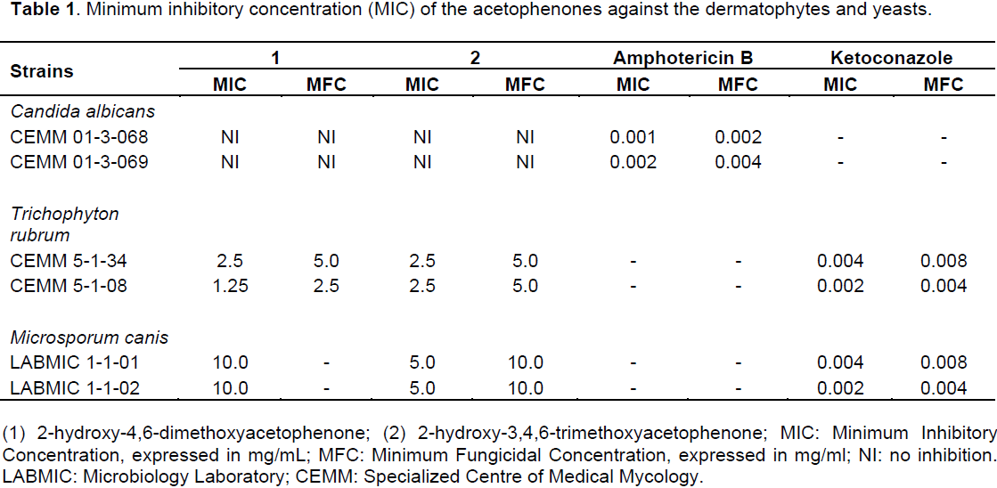
The data presented in Table 1 shows that the acetophenones tested have similar antifungal activity against the dermatophytes. 2-hydroxy-4,6-dimethoxy-acetophenone was active against T. rubrum, with MICs ranging from 1.25 to 2.5 mg/mL and MFCs of 2.5 to 5.0 mg/mL for the two strains tested, while 2-hydroxy-3,4,6-trimethoxyacetophenone presented MIC of 2.5 mg/m and MFC of 5.0 mg/mL against the same strains.
For the strains of M. canis, 2-hydroxy-4,6-dimethoxy-acetophenone presented MIC of 10.0 mg/mL at the highest concentration tested, but did not present MFC. The yeast strains were not sensitive to either of the two compounds.
Synergistic effect of ketoconazole
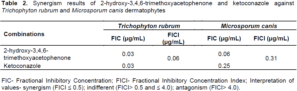
To assess the possibility of a more powerful antifungal effect of acetophenone, pooled samples were tested in combination with the commercial antifungal agent. Table 2 demonstrates a synergistic effect of ketoconazole in combination with acetophenone, with MIC values just below four-fold of the individual compounds.
The startling combination effects of ketoconazole and the compound were confirmed in this test, with FICI of 0.06 µg/mL against T. rubrum and FICI of 0.31 µg/mL against M. canis, resulting in synergic effects.
Meyer et al. (1982) established a relation between the degree of toxicity and the mean lethal dose (LC50) of plant substances against larvae of A. salina L., and since then the benchmark has been that LC50 values <1000 µg/mL indicate that a substance is bioactive. This result corroborates the antifungal properties of the compounds.
As a result of the resistance developed by micro-organisms to drugs currently used to treat fungal infections, there is the need to find new compounds with biological activity, particularly those of plant origin. The two compounds tested here; 2-hydroxy-4,6-dimethoxy-acetophenone and 2-hydroxy-3,4,6-trimethoxy-acetophenone, presented antifungal activity against the dermatophyte strains investigated.
Despite the large number of articles reporting experiments to find compounds with antifungal activity, only a few have reported the action mechanism of the compounds tested (Zacchino, 2001). Therefore, the broth microdilution method was used in this study, but it was not possible to determine the action mechanism of the acetophenones against the fungal strains studied. However, it is believed in this study that it could be related to the synthesis of ergosterol, since there are reports in the literature of acetophenones that have hypocholesterolemic activity associated with synthesis of this sterol (Piyachaturawat et al, 2002).
Despite having activity against strains of dermatophytes, the compounds were not active against strains of C. albicans. Resistance of Candida strains is related to several virulence factors, which comprises adhesion and biofilm formation, dimorphism, cell wall composition and secretion of hydrolytic enzymes such as proteases and phospholipases (Giolo and Svidzinski, 2010; Sardi et al., 2013).
Studies of antifungal activities of the acetophenones from plants are still scarce in the literature. Gul et al. (2002) reported good antifungal activity of acetophenone-derived bis Mannich bases against the dermatophyte species of T. rubrum and M. canis, with potential for developing novel antifungal agents.
In a study of the antifungal activity of acetophenone derivatives, 2,3,4 trihydroxyacetophenone (Gala) was the compound that best inhibited the filamentous fungi, with MIC of 31.2 μg/mL, indicating greater activity than fluconazole against M. canis. On the other hand, 2,4,6-trihydroxyacetophenone (THA) was the least active compound against the yeast C. albicans, presenting MIC of 125μg/mL (Rebelo, 2005). The results of this study demonstrated the oxidative potential of THC besides the antioxidant activity in vitro of the two acetophenones tested.
The oxidative potential of THA was also demonstrated by Ferreira (2005) in an investigation of the oxidative activity of synthetic analogs of acetophenone, where THA and Gala also showed important oxidative potential, in vitro and in vivo. THA also presented strong hypertriglyceridemic activity, acting as an excellent inhibitor of absorption of triglycerides in animals treated with olive oil.
The activity in vitro of compounds with little or no antifungal activity has been tested in combination with antifungal drugs (Khan and Ahmad, 2012). Therefore, the interaction between the compounds and antifungal drugs may prove to be an alternative for the treatment of fungal infections and antifungal resistance (Vandeputte et al., 2012). The compound 2-hydroxy-3,4,6-trimethoxy-acetophenone in combination with the antifungal ketoconazole in vitro had a synergistic effect, thus enhancing the antifungal activity of the compound, suggesting future pharmacological use as an adjuvant for these drugs (Guerra et al., 2015).
The authors have not declared any conflict of interests.
The authors are grateful to the Master’s Program in Natural Resources of State University of Ceará, the Microbiology Laboratory of Vale do Acaraú State University, the financial support from FUNCAP, CNPq and Universal nº 447 291/2014.9.
REFERENCES
|
Araújo-Junior VT, Silva MS, Cunha EVL, Agra MF, Silva-Filho RN, Barbosa-Filho JM, Braz-Filho R (2004). Alkaloids and diterpenes from Croton moritibensis. Pharm. Biol. 42(1):62-67.
Crossref
|
|
|
|
Barreto MB, Gomes CL, Freitas JVB, Pinto FGL, Silveira ER, Gramosa NV (2013). Flavonoids and terpenoids of Croton muscicarpa (Euphorbiaceae). Quim Nova 36(5):675-679.
Crossref
|
|
|
|
|
Bonifait L, Marquis A, Genovese S, Epifano F, Grenier D (2012). Synthesis and antimicrobial activity of geranyloxy- and farnesyloxy-acetophenone derivatives against oral pathogens. Acta Farm. Bonaer. 83(6):996-999.
Crossref
|
|
|
|
|
Brito EH, Fontenelle ROS, Brilhante RS, Cordeiro RA, Soares Júnior FA, Monteiro AJ, Sidrim JJ, Rocha MF (2007). Phenotypic characterization and in vitro antifungal sensitivity of Candida spp. and Malassezia pachydermatis strains from dogs. Vet. J. 174(1):147-153.
Crossref
|
|
|
|
|
Carneiro VA, Santos HS, Sousa AFV, Bandeira PN, Albuquerque MR, Pereira MO, Henriques M, Cavada BS, Teixeira EH (2011). Casbane Diterpene as a Promising natural antimicrobial agent against biofilm-associated Infections. Molecules 16(1):190-201.
Crossref
|
|
|
|
|
Catalán CAN, Heluani CS, Kotowicz C, Gedris TE, Herz W (2003). A linear sesterterpene, two squalene derivatives and two peptide derivatives from Croton hieronymi. Phytochemistry 64(2):625-629.
Crossref
|
|
|
|
|
Cechinel Filho V, Miguel OG, Calixto JB (1995). Antispasmodic activity of Xanthoxyline derivatives structure activity relationships. J. Pharm. Sci. 84(4):473-475.
Crossref
|
|
|
|
|
Clinical and Laboratory Standards Institute (CLSI) (2002). Reference Method for Broth Dilution Antifungal Susceptibility Testing of Filamentous Fungi: Approved Standard. CLSI Document M38-A: Wayne, PA, USA: Clinical and Laboratory Standards Institute, 2002.
|
|
|
|
|
Clinical and Laboratory Standards Institute (CLSI) (2008). Reference Method for Broth Dilution Antifungal Susceptibility Testing of Yeasts; Approved Standard-Third Edition. CLSI Document M27-A3. Wayne, PA, USA: Clinical and Laboratory Standards Institute, 2008.
|
|
|
|
|
Eksi F, Gayyurhan FD, Balci I (2013). In vitro susceptibility of Candida species to four antifungal agents assessed; by the reference broth microdilution method. Sci. World J. 2013:236903.
Crossref
|
|
|
|
|
Ferreira EA (2005). Assessment of potential antioxidant and hypertriglyceridemia of synthetic analogues from acetophenone. Thesis: Department of Pharmacy, Federal University of Santa Catarina, Florianópolis, 2005.
|
|
|
|
|
Guerra FQS, Araújo RSA, Sousa JP, Pereira FO, Mendonça-Junior FJB, Barbosa-Filho JM, Lima EO (2015). Evaluation of antifungal activity and mode of Action of new coumarin derivative, 7-hydroxy-6-nitro-2H-1-benzopyran-2-one, against Aspergillus spp. J. Evid. Based Complementary Altern. Med. 2015:925096.
|
|
|
|
|
Gul HI, Ojanen T, Hänninen O (2002). Antifungal evaluation of bis mannich bases derived from acetophenones and their corresponding piperidinols and stability studies. Biol. Pharm. Bull. 25(10):1307-1310.
Crossref
|
|
|
|
|
Guo J, Brosnan B, Furey A, Arendt EK, Murphy P, Coffey A (2012). Antifungal activity of lactobacillus against Microsporum canis, Microsporum gypseum and Epidermophyton floccosum. Bioeng. Bugs 3(2):104-113.
|
|
|
|
|
Havlickova B, Czaika VA, Friedrich M (2008). Epidemilogical trends in skin mycoses worldwide. Mycoses 51(4):2-15.
Crossref
|
|
|
|
|
Johnson MD, MacDougall C, Ostrosky-Zeichner L, Perfect JR, Rex JH (2004). Combination antifungal therapy. Antimicrob. Agents Chemother. 48:693-715.
Crossref
|
|
|
|
|
Khan MSA, Ahmad I (2012). Antibiofilm activity of certain phytocompounds and their synergy with fluconazole against Candida albicans biofilms. J. Antimicrob. Chemother. 67(3):618-621.
Crossref
|
|
|
|
|
Maciel MAM, Dantas TNC, Camara JKP, Pinto AC, Veiga Jr. VF, Kaiser CR, Pereira NA, Carneiro CMTS, Vanderlinde FA, Lapa AJ, Agner AR, Cólus IMS, Echevarria-Lima J, Grynberg NF, Esteves-Souza A, Pissinate k, Echevarria A (2006). Pharmacological and biochemical profiling of lead compounds from traditional remedies: the case of Croton cajucara. Adv. Phytomed. 2:225-253.
Crossref
|
|
|
|
|
Malaviya R, Malaviya R, Uckun FM (2010). Anti-inflammatory activity of 2,4, 6-trihydroxy-alpha-p-methoxyphenyl-acetophenone (compound D-58). Dermatology 201(4):337-342.
Crossref
|
|
|
|
|
Mattei AS, Beber MA, Madrid IM. Dermatophytosis in small animals. Microbiol. Infect. Dis. 2(3):1-6.
|
|
|
|
|
Mclaughlin JL (1991). Crown gall tumours on potato discs and brine shrimp lethality: two simple bioassays for higher plant screening and fractions. In: Dey PM, Harbone JB, eds. Methods in Plant Biochemistry. New York, NY, Academic Press; 1991.
|
|
|
|
|
Mehraboni M, Kazemi A, Mousavi SA, Rezaifar M, Alikhah H, Nosky A (2013). Evaluation antifungal of Myrtus communis L. by bioautografy method. Jundishapur J. Microbiol. 6(8):e8316.
|
|
|
|
|
Meyer BN, Ferrigni NR, Putnam, JE, Jacobsen, LB, Nichols DE, McLaughlin JL (1982). Brine shrimp: a convenient general bioassay for active plants constituents. Planta Med. 45(5): 31-34.
Crossref
|
|
|
|
|
Negri M, Silva S, Henrique M, Oliveira R (2012). Insights into Candida tropicalis nosocomial infections and virulence factors. Eur. J. Clin. Microbiol. Infect. Dis. 31(7):1399-1412.
Crossref
|
|
|
|
|
Niero R, Amaral FL, Pizzolatti MG, Calixto JB, Cechinel Filho V, Monache FD, Yunes RA (1996). Isolation of triterpenes and an acetophenone derivative with antispasmodic activity from Euphorbia milli Desmoul. ex Boiss (Euphorbiaceae). Acta Farm. Boanaer. 15(4):239-242.
|
|
|
|
|
Odds FC (2003). Synergy, antagonism, and what the chequerboard puts between them. J. Antimicrob. Chemother. 52(1):1.
Crossref
|
|
|
|
|
Oliveira MT, Teixeira AMR, Coutinho HDM, Menezes IR, Sena DM Jr, Santos HS, de Mesquita BM, Albuquerque MR, Bandeira PN, Braz-Filho R (2014). Identification and modulatory activity assessment of 2-hydroxy-3,4,6-trimethoxyacetophenone isolated from Croton anisodontus Mull. Arg (Euphorbiaceae). Nat. Prod. Commun. 9(5):665-668.
|
|
|
|
|
Peres NTA, Rossi A, Maranhão FCA, Martinz-Rossi NM (2010). Dermatophytes: host-pathogen interaction and antifungal resistance. An Bras. Dermatol. 85(5):657-67.
|
|
|
|
|
Pisco L, Kordian M, Peseke K, Feist H, Michalik D, Estrada E, Carvalho J, Hamilton G, Rando D, Quincoces J (2006). Synthesis of compounds with antiproliferative activity as analogues of prenylated natural products existing in Brazilian propolis. Eur. J. Med. Chem. 41(3):401-407.
Crossref
|
|
|
|
|
Piyachaturawat P, Tubtim C, Chuncharunee A, Kumaratat P, Suksamrarn A (2002). Evaluation of the acute and subacute toxicity of a choleretic phloracetophenone in experimental animals. Toxicol. Lett. 129(1-2):123-132.
Crossref
|
|
|
|
|
Posteraro B, Tumbarello M, Sorda M, Spanu T, Trecarichi EM, Bernardis F, Scoppettuolo G, Sanguinetti M, Fadda G (2006). Azole resistance of Candida glabrata in a case of recurrent fungemia. J. Clin. Microbiol. 44(8):3046-3047.
Crossref
|
|
|
|
|
Rebelo JM (2005). Evaluation of antioxidant and antifungal activity of synthetic analogues of acetophenone and pro-oxidant and anti-tumor of synthetic chalcones. Thesis: Department of Biotechnology, Federal University of Santa Catarina, Florianópolis, 2005.
|
|
|
|
|
Risco E, Ghia F, Vila R, Iglesias J, Alvarez E, Canigueral S (2003). Immunomodulatory activity and chemical characterisation of sangre de Drago (Dragon's blood) from Croton lechleri. Planta Med. 69(9):785-794.
Crossref
|
|
|
|
|
Sá NC, Cavalcante TTA, Araújo AX, Santos HS, Albuquerque MR, Bandeira PN, da Cunha RM, Cavada BS, Teixeira EH (2012). Antimicrobial and antibiofilm action of Casbane Diterpene from Croton nepetaefolius against oral bacteria. Arch. Oral Biol. 57(5):550-555.
Crossref
|
|
|
|
|
Sala A, Recio MC, Giner RM, Má-ez S, Ríos JL (2001).New acetophenone glucosides isolated from extracts of Helichrysum italicum with antiinflammatory activity. J. Nat. Prod. 64(10):1360-1362.
Crossref
|
|
|
|
|
Salatino A, Salatino MLF, Negri G (2007). Traditional uses, chemistry and pharmacology of Croton species (Euphorbiaceae). J. Braz. Chem. Soc. 18(1):11-33.
Crossref
|
|
|
|
|
Santos HS, Rodrigues MFM, Lemos TLG, Queiroz MFJ, Braz-Filho R (2008). Casbane diterpenes and acetophenones of Croton nepetaefolius (Euphorbiaceae). Quim Nova 31(3):601-604.
Crossref
|
|
|
|
|
Saúde-Guimarães DA, Faria AR (2007). Natural compounds with anti-Trypanossoma cruzi activity. Braz. J. Pharmacog. 17(3):455-465.
|
|
|
|
|
Seebacher C, Bouchara JP, Mignon B (2008). Uptade of the epidemiology of dermatophyte infections. Mycopathologia 166(5-6):335-352.
Crossref
|
|
|
|
|
Sousa AH, Junior JNS, Guedes MLS, Braz-Filho R, Costa-Lotufo LV, Araújo A J, Silveira ER, Lima MAS (2015). New Terpenoids from Croton limae (Euphorbiaceae). J. Braz. Chem. 26(8):1533-1741.
|
|
|
|
|
Vandeputte P, Ferrari S, Coste AT (2012). Antifungal resistance and new Strategies to control fungal infections. Int. J. Microbiol. 2012:713687.
Crossref
|
|
|
|
|
Vasconcellos C, Pereira CQM, Souza MC, Pelegrini A, Freitas RS, Takahashi JP (2013). Identification of fungi species in the onychomycosis of institutionalized elderly. An Bras Dermatol. 88(3):377-380.
Crossref
|
|
|
|
|
Yenisehirli G, Bulut Y, Günda YE (2007). Antifungal susceptibility of Candida albicans isolates recovered from blood cultures of intensive care unit patients. Ankem Dergisi 21(3):146-149.
|
|
|
|
|
White TC, Oliver BG, Hen, MR (2008). Generating and testing molecular hypotheses in the dermathophytes. Eukaryot. Cell 7(8):1238-1245.
Crossref
|
|
|
|
|
Zacchino AS (2001). Strategies for discoveries of new antifungal agents. In: Yunes RA, Carlixto JB, eds. Medicinal plants on the perspective of modern medicinal chemistry. Chapecó, SC: Argos; 2001.
|
|
|
|
|
Zou GA, Su ZH, Zhang HW, Wang Y, Yang JS, Zou ZM (2010). Flavonoids from the stems of Croton caudatus Geisel. var. tomentosus Hook. Molecules 15(3):1097-1102.
Crossref
|
|