Full Length Research Paper
ABSTRACT
The main sources of human exposure to aluminum are found in the extensive presence of the metal in the environment and its growing industrial applications. The present study was carried out to determine the effectiveness of the Thymus vulgaris L. extract in alleviating aluminum chloride (AlCl3) toxicity on biochemical and antioxidant parameters. The experiment’s rats were divided into five groups: a control group; an intoxicated group (300 mg AlCl3/kg bw); and three other groups which were given AlCl3 (300mg/kg/bw) then T. vulgaris. L extract (T.v), Malic Acid (MA) and Vitamin E (Vit E) at a concentration of 150mg/kg/bw. Each group was given its respective dosage, daily for 90 days. The results showed a significant decrease in the body/liver/kidney weights, the plasma total protein (T. Protein) and albumin (Alb) levels (p≤0.05) in the intoxicated rats group. However, a significant increase in the plasma uric acid (Uac), alkaline (AlP) and acid phosphatase (AcP) levels was noted (p≤0.05) in the same group. The amount of aluminum, TBARS and nitrate/nitrite (NO) in the liver and kidney tissues of the rats treated with AlCl3 was also found to have increased (p≤0.05), while the levels of glutathione peroxidase (GSH-Px) and glutathione transferase (GSH-St) have decreased significantly (p≤0.05). These altered parameters were restored in the rats treated with the T. vulgaris L. extract. Therefore, due to these beneficial effects, T. vulgaris L. could potentially be used to antagonize AlCl3 toxicity.
Key words: Thymus vulgaris L., aluminum chloride, oxidative stress, malic acid, vitamin E.
INTRODUCTION
Aluminum is a metal used more and more in our daily routine in spite of its toxic impact its biological functions remain unknown. Al has been considered as a toxic agent in many pathological processes such as neurodegenerative diseases (Exley, 2016), mycrocytic anaemia and osteoporosis (Chappard et al., 2016). Once absorbed, Al gathers necessarily in the bones, brain, liver and kidneys (Kinawy, 2019). This metal was considered hurtless since its presence in the trivalent form meant that it would easily attach to ions and forms colloidal polymeric particles. This form of particles is insoluble as a result it was believed that the absorption of the metal would be restricted (Bondy, 2010). In addition, toxic impacts of Al have led to histopathological alterations in the kidney and liver (Ghorbel et al., 2015). The molecular mechanisms include membrane function disruption, oxidative stress induction and disruption of several metals’ metabolism (Hasona and Ahmed, 2017). Therefore, the estimation of antioxidant defense has become an important aspect of investigation of Al toxicity.
There is a growing tendency towards using phytotherapy owing to the general belief that it has no side effects compared to chemotherapy (Geleta et al., 2016). Many natural product extracts have been found to have a variety of pharmacological and antioxidant effects. Thymus vulgaris (common thyme), locally known in Algeria as “Zaatar”, belonging to the Lamiaceae family, is a perennial herb indigenous in North Africa, Central and Southern Europe. Common Thyme’s stems are branching and of a silver-grey color. Its leaves are small, oval and coiled at the edges and very fragrant while its flowers appear from May to September and are of a white or pale pink color. T. vulgaris L. has been commonly used since ancient times in the treatment of burns and poisoning caused by snakes and scorpions. It is an aromatic medicinal herb which is widely used in traditional folk medicine for its antimicrobial effects (Hosseinzadeh et al., 2015), anti-inflammatory and antalgic effects, antioxidant benefits (Tural and Turhan, 2017) and its antifungal properties (Benabed et al., 2018). It is also recognized for its antispasmodic, antiseptic, anthelmintic, diuretic and sedative properties (Hosseinzadeh et al., 2015). It is these various benefits of T. vulgaris L. that form the motivation behind the current study which aims to evaluate the protective effects of T. vulgaris L. against AlCl3 toxicity in rats.
Various acute and chronic metal intoxications treatments are available. These include chelating agents which have been used clinically as antidotes that bind and enhance the excretion of toxic elements. The chelating agent most commonly used in Al disorders is desferrioxamine (DFO), which has a great ability to decrease the Al body toxicity by increasing its excretion (Kruck et al., 2004). However, DFO therapy is associated with toxic side effects, is very expensive and is only efficient intravenously or subcutaneously. Therefore, alternative molecules with the same efficiency as DFO, but ones which can be orally administered were needed. One such alternative is the Malic acid chelator which is a non-toxic and natural compound containing dicarboxylic acid, and magnesium. The chelation abilities of MA-mg against AlCl3 toxicity were assessed when it was administered to mice exposed to Al at about one-fourth of the LD50 level. Compared to other chelators, the MA-mg product showed a better therapeutic effectiveness.
Vitamin E (Vit E) (alpha-tocopherol) is a powerful naturel antioxidant. It acts in synergy with other molecules like vitamin C, selenium and zinc. A good intake of vitamin E can neutralize the excess of free radicals and therefore prevent cellular activity disruption which is normally caused by exposure to various stressors. The antioxidant function of this micronutrient could, at least in part, enhance immunity by maintaining the functional and structural integrity of important immune cells (El-Demerdash, 2004; Kutlbay et al., 2007).
The present study was carried out to examine the effects of AlCl3 poisoning on the biochemical and antioxidant parameters of the liver and kidney tissues of rats. Due to the health problems caused by AlCl3 and many other environmental pollutants, various investigations have been undertaken in this study to evaluate the chelator effects offered by the natural Thyme plant and the Malic acid as well as the relative antioxidant potential offered by Vit E. The present study however, focuses on determining the efficiency of these treatments especially that of T. vulgaris L. aqueous extract in antagonizing the biochemical alterations and oxidative stress leading to liver and renal dysfunction in male rats.
MATERIALS AND METHODS
Collection of plant material
T. vulgaris L. (Lamiaceae) was collected in June 2014 from the Mostaganem region in the north west of Algeria. The plant’s identification was confirmed at the department of Botany of Ahmed Ben Bella University 1 (Oran, Algeria) where a specimen (voucher No. LB 2368) was kept.
Preparation of T. vulgaris L. extracts
The aerial parts of the plant (leaves, flowers and stems) were air-dried in the dark away from humidity at room temperature and then ground to fine powder. The powdered aerial parts of T. vulgaris L. (5 g) were then extracted with boiled distilled water (500 ml) for 30 min. This water extract was then filtered, lyophilized and stored at -20°C until use. In order to extract the adequate amount of extract for experimental animal, the procedure of preparation was repeated multiple times. The yield of extraction is 20.22%.
Animals and experimental design
Male Wistar rats of four weeks old weighing (70 ± 10 g) were housed in standard cages in groups of six rats each. All groups were kept in a 12 h light/12 h dark cycle, at a room temperature of (22 ± 2°C) and were fed ad libitum. All experiments reported in this study were carried out in accordance with current guidelines for the care and use of laboratory animals (8th edition, 2011). All animals within each treatment group were given their respective diet for 90 days as follows:
(i) Group 1 (C): Untreated control group.
(ii) Group 2 (AlCl3): The rats were orally given a solution composed of AlCl36H2O which was dissolved in distilled water. The daily dose given was 300 mg/kg bw.
(iii) Group 3 (AlCl3 + T. vulgaris L.): During the first 45 days, the rats were given to drink by feeding bottle the same AlCl36H2O solution as Group 2 at a daily dose of 300 mg/kg bw. This was then followed by giving the rats a treatment solution of T. vulgaris L. aqueous extract at a daily dose of 150 mg/kg bw for the following 45 days for a total duration of 90 days.
(iv) Group 4 (AlCl3 + MA): The rats in this group were given to drink the same AlCl36H2O solution for 45 days which was then followed by a Malate Magnesium chelator solution for 45 days at a concentration of 150 mg/Kg bw.
(v) Group 5 (AlCl3 + Vit E): The rats in this group were given to drink the same AlCl36H2O solution for 45 days followed by a Vitamin E solution for 45 days at a dose of 150 mg/Kg bw.
T. vulgaris L. (150 mg/Kg) is a tolerated dose and without toxic effect because it represents the (1/30) of LD50 (lethal dose of 50% of population) of alcoholic extract which is 5 g/kg of body weight used in the study of El-Newary et al., 2017. In another study (Geleta et al., 2016) researchers tested the dose (10.000 mg/kg) of aqueous extract of T. shimperi on acute intoxication also tested the dose of (600 mg/kg) in subchronic toxicity and observed their non-toxicity. Therefore the dose chosen in this study is lower than the LD50 previously noted. The animals were observed daily for any signs of toxicity and their body weight was recorded on a weekly basis throughout the experimental period.
Blood collection and tissue sample preparation
At the end of the experimental period and after the administration of the last dose, the rats were left to rest overnight and were sacrificed under pentobarbital anesthesia the following day. The rats’ blood samples were then collected, and the liver and kidney organs were harvested, rinsed with a saline solution (0.9% NaCl) and then weighed. The organs’ weight ratios were estimated and their relative weight calculated as g/100 g BW. To evaluate the oxidative status, the liver and kidney were homogenized in suitable buffers: in 1.15% KCl for thiobarbituric acid reactive substances (TBARS), in a 0.1 M phosphate buffer (pH 7.2) for glutathione transferase (GSH-ST) and glutathione peroxidase (GSH-PX), in a PBS (Phosphate buffered saline, PH 7.4) solution for nitrite estimation In addition, some portions of the liver and kidney were digested with nitric acid to determine the aluminum concentration.
Biochemical parameters
Alkaline phosphatase (AlP), acid phosphatase (AcP), uric acid (Uac) and albumin (Alb), were estimated in serum using Chrono-Lab kits (Spain). Total protein was measured by using bovine serum albumin as a standard (Lowry et al., 1951) Folin and Ciocalteus Phenol reagents were used to develop the blue color that was measured spectrophotometrically at 750 nm.
Lipid peroxidation (TBARS)
Lipid peroxidation in the liver and kidney tissues was estimated by measuring the formed malondialdehyde (MDA) using thiobarbituric acid reactive substances, according to the spectrophotometric method of (Okhawa et al., 1979). Aliquots (0.2 ml) of hepatic or renal homogenates were added to 0.2 ml of 8.1% (w/v) sodium dodecyl sulfate, 1.5 ml of 20% (w/v) acetic acid buffer (pH 3.5), and 1.5 ml of 0.8% (w/v) thiobarbituric acid. After completing the volume with 4 ml of distilled water, the mixture was heated for 1h at 90°C then cooled and centrifuged at 4000 r/min for 10 min. TBARS were measured at 532 nm and expressed as MDA equivalents (nmol/g of Protein) using a molar extinction co-efficient: 1.56 ×10⁵mol/L/cm.
Glutathione transferase (GSH-ST)
Glutathione transferase (GSH-ST) activity in the liver and kidney tissues was assessed using an assay kit provided by Cayman (Chemical, USA).
Glutathione peroxidase (GSH-Px)
Glutathione peroxidase (GSH-Px) activity in the liver and kidney tissues was assessed by the method of Rotruck et al. (1973). Briefly, the reaction mixture contained 0.2 ml of Tris–HCl buffer (0.4 mol/L, pH 7.0), 0.2 ml of reduced GSH (1 mmol/L), 0.1 mL of Sodium Azide (10 mmol/L), 0.1 ml of H2O2 (1 mmol/L) and 0.2 ml of tissue homogenates. After incubation of the mixture at 37°C for 10 minutes, the reaction was stopped by the addition of 0.4 ml of 10% trichloroacetic acid, and tubes were subjected to centrifugation at 2400 r/min for 10 min. The supernatant (0.2 ml) was then added with 0.1 ml of Ellman's reagent (0.0198 g of DTNB prepared in 0.1% sodium citrate). The Absorbance was recorded at 340 nm.
Nitrite (NO)
Nitrate Nitrite activity in the liver and kidney tissues was assessed using an assay kit provided by Cayman (Chemical, USA).
Aluminum Measurement
The concentration of AlCl3 was estimated in the liver and kidney organs by using an atomic absorption spectrometer (Shimadzu, AA6200) following a wet acid digestion method as modified for dry-weight samples (Van Ginkel et al., 1990). A fraction of each organ (approximately 100 mg) was heated at 60°C for 24 h to obtain a constant dry weight. This was then placed in weighed flasks and digested with a nitric acid solution (65°) for 8 h. To get a final volume of 4 ml, a nitric acid solution (1% concentration) was then added to determine the aluminum concentration in the sample. The atomic absorption signal was measured by integrating the total absorption profile at 309.3 nm with a spectral bandwidth of 0.5 nm. All the analyses were performed in triplicate, the limit detection to Al is of 0.02 mg/L and the results were expressed in µg/g tissue wet.
Statistical analysis
The results are expressed as a mean ± standard error of the mean (SEM). Data comparison was determined by one-way ANOVA followed by Tukey post hoc analysis and the results were considered statistically significant when p < 0.05.
RESULTS
Throughout the study, some clinical signs of toxicity were observed in the AlCl3-treated rats at the dose previously indicated. These included; fur loss, urine coloration, as well as a lack of activity (dullness, spirit depression) when compared to the rats in the control group. However, no rat deaths were recorded during the experimental period.
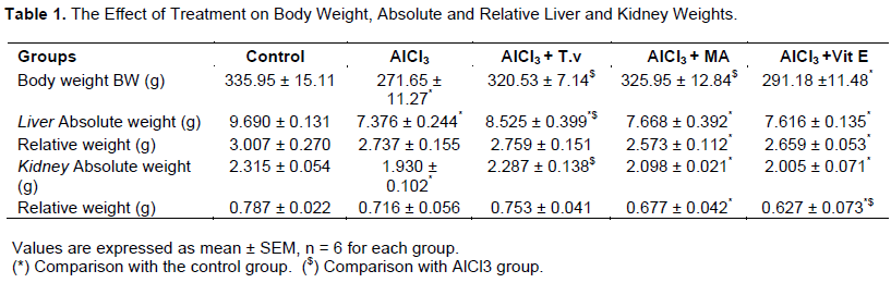
Effect of treatment on body, liver and kidney weights
At the end of the experiment, AlCl3 administration in rats revealed a significant decrease in body weight levels by 19.13% when compared to the control group. A significant decrease (p≤0.05) is also observed in the absolute liver and kidney tissue weights when compared to the control group as shown in Table 1. However, the group of rats treated with T. vulgaris L. showed considerably less weight loss at the end of the experiment (p≤0.05) when compared to the intoxicated group. A similar improvement, albeit to a lesser extent than the one recorded in the T. vulgaris L-treated group, was also observed in the MA and Vit E treated groups.
Effect of treatment on serum biochemical parameters in aluminum-induced hepatic and renal toxicity in rats
Treatment with AlCl3 increased significantly the plasma AlP level by 32.34% when compared to the control group (Table 2). In addition, it was found that the concentration of plasma AcP and Uac has significantly increased by 3-fold in the rats treated with AlCl3. Plasma Alb and T. Protein, however, decreased significantly by (56.68 and 51.18% respectively) in the intoxicated rats group.

Examining the groups treated with either T. vulgaris L. or MA, a significant reduction in the plasma AlP (T. vulgaris L. 32.92%, MA: 22.95%) and AcP (T. vulgaris L. 57.70%, MA: 40.53%) was observed when compared to the intoxicated group. In addition, the Vit E treated group showed a low decrease in the same parameters that soared in the intoxicated group. The concentration of Uac was also significantly reduced by the T. vulgaris L. and Vit E treatments (49%, 28.14% respectively). The effect of the MA treatment on this parameter was found to be smaller (22.25%) in comparison. These results were accompanied by an increase in Alb and T. Protein concentrations in the T. vulgaris L. treated group (47.50 and 45.24% respectively) when compared to those of the intoxicated group. MA and Vit E treatments only slightly improve these parameters.
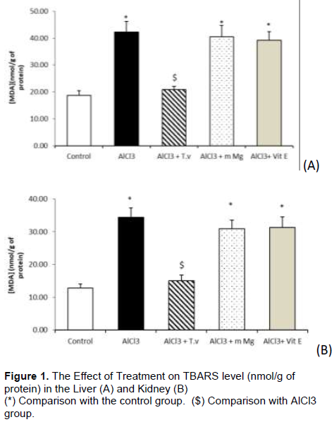
Effect of treatment on lipid peroxidation on aluminum-induced hepatic and renal toxicity in rats
As shown in Figure 1, a significant increase in the TBARS level (55.67% in the liver tissue and 62.64% in the kidney tissue) was observed in the intoxicated rats when compared to the control groups. The administration of T. vulgaris L. decreased the TBARS production significantly by a rate of 50.64% in the liver and 56.3% in the kidney when compared to the intoxicated rats. Malate and Vitamin E treatments showed less efficient results in term of restoring normal values of TBARS.
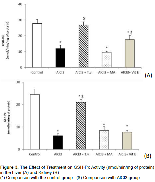
Effect of treatment on antioxidant parameters on aluminum-induced hepatic and renal toxicity in rats
The results obtained showed that GST and GPX levels (Figures 2 and 3) were significantly decreased in the liver (62.87 and 57.23% respectively) and kidney (73.23 and 75.18% respectively) of the rats treated with AlCl3. The concentration of NO was also significantly increased (p≤0.05) as shown in Figure 4. However, liver and kidney GST and GPX levels increased by 2 and 3-fold respectively in the groups treated with T. vulgaris L. A significant increase (p≤0.05) in liver GST and GPX levels was also respectively noted in the groups treated with MA and Vit E when compared to the AlCl3-intoxicated group. In addition, treatment with T. vulgaris L. caused a significant decrease in NO concentration (67.46 and 41.96%) in the liver and kidney tissues respectively. Only a slight reduction in liver and kidney (p≤0.05) NO concentration resulted from the Vit E treatment when compared to the intoxicated group.


Effect of treatment on liver and kidney aluminum content
The AlCl3 concentration in the liver and kidney of rats was measured after 3 months of oral AlCl3 exposure and the results are presented in Table 3. The concentration of AlCl3 in the liver and kidney of the intoxicated group was higher by 3 and 5 fold respectively when compared to the control group. However, this concentration was significantly decreased in liver and kidney of the group treated with T. vulgaris L. (32.42 and 59.28%) when compared to the intoxicated group.

DISCUSSION
Aluminum absorbed by the human body via the gastro- intestinal and the respiratory tracts, is known to disrupt the pro-oxidant and antioxidant balance of tissues, leading to various biochemical and physiological dysfunctions (Exley, 2004; Nehru and Bhalla, 2006). In this study, Aluminum Chloride was chosen over other Aluminum compounds because the stomach already contains and utilizes Chloride. Therefore, this form of Aluminum can be introduced with minimal change to gastric fluid composition. The focus of the present study is on the liver and kidney organs where metals are usually accumulated and where toxic effects can be expected.
The results of the study have indicated that body, liver and kidney weights gain in the rats treated with AlCl3 was markedly less (p≤0.05) when compared to the normal control group. This demonstrates that Al administration has a detrimental effect on the rats’ body weight. This finding agrees with the results of other studies where balgoon (2019) noticed a significant reduction of about 3.51% in weight gain in AlCl3-treated rats. Similar results were also obtained by (Singla and Dhawan, 2013). A significant decrease in absolute and relative liver and kidney weights was observed in AlCl3-treated rats which is in agreement with previous reports by (Yeh et al., 2009; Paz et al., 2017). However, the current study has found that rats treated with T. vulgaris L. showed a significant improvement in body, liver and kidney weights gain. In the study of Manafi et al. (2014) broilers intoxicated with aflatoxin and treated with Thyme essence showed a significant gain in body weight revealing the same beneficial impact.
Several studies have reported that Al accumulates in mammalian tissues such as brain, bone, liver and kidney (Belaïd-Nouira et al., 2013). As a result, Al causes alterations in the biliary secretory function and an increase in oxidative stress in hepatic tissues (Gonzalez et al., 2004). Such accumulation might be the result of the higher affinity of Al for transferrin, which, in turn, might also explain the interference it causes with iron metabolism (Crichton et al., 2002). The present study results are also consistent with recent findings that showed that chronic Al consumption causes significant plasmatic increases in the activities of AlP and AcP enzymes which could be due to severe damage in the tissue membranes (Esmaeili et al., 2000; Al-Qhtani and Farran, 2017). Moreover, Rahman et al. (2000) suggested that the decrease in the activities of AlP and AcP in different tissues might be due to the increased permeability of the plasma membrane or the cellular necrosis of the liver, kidneys and lungs. In addition, Ochmanski and Barabasz (2000) proposed that the salts of Al could potentially enter inside the cells and might directly bind to the DNA and RNA helices. This prevents their replication and then inhibits the activities of AlP and AcP. In this study, it was shown that an administration of the Thymus vulgaris L. aqueous extract has led to a marked reduction in the elevated activities of AlP and AcP in AlCl3 treated rats. In the study of Abd El Kader and Mohamed (2012) rats intoxicated with paracetamol and treated with thyme showed the same effect.
The observed decline in the level of protein in AlCl3 treated rats in this study agrees with the findings of Kinawy (2019) and might be due to changes in the metabolism of proteins, particularly albumin. On one hand, this could be attributed to malnutrition but on the other hand, it could be due to a reduction in the protein synthesis in the liver (Tripathi et al., 2009). The same results also revealed that AlCl3 intoxication caused a significant decrease (p< 0.05) in plasma T. Protein and Alb (Massoud et al., 2016), while an increase in AlP parameter was observed. Furthermore, aluminum may promote proteinuria by causing a nephritic syndrome or chronic glomerulonephritis.
The accumulation of Al in the kidney is usually accompanied by renal failure (Ecelbarger et al., 1994). Moreover, Al accumulation in the kidney promotes degeneration in renal tubular cells, inducing nephrotoxicity (Somova et al., 1997; Mohamed et al., 2017). In addition, renal functions can also be altered in the early stages of hepatic damage before the formation of ascites (Monasterolo et al., 1993). Uric acid is the major product of purine bases, adenine and guanine. It is partially considered as a significant marker of renal function. The Plasma Uac (Uric acid) elevation recorded in this study after AlCl3 exposure is in agreement with the majority of researches (Sivakumar et al., 2014; Al-Qhtani and Farran, 2017), and is also supported by the findings of Rudenko et al. (1998) who reported that AlCl3 intensifies the acid–secretion function of the kidney and changes the transport of sodium.
It has been suggested that the toxic effects associated with aluminum are due to the generation of reactive oxygen species, which results in the oxidative deterioration of cellular lipids, proteins and DNA. In the present experiment, we observed a significant increase in liver and kidney lipid peroxidation (TBARS) after AlCl3 exposure. These results are similar to previously published findings demonstrating that aluminum salts intake promoted lipid peroxidation in liver and kidney tissues (Belaïd-Nouira et al., 2013; Tahari et al., 2016). It was also found out that the consumption of T. vulgaris L. induces a significant decrease (p≤0.05) in TBARS level.
Our study demonstrated that AlCl3 administration caused a reduction in liver and kidney glutathione transferase (GSH-St) and glutathione peroxidase (GSH-PX) activities. The results are in accordance with Newairy et al. (2009). Furthermore, El-Sayed et al. (2011) reported a significant decrease in both enzymes in the liver of about (56 and 80%) respectively. A reduction of the kidney’s GSH-Px activity was also noticed by Mahieu et al., 2009.
Nitrite resulted from the oxidation of NO, so Nitrite estimation is indirectly that of nitric oxide (NO) content in the biological samples. It has been suggested that Nitric oxide plays multiple roles in Al intoxication (Guo et al., 2001). The present investigation showed that AlCl3 toxicity excessively increases NO products in the liver and kidney tissues which agree with many studies (Guo et al., 2005).
Recent studies demonstrated that aluminum may alter the activity of tissue antioxidative enzymes as TBARS and NO activity (Taïr et al., 2016; Benyettou et al., 2017). Our data support these results and the levels of these parameters were found to be elevated in AlCl3-treated rats. However, GSH-Px and GSH- St activity were decreased in the liver and kidney tissues. These observations are similar to previously published findings demonstrating that the aluminum intake promoted oxidative stress while the extract plants administration restored it (Kutlubay et al., 2007; Joshi et al., 2013).
Our results have indicated that a significant accumulation of AlCl3 occurs in the liver and kidney tissues of AlCl3-intoxicated rats. The same results were observed in other studies (Yeh et al., 2009) where AlCl3 accumulation increased by 3 times in the liver and 6 times in the kidney (Missel et al., 2005; Cheng et al., 2014). However, the present study has shown that following T. vulgaris L. extract treatment; a significant decrease in AlCl3 accumulation in the liver and kidney tissues was recorded.
It is well documented that the leaves and flowers of plants contain numerous aromatic chemical products which prevent the cellular damage caused by free radical molecules, ions or atoms (Dapkevicius et al., 2002; Al-Asmari et al., 2017). T. vulgaris L. is a famous herb cultivated all over the world, used essentially in food as a flavor and in medicine as an anti-microbial, anti-poisonous and deworming agent.
Intoxicated rats treated with thyme extract have shown low AlP and AcP levels in plasma which indicates the thyme’s hepatoprotective impact. T. vulgaris L. aqueous extract also elevated the total serum protein, Alb and Uac levels. This explains the healing effect of thyme on aluminum toxicity and confirmed its nephroprotective effect as stated by El-Newary et al. (2017). This positive effect could be due to its secondary metabolites composition including alkaloids, polyphenols and flavonoids. The main compounds of T. vulgaris L. are the natural terpenoid thymol (12-61%) and its phenol isomer carvacrol (0.4-20.6%) which revealed a high antioxidative, antispasmodic and antibacterial effects (Amiri, 2012). In addition, T. vulgaris L. Contains high levels of vitamins B3, C and E. It is also; very rich in potassium (K), Magnesium (Mg) and others trace elements (Komaki et al., 2016). Due to this composition, thyme is considered as a high regulator of homeostasis. This study also proclaimed that intoxicated rats given aqueous thyme extract showed remarkable lowered levels of MDA and NO and elevated levels of GSH-St and GSH-px in renal and hepatic tissues compared to control group. This implies the efficient and protective impact of thyme which is probably linked to its capacity to reduce oxidative stress as free radical scavenger or as a quencher of singlet oxygen formation preventing peroxidation of lipids.
El-Newary et al. (2017) found out that dietary supplementation of T. vulgaris L. reduced all the above measured parameters in the liver and kidney of rats. These results highlighted the benefit of T. vulgaris L. as a dietary antioxidant.
Other reports indicate that the essential oil of T. vulgaris L. has a potential antioxidant activity and a protective effect against toxicity of aflatoxins (El-Nekeety et al., 2011). The properties of the T. vulgaris L. constituents as polyphenols (Thymol and Carvacrol) and flavonoids (caffeic acid, rosmarinic acid, apigenine, thymonine and luteolin) protect the cellular membranes’ integrity (Ündeger et al., 2009; Popovic-Milenkovic et al., 2014) from AlCl3-induced oxidative damage and repair the antioxidant system. Moreover, other authors stated that thymol was found to decrease ROS production in dependent concentration and inhibit hepatotoxicity induced by CCl4 in male Swiss albino mice as assessed by lipid peroxidation and by histopathological examination (Alam et al., 1999).
T. vulgaris L. has noticeably decreased the aluminum content in the liver and kidney. Some authors ascertained the fact that phenolic compounds were not able to chelate metal ions. Melidou et al. (2005) found that intracellular binding of iron is responsible for the protection offered by flavonoids against H2O2-induced DNA damage. On the other hand, complexation of plant extracts with metal ions might diminish the absorption of metal ions leading to chelating and blocking metal-mediators generating the ROS (Yang et al., 2013).
In the current study, it was also found that MA and Vit E treatments only partially alleviated the harmful effects of AlCl3 toxicity as observed in previous research by El-Demerdash (2004) and Masoud et al. (2016). These results also concur with previously reported data indicating that administration of Aluminum with some plant preparations such as the Crocus sativus L. extract (Shati and Alamri, 2010), the Zingiber Officinale extract (Hasona and Ahmed, 2017) or Propolis (Turkez et al., 2010) or Selenium antioxidant products showed a recovery from hepatic damage (Viezeliene et al., 2011). Mahieu et al. (2009) and Yeh et al. (2009) have also revealed the beneficial effects of Melatonin and Taurine antioxidants against AlCl3 nephrotoxicity.
CONCLUSION
Aluminum has adverse effects on human health and special attention must be paid to the sources of Al in food, water and personal care products. From the results presented in this study, it can be seen that exposure to AlCl3 is capable of inducing significant harmful changes in some biochemical characteristics and redox status in liver and kidney tissues. However, this study has also demonstrated the therapeutic effects of the T. vulgaris L. plant in minimizing Aluminum liver and kidney toxicity. This treatment resulted in a significant improvement in body, liver and kidney weights and the plasma biochemical parameters (T. Protein, Alb, AlP, AcP and Uac). In addition, the T. vulgaris L. treatment led to a more efficient recovery from the oxidative tissue damage caused by Al intoxication (TBARS, GSH-Px, GSH-St and NO) when compared to the Malate and Vitamin E treatments. Therefore, using T. vulgaris L. extract could be a protective and preventative method in overcoming the detrimental effects of Aluminum toxicity.
CONFLICT OF INTERESTS
The authors have not declared any conflict of interests.
ACKNOWLEDGEMENT
The authors are grateful to Mrs Mokrane Amina and Mr Mokrane Ali for their help in language correction of this manuscript.
REFERENCES
| Abd El Kader MA, Mohamed NZ (2012). Evaluation of protective and antioxidant activity of thyme (Thymus Vulgaris) extract on paracetamol-induced toxicity in rats. Australian Journal of Basic and Applied Sciences 6(7):467-474. | ||||
|
Alam K, Nagi MN, Badary OA, AL-Shabanah OA, Al-Rikabi AC, Al-Bekairi AM (1999). The protective action of thymol against carbon tetrachloride hepatotoxicity in mice. Pharmacological Research 40(2):159-163. Crossref |
||||
|
Al-Asmari AK, Athar T, Al-Faraidy AA, Almuhaiza MS (2017). Chemical composition of essential oil of Thymus vulgaris collected from Saudi Arabian market. Asian Pacific Journal of Tropical Biomedicine 7(2):147-150 Crossref |
||||
| Al-Qhtani SA, Farran SK (2017). The Protective and therapeutic effect of resveratrol in improving renal and hepatic failure induced by aluminum chloride in experimental animals. Journal of American Science 13(10). | ||||
|
Amiri H (2012). Essential oils composition and antioxidant properties of three Thymus species. Evidence-Based Complementary and Alternative Medicine 2012: 8. Crossref |
||||
|
Balgoon MJ (2019). Assessment of the protective effect of Lepidium sativum against aluminum-induced liver and kidney effects in albino rat. BioMed Research International . Crossref |
||||
|
Belaïd-Nouira Y, Bakhta H, Haouas Z, Flehi-Slim I, Ben Cheikh H (2013). Fenugreek seeds reduce aluminum toxicity associated with renal failure in rats. Nutrition Research and Practice 7(6):466-474. Crossref |
||||
|
Benabed KH, Gourine N, Ouinten M, Bombarda I, Yousfi M (2018). chemical composition, antioxidant and antimicrobial activities of the essential oils of three algerian lamiaceae species. Current Nutrition and Food Science 13(2):97-109. Crossref |
||||
|
Benyettou I, Kharoubi O, Hallal N, Benyettou HA, Tair K, Belmokhtar M, Aoues A, Ozaslan M (2017). Aluminium-induced behavioral changes and oxidative Stress in developing rat Brain and the possible ameliorating role of omega-6/omega-3 ratio. Journal of Biological sciences 17(3): 106-117. Crossref |
||||
|
Bondy SC (2010). The neurotoxicity of environmental aluminum is still an issue. Neurotoxicology 31(5):575-581. Crossref |
||||
|
Chappard D, Bizot P, Mabilleau G, Hubert L (2016). Aluminum and bone: Review of new clinical circumstances associated with Al(3+) deposition in the calcified matrix of bone. Morphologie 100(329):95-105. Crossref |
||||
|
Cheng D, Zhu C, Wang C, Xu H, Cao J, Jiang W (2014). Hepatoprotective effects of apple polyphenol extract on aluminum-induced liver oxidative stress in the rat. Canadian Journal of Physiology and Pharmacology 92:109-116. Crossref |
||||
|
Crichton S, Wilmet R, Legssyer l (2002). Molecular and cellular mechanisms of iron homeostasis and toxicity in mammalian cells. Journal of Inorganic Biochemistry 91(1):9-18. Crossref |
||||
|
Dapkevicius A, van Beek TA, Lelyveld GP, van Veldhuizen A, de Groot Ae, Linssen JPH, Venskutonis R (2002). Isolation and structure elucidation of radical scavengers from Thymus vulgaris leaves. Journal of Natural Products 65(6):892-896. Crossref |
||||
|
Ecelbarger CA, MacNeil GG, Greger JL (1994). Aluminum retention by aged rats fed aluminum and treated with desferrioxamine. Toxicology Letters 73(3):249-57. Crossref |
||||
|
El-Demerdash FM (2004). Antioxidant effect of vitamin E and selenium on lipid peroxidation, enzyme activities and biochemical parameters in rats exposed to aluminum. Journal of Trace Elements in Medicine and Biology 18(1):113-121. Crossref |
||||
|
El-Nekeety AA, Mohamed SR, Hathout AS, Hassan NS, Aly SE, Abdel-Wahhab MA (2011). Antioxidant properties of Thymus vulgaris oil against aflatoxin-induce oxidative stress in male rats. Toxicon 57(7-8):984-991. Crossref |
||||
|
El-Newary SA, Shafie NM, Omer EA (2017). The protection of Thymus vulgaris leaves alcoholic extract against hepatotoxicity of alcohol in rats. Asian Pacific Journal of Tropical Medicine 10(4):361-371. Crossref |
||||
|
El-Sayed WM, Al-Kahtania MA, Abdel-Moneima AM (2011). Prophylactic and therapeutic effects of taurine against aluminum-induced acute hepatotoxicity in mice. Journal of Hazardous Materials 192(2):880-886. Crossref |
||||
| Esmaeili MA, Sonboli A, Kanani MR, Sadeghi H (2000). Salvia sahendica prevents tissue damages induced by alcohol in oxidative stress conditions: Effect on liver and kidney oxidative parameters. Journal of Medicinal Plants Research 3(4):276-283. | ||||
|
Exley C (2004). The pro-oxidant activity of aluminum. Free Radical Biology and Medicine 36(3):380-387. Crossref |
||||
|
Exley C (2016). The Toxicity of aluminum in humans. Morphologie 100(329):51-55. Crossref |
||||
|
Geleta B, Debelo N, Afework M, Debella A, Makonnen E, Ergete W (2016). Assessment of Hematological, Biochemical and Histopathological effects of Acute and Sub-chronic administration of the Aqueous leaves extract of Thymus schimperi in Rats. Journal of Clinical Toxicology 6(2):286. Crossref |
||||
|
Ghorbel I, Khemakhem M, Boudawara O, Marrekchi R, Jamoussi K, Ben Amar R, Boudawara T, Zeghal N, Grati Kamoun N (2015). Effects of dietary extra virgin olive oil and its fractions on antioxidant status and DNA damage in the heart of rats co-exposed to aluminum and acrylamide. Food and Function 6(9): 3098-3108. Crossref |
||||
|
Gonzalez MA, Roma M, Bernal C, Alvarez ML, Carrillo MC (2004). Biliary Secretory function in Rats Chronically Intoxicated with Aluminum. Toxicological Sciences 79(1):189-195. Crossref |
||||
|
Guo CH, Huang CJ, Chen ST, Hsu GSW (2001). Serum and testicular testosterone and nitric oxide products in aluminum-treated mice. Environmental Toxicology and Pharmacology 10(1-2):53-60. Crossref |
||||
|
Guo CH, Lin CY, Yeh MS, Hsu GSW (2005). Aluminum-induced suppression of testosterone through nitric oxide production in male mice. Environmental Toxicology and Pharmacology 19(1):33-40. Crossref |
||||
|
Hasona NA, Ahmed MQ (2017). Antioxidant and ameliorative effects of Zingiber Officinale against aluminum chloride toxicity. Science International 5(3):96-104. Crossref |
||||
| Hosseinzadeh S, Kukhdan AJ, Hosseini A, Armand R (2015). The application of Thymus vulgaris in traditional and modern medicine: A Review. Global Journal of Pharmacology 9(3):260-266. | ||||
| Joshi DK, Choudhary M, Tripathi S, Singh Negi MP, Mahdi AA (2013). Age dependent relative risk of aluminum toxicity: levels of metals and enzymatic and non-enzymatic antioxidants status in liver, kidney and brain of aluminum treated young and old rats. International Journal of Biological and Pharmaceutical Research 4(3):176-185. | ||||
|
Kinawy AA (2019). Potential toxicity of aluminum and fluoride on some biochemical aspects of male rat's offspring. The Journal of Basic and Applied Zoology 80:18. Crossref |
||||
|
Komaki A, Hoseini F, Shahidi S, Baharlouei N (2016). Study of the effect of extract of Thymus vulgaris on anxiety in male rats. Journal of Traditional and Complementary Medicine 6(3): 257-261. Crossref |
||||
|
Kruck TP, Cui JG, Percy ME, Lukiw WJ (2004). Molecular shuttle chelation: the use of Ascorbate, Desferrioxamine and Ferelax-G in combination to remove nuclear bound aluminum. Cellular and Molecular Neurobiology 24(3):443-459. Crossref |
||||
| Kutlubay R, Oguz OE, Abban G, Turgut S (2007). Amelioration of aluminum-induced liver damage by vitamin E. Saudi Medical Journal 28 (2):197-200. | ||||
| Lowry OH, Rosebrough NJ, Farr AL, Randall RJ (1951). Protein measurement with the Folin phenol reagent. The Journal of Biological Chemistry 193(1):265-275. | ||||
|
Mahieu S, Contini MDC, González M, Millen N (2009). Melatonin reduces oxidative damage induced by Aluminum in rat kidney. Toxicology Letters 190(1):9-15. Crossref |
||||
| Manafi M, Hedayati M, Yari M (2014). Aflatoxicosis and herbal detoxification: The effectiveness of Thyme essence on performance parameters and antibody titers of commercial broilers fed aflatoxin B1. Research in Zoology 4(2):43-50. | ||||
| Massoud AA, Hegazi MM, Hashem AA, Basuony NS (2016). Protective role and ameliorating effect of vitamin E in hepatic and renal functions of mice treated with aluminum chloride. Egyptian Journal of Experimental Biology 12(1):23-30. | ||||
|
Melidou M, Riganakos K, Galaris D (2005). Protection against nuclear DNA damage offered by flavonoids in cells exposed to hydrogen peroxide: the role of iron chelation. Free Radical Biology and Medicine 39(12):1591-1600. Crossref |
||||
|
Missel JR, Schetinger MR, Gioda CR, Bohrer DN, Pacholski IL, Zanatta N, Martins MA, Bonacorso H, Morsch VM (2005). Chelating effect of novel pyrimidines in a model of aluminum intoxication. Journal of Inorganic Biochemistry 99(9):1853-1857. Crossref |
||||
| Mohamed HH, Abd Ali IK, Reshag AF (2017). Histological changes in the liver, kidney and spleen of White Albino Rat after Aluminum Chloride administration. The Iraqi Journal of Veterinary Medicine 41(2):1-6. | ||||
|
Monasterolo L, Peiretti A, Elias MM (1993). Rat renal functions during the first days post-bile duct ligation. Renal Failure 15(4):461-467. Crossref |
||||
|
Nehru B, Bhalla P (2006). Reversal of an aluminum induced alteration in redox status in different regions of rat brain by administration of centrophenoxine. Molecular and cellular biochemistry 290(1-2):185-191. Crossref |
||||
|
Newairy AS, Salama AF, Hussien HM, Yousef MI (2009). Propolis alleviates aluminum-induced lipid peroxidation and biochemical parameters in male rats. Food and Chemical Toxicology 47(6):1093-1098. Crossref |
||||
| Ochmanski W, Barabasz W (2000). Aluminum-occurrence and toxicity for organisms. Przeglad Lekarski 57(11):665-668. | ||||
|
Okhawa H, Ohishi N, Yagi K (1979). Assay for lipid peroxides in animal tissues by thiobarbituric acid reaction. Analytical Biochemistry 95(2):351-358. Crossref |
||||
|
Paz LNF, Moura LM, Feio DCA, Cardoso MDSG, Ximenes WLO, Montenegro RC, Alves APN, Burbano RP, Lima PDL (2017). Evaluation of in vivo and in vitro toxicological and genotoxic potential of aluminum chloride. Chemosphere 175:130-137. Crossref |
||||
| Popovic-Milenkovic MT, Tomovic MT, Brankovic SR, Ljujic BT, Jankovic SM (2014). Antioxidant and anxiolytic activities of Crataegus nigra Wald. et Kit. Berries. Acta Poloniae Pharmaceutica 71(2):279-285. | ||||
|
Rahman MF, Siddiqui MK, Jamil K (2000). Acid and alkaline phosphatase activities in a novel phosphorothionate (RPR-11) treated male and female rats. Evidence of dose and time-dependent response. Drug and Chemical Toxicology 23(3):497-509. Crossref |
||||
|
Rotruck JT, Pope AL, Ganther HE, Swanson AB, Hafema, DG, Hoekstra WG (1973). Selenium: biochemical role as a component of glutathione peroxidase. Science 179(4073): 588-590. Crossref |
||||
| Rudenko SS, Bodnar BM, Kukharchuk OL, Mahalias VM, Rybshchka MM, Ozerova IO, Chala KM, Khalaturnik MV (1998). Effect of selenium on the functional state of white rat kidney in Aluminum cadmium poisoning. Ukrainskii Biokhimicheskii Zhurnal 70(6):98-105. | ||||
| Shati AA, Alamri SA (2010). Role of saffron (Crocus sativus L.) and honey syrup on aluminum-induced hepatotoxicity. Saudi Medical Journal 31(10):1106-1113. | ||||
|
Singla N, Dhawan DK (2013). Zinc protection against aluminum induced altered lipid profile and membrane integrity. Food and Chemical Toxicology 55:18-28. Crossref |
||||
|
Sivakumar S, Khatiwada CP, Sivasubramanian J, Raja B (2014). FTIR study of protective action of deferoxamine and deferiprone on the kidney tissues of aluminum loaded mice. Spectrochimica Acta Part A: Molecular and Biomolecular Spectroscopy 118:488-497. Crossref |
||||
| Somova LI, Missankov A, Khan MA (1997). Chronic aluminum intoxication in rats: dose-dependent morphological changes. Methods and Findings in Experimental and Clinical Pharmacology 19(9):599-604. | ||||
|
Tahari FZ, Lablack M, Ait hamadouche N, Tahari Z, Aoues A (2016). Protective effect of Haloxylon salicornicum on hepatic and renal functions of Wistar rats exposed to aluminum. African Journal of Biotechnology 15(9):293-302. Crossref |
||||
|
Taïr K, Kharoubi O, Taïr OA, Hellal N, Benyettou I, Aoues A (2016). Aluminium-induced acute neurotoxicity in rats: Treatment with aqueous extract of Arthrophytum (Hammada scoparia). Journal of Acute Disease 5(6):470-482. Crossref |
||||
|
Tripathi S, Mahdi AA, Nawab A, Chander R, Hasan M, Siddiqui MS, Mahdi F, Mitra K, Bajpai VK (2009). Influence of age on aluminum induced lipid peroxidation and neurolipofuscin in frontal cortex of rat brain: A behavioral, biochemical and ultrastructural study. Brain Research 1253:107-116. Crossref |
||||
|
Tural S, Turhan S (2017). Antimicrobial and antioxidant properties of thyme (Thymus vulgaris L.), rosemary (Rosmarinus officinalis L.) and laurel (Lauris nobilis L.) essential oils and their mixtures. GIDA 42(5):588-596. Crossref |
||||
|
Ündeger Ü, Bas aran A, Degen GH, Bas aran N (2009). Antioxidant activities of major thyme ingredients and lack of (oxidative) DNA damage in V79 Chinese hamster lung fibroblast cells at low levels of carvacrol and thymol. Food and Chemical Toxicology 47(8):2037-2043. Crossref |
||||
|
Van Ginkel MF, van der Voet GB, de Wolf FA (1990). Improved method of analysis for aluminum in brain tissue. Clinical Chemistry 36(4):658-661. Crossref |
||||
|
Viezeliene D, Jansen E, Rodoviciusc H, Kasauskas A, Ivanov L (2011). Protective effect of selenium on aluminum-induced oxidative stress in mouse liver in vivo. Environmental Toxicology and Pharmacology 31(2):302-306. Crossref |
||||
|
Yang UJ, Park TS, Shim SM (2013). Protective effect of chlorophyllin and lycopene from water spinach extract on cytotoxicity and oxidative stress induced by heavy metals in human hepatoma cells Journal Toxicology and Environmental Health - Part A 76(23):1307-1315. Crossref |
||||
|
Yeh YH, Lee YT, Hsieh HS, Hwang DF (2009). Effect of taurine on toxicity of aluminum in rats. e-SPEN. European e-Journal of Clinical Nutrition and Metabolism 4(4):187-192. Crossref |
||||
Copyright © 2024 Author(s) retain the copyright of this article.
This article is published under the terms of the Creative Commons Attribution License 4.0


