Full Length Research Paper
ABSTRACT
Titanium dioxide nanoparticles are extensively used metal in cosmetics, sunscreens, food products, paints and drugs, producing oxidative stress and deoxyribonucleic acid damage. This study aimed to improve the overall (TiO2NP) use and decrease its related health hazards through the following objective among albino rates: To assess the TiO2NPs oral administration pulmonary induced toxic effect and to study the beneficial role of Idebenone as a novel agent protecting the human body against this toxic effect. An experimental trial conducted on 75 adult albino rats of both sex, rats were classified into; Group I, II: Negative, positive controls. Group III: Rats gavaged orally with 200 mg/kg b.w. Idebenone. Group IV: Rats received 250 mg/kg b.w. Titanium dioxide nanoparticles. Group V: Rats gavaged orally with Idebenone then Titanium dioxide nanoparticles with previous doses. The blood samples were withdrawn for estimating nitric oxide, reduced glutathione levels. Broncho-alveolar lavage fluid was collected for estimating oxidative stress markers and inflammatory cytokines (IL-6, TNF-α). Then histological examination and Comet assay from the lung tissues were done. The collected data were coded and analyzed using the suitable tests by the SPSS program. Titanium dioxide nanoparticles significantly decreased and increased in values of blood reduced glutathione and nitric oxide, respectively. In addition, it caused significant increase broncho-alveolar lavage fluid oxidative stress markers and inflammatory cytokines with marked histo-pathological changes in the lung cells were observed with DNA damage. Upon supplementation of Idebenone with titanium dioxide nanoparticles produced normalization of the oxidative stress markers and partial protection of pulmonary histological changes with moderate protective effects against DNA damage. Titanium dioxide nanoparticles exposure causes toxic effects on the lung and administration of Idebenone offers protection against its damaging effects.
Key words: Idebenon, nanotoxicity, titanium dioxide.
INTRODUCTION
Titanium dioxide (TiO2) is the commonest (Ti) compound (Robert, 2013), formed from (Ti) natural oxidization, and founded in four polymorphs: anatase, rutile, brookite, and TiO2 (B) (Vittoriadiamanti, 2013). Only the anatase and rutile are interesting for practical commercial applications (Laurence, 2014). Refine titanium dioxide chemical is a delicate, white powder that produces a coruscate, white pigments (Baan, 2007; Mengjia et al., 2018).
TiO2 is extensively used in everyday life, especially as white pigment in painting, food additives and cosmetic industries, thanks to its high refractive index (Vittoriadiamanti, 2013). And also used in dyes and varnishes, textiles, paper, plastics, adhesives, plastics and rubber, printing inks, coated fabrics and textiles, also ceramics, roofing materials, toiletry, perfumery, detergent, toothpaste, soap, water disinfectant agents, drugs, photocatalytic, and antimicrobial compound (Jotham and Xuelia, 2018).It is the active ingredient in sunscreen by blocking absorption of the sun’s ultraviolet light (Chen et al., 2009).
Titanium dioxide (TiO2) is used widely in biomedical applications and implanted devices( dental implants, joint replacements, cardiovascular stents, and spinal fixation devices) because of its excellent is a biocompatible compound with bioactive and bacteriostatic properties, blood compatibility, corrosion resistance, and negative surface charge in physiological solution that resulting in the nucleation and growth of CaP phase and accelerated osteointegration. However, these implants release titanium under mechanical stress or altered physiological conditions such as low pH in nanometer size range (Vamanu et al., 2008; Shtansky et al., 2015)
It’s widely used as a powder and increasingly in a Nano particulate form, which considered nontoxic at the normal employed concentrations (Robert, 2013), The nanotechnology exponential evolution in the areas of its use, makes it coveted for commercial and medical applications (Shukla et al., 2011).
Titanium dioxide nanoparticles (TiO2NP) enters the human body through inhalation, skin and eye contacts to be finally deposited in the lung acidic fluid lining, where it dissolution, leading to transient increases in Ti ions that permeates pulmonary cells nuclei and may directly embarrass the structure and function of genomic DNA producing severe pulmonary toxicity by oxidative stress and DNA damage (Liang et al., 2011). These pulmonary toxic effects are dose- and size-dependent, the smaller nanoTiO2 particles (20 nm) the greater pulmonary inflammatory response in rats and mice (Wang et al., 2009) and causes inflammation, cell death and DNA damage by oxidative stress are caused by reactive oxygen species (ROS) under the presence of sunlight or UV light in mammalian cells, and reactive nitrogen species (RNS) accumulation (Liu et al., 2012; Imran, 2012). Lipid per oxidation, and genes expression
modifications involved in the oxidative or detoxification processes after NPs exposure. On the other hand, Idebenone is a synthetic product similar to coenzyme Q10. It works as an antioxidant and protects cells from oxidative damage (Azim et al., 2015).
In addition, TiO2NP absorbed through gut because of its small dimension, and then transported to the blood causing adverse biological interactions in several organs. Moreover, it has been classified by the International Agency for Research on Cancer as Group 2B carcinogen, a “possible carcinogen to humans”. Different toxic effects on the lung functions were reported with various routes of exposure, such as the intragastric, intraperitoneal and intratracheal; dermal (Liang et al., 2011).
The pulmonary toxic effect after oral exposure was rarely studied. We are in need to discover a newly protective agent to neutralize the toxic effect of these nanoparticles that daily exposed. Because of this and due to their potential hazards and its wide utilization so its use is a great matter among health and environmental researchers so, this study was conducted aimed to improve the overall (TiO2NP) use and decrease its related health hazards through the following objective among albino rats: to assess the TiO2NPs oral administration pulmonary induced toxic effect and to study the beneficial role of Idebenone as a novel agent protecting the human body against this toxic effect.
MATERIALS AND METHODS
An experimental clinical trial conducted on 75 adult albino rats of both sex from Faculty of Medicine Animal house at Zagazig University who received human care in compliance with the animal guidelines and ethical regulations in accordance with the guide for the care and use of laboratory animals (Institute of Laboratory Animal Resources et al., 1996).
The experiment phases
Fourteen days prior to the experiment, all animals were left to acclimatize and placed in plastic cages free from any source of chemical contamination under controlled conditions with an ambient range of temperature (22±2ËšC), relative humidity 50±5% and a 12 h light cycle with free access to tap water and balanced food (ad libtium) given before and during drug administration.
Preparation of the chemicals
Titanium dioxide nanoparticles (TiO2NPs)
White odourless fine powder manufactured by SigmaË—Aldrisch chemical company, Germany was purchased from sigma Egypt. Its particle size 35 to 65 m2/g surface area and purity ≥99.5% trace metals basis.
Gum acacia
It is presented in a powder form and prepared by dissolving 10 g in 100 ml boiled distilled water. It was obtained from El-Nasr Pharmaceutical Chemicals Company, Egypt.
Idebenone
Capsules (45mg) were purchased from Sigma-Aldrich Co (St. Louis, MO, and USA) dissolved in distilled water.
Rats were divided into five groups as follow; Group I (negative control) (n=15): each rat received regular diet and distilled water for 30 days to measure the basic parameters; Group II (positive control) (n=15): each rat orally gavaged 1 ml of 5% gum acacia solution(solvent of titanium dioxide) once daily; Group III (Idebenone treated group) (n=15): each rat orally gavaged(200 mg/Kg) daily disolved in 1 ml distilled water (Gunnar et al., 2009). The low therapeutic dose in human is 5 mg/Kg (Schulza et al., 1984) and high therapeutic dose is 2 tablets three times per day (Oscar et al., 2017); Group IV (Titanium Dioxide nanoparticles treated group (TiO2NPs): (n=15); rats were orally gavaged with 250 mg/kg body weight Titanium dioxide nanoparticles (1/10 LD50) dissolved in 1ml of 5% gum acacia solution once daily (Rizk et al., 2017). This dose tested by Rizk et al. (2017) to produce cytotoxicity. Daily exposure of TiO2 NPs in food reaches as high as 0.5 to 9 g/kg (Weir et al., 2012).
Group V (TiO2NPs + Idebenone treated group: (n=15); rats were treated with (250 mg/kg/day) TiO2NPs along with idebenone (200 mg/kg/day) both dissolved as before, gavaged once daily for 30 days.
Biochemical analysis (At the end of the experiment)
Rats were anaesthetized with ether then venous blood samples were collected from the retro orbital plexuses by micro capillary glass tubes under light ether anaesthesia (Johnson, 2007). Serum samples were obtained from centrifuged blood and maintained at -20°C to be used for estimation of reduced glutathione (GSH) by the method of Moron et al. (1979) and nitric oxide (NO), according to the method of Montgomery and Dymock (1961).
Preparation of bronchio-alveolar fluids (BALF)
The rat’s lung and trachea were bared by dissection, and then the left lung was tentatively clamped. The right lung was lavaged with 6 ml of warm normal saline, then, the BALF were centrifuged at 400 g for 10 min. The concentrations of lactate dehydrogenase (LDH), superoxide dismutase (SOD), and malondialdehyde (MDA) in BALF were analyzed using biochemical analysis kits (Shangbo, Beijing, China). SOD was determined by Kakkar et al. (1984) method, LDH by modified IFCC method in which rate of oxidation of NADH to NAD was measured as a decrease in absorbance that was proportional to the LDH activity in the sample (Varley et al., 1980).The reactions were measured using a UV/Vis spectrometer (Schwarze el al., 2006).
Histopathological examination and comet assay
The lungs were removed and macroscopically inspected to determine any obvious abnormalities, then divided into two parts, the first part was fixed in 10% formalin for histopathological examination by light microscope according to Bancroft and Gamble (2002) as the followings, thick paraffin sections were placed on gelatin-coated slides and dried. Graded alcohol was used to hydrate the sections that stained with Hematoxylin stain for 2 mins and Eosin stain for 30 secs. The second part was put in saline for Comet assay to investigate DNA damage according to the method of Singh et al. (1988) and Khan et al. (2015) in Animal Reproductive Research Institute (ARRI) of Agricultural Research Centre of Ministry of Agriculture and Land Reclamation (Elharam, Giza).
Comet assay
Preparation of base slides
Low melting point agarose (LMPA 0.5%) and normal melting agarose (NMA 1.0%) were prepared. While NMA is hot, clean dry slides were dipped up to one-third of frosted area, removed and laid to dry.
Lung cell isolation
A small piece of the lung was placed in 1 ml cold Hank's Balanced Salt Solution "HBSS" containing 20 mM EDTA/10% Dimethylsulfoxide "DMSO", minced into fine pieces, let settle, removed and 5 to 10 ul mixed with 75 ul LMPA, and processed accordingly.
Electrophoresis of micro gel slides
Slides were arranged on the horizontal gel box beside each other. Electrophoresis buffer covers the slides for 20 mins to allow expression of alkali-labile DNA damage. The power was turned on and the current was 300 mA to electrophorese the slides for 30 mins. Then the power was turned off and the slides were stained with 80 ul IX Ethidium bromide "EtBr” leaved for 5 min and then dipped in chilled distilled water to remove excess stain. The slides were scored immediately.
Evaluation of DNA damage
For ideation of DNA damage, observations are made of EtBr-stained DNA using a 40× objective on a fluorescent microscope. A Komet 5 image analysis software developed by Kinetic Imaging, Ltd. (Liverpool, UK) linked to a CCD camera that was used to assess the quantitative and qualitative extent of DNA damage in the cells by measuring the length of DNA migration (tail length) and the percentage of migrated DNA in the tail (tail DNA%). Finally, the program calculates tail moment (correlation between tail length and tail DNA %). Generally, images of 100 (50×2) randomly selected cells are analyzed per sample. The mean value (for 100 cells) was calculated.
Statistical analysis
All the collected data were coded and analysed using SPSS version 20.0. The quantitative data were summarized and expressed as mean±standard deviation (X±SD), and analysed using by one-way analysis of variance (ANOVA) test, followed by least significant difference (LSD). P-value was set at <0.05 for significant results, <0.01 for high significant result and <0.001 for very high significant expressed.
RESULTS
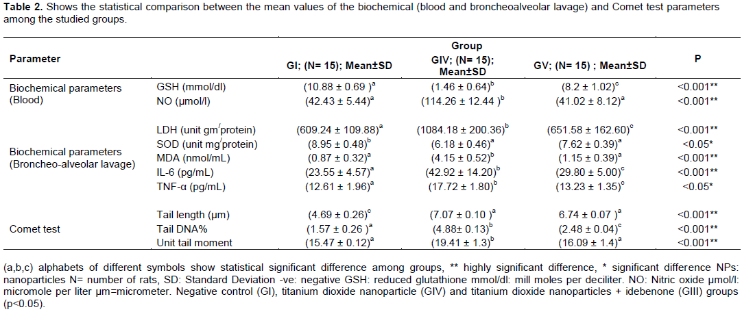
The studied 75 rates show no significant difference as regards age and sex. There was no statistical significant difference as regards the biochemical (blood and broncheo-alveolar lavage) and Comet test parameters among negative control (GI) positive control GII and idebenone groups (GIII) (Table 1 and Figure 1). While there were statistical significant difference as regards the biochemical (blood and broncheo-alveolar lavage) and Comet test parameters among in negative control (GI), titanium dioxide nanoparticle (GIV) and titanium dioxide nanoparticles + idebenone (GIII) groups (Table 2 and Figure 2).


Single cell gel electrophoresis
No significant differences regarding mean values of comet tail length, percentage of tail DNA (tail DNA%) and tail moment among –ve,+ve controls and idebenone groups (p>0.05) by ANOVA test, so we used negative control group (I) as a standard reference for comparison with other treated groups (Table 1). Table 2 showed significant difference among –ve control, TiO2NPs and TiO2NPs+ idebenone regarding comet tail length (μm), tail DNA% and unit tail moment (P<0.001) by ANOVA test. Least significant test revealed significant increase in unit tail moment in both TiO2NPs and TiO2NPs+ idebenone groups when compared with control group (p<0.001) while no significant difference in TiO2NPs+idebenone when compared with TiO2NPs was detected(P>0.05) . (Figure 3) showed normal lung nuclei and undamaged cells in control group (fig1a) while abnormal tailed nuclei and damaged cells in TiO2NPs group (Figure 1b and c) and TiO2NPs+idebenone groups (Figure 1d) less number of abnormal tailed nuclei and damaged cells were detected.
Histopathological
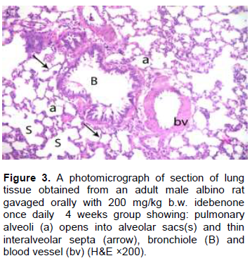
The light microscopical examination of hematoxylin and eosin (H&E) stained sections from the lung of control and idebenone groups, showed normal spongy histological appearance with numerous alveoli connected together with alveolar pores and opens into alveolar sacs, thin inter alveolar septa, bronchioles and blood vessels (Figures 4 and 5). while diffuse alveolar damage with marked consolidation of lung tissue, collapsed alveoli, marked thickening of interalveolar septa and extra-vasations of RBCs in the alveolar lumen, hyperemic foci, thickened wall pulmonary blood vessels, heavy infiltration with chronic inflammatory cells mainly lymphocytes and partial shedding of mucosal lining of bronchioles were detected in TiO2ONPs intoxicated group (Figures 6 to 8). In TiO2NPs+ idebenone treated group lung sections showed normal alveoli with some collapsed alveoli scattered in-between, mild thickening of interalveolar septa and inflammatory cellular infiltration (Figure 9)


DISCUSSION
Intentional and unintentional exposures to titanium dioxide nanoparticles are by oral, ingestion, inhalation, dermal routesand by intravenous injection. It is one of the most widely utilized nanoparticles as its particles are small in size makes nanotechnology so useful in medicine and industry (Pileni, 2001).
Rosa et al. (2010) reported that the more decrease in the dimensions of nanoparticles the more thawing rate with more toxic effects. The extensive use of nano-particles causes potential risks for human and environmental biological system. It has great hazards to human health (Attia et al., 2013). The nanosized TiO2 is used in numerous applications as colour of foods, cosmetics and environmental decontaminant of air, soil and water (Matt et al., 2014). TiO2NPs can accumulate in body tissues causing inflammation, cell death, apoptosis, finally organ injury. Additionally, TiO2NPs induce reactive oxygen species (ROS) production leading to DNA damage (Gao et al., 2013).
In the present study the administration of TiO2NP showed significant decrease in serum oxiditave marker (GSH). This observation was in line with the results of Shukla et al. (2011) who reported that in vitro studies on human colon carcinoma cell line exposed to TiO2NPs resulted in decreased viability, increased hydrogen peroxide free radicles and decreased glutathione (GSH) levels. This GSH level decrease can be due to either direct action of TiO2NPs on its synthesis or interference with its action in scavenging free radicals.
The results of present study showed highly significant increase in mean values of serum NO. This result was in accordance with Gillis et al. (2014). High amounts of NO are released from the inducible nitric oxide synthase (iNOS) enzyme isoform evoked by inflammatory stimuli from variety of cell types. Oxidative stress and the rising levels of free radicals have a strong relation with the increased levels of inducible nitric oxide synthase (iNOS) protein in the lung with production of NO as a compensative mechanism (Porter et al., 2006).
Increased serum nitric oxide levels in TiO2NPs intoxicated rats suggest that these nanoparticles can induce oxidative stress and increase pro inflammatory mediators (Kunal et al., 2009). Idebenone, a short chain benzoquinone related to coenzyme Q10 in its structure, is a potent antioxidant, electrons carriers and free radicals scavenger protecting bio membranes from oxidative damage induced by pollutants (Al-Rasheed et al., 2013).
The present study showed non-significant difference in mean values of serum NO in TiO2NPs + Idebenone groups when compared with vet control group, but there was highly significant decrease in mean values of serum NO in TiO2NPs + idebenone group when compared with TiO2NPs intoxicated group. These results coincide with Suno et al. (2015) who reported that Idebenone has a potential preventive effect against lung inflammation caused by TiO2NPs. The inhibition of the phagocytes production of reactive oxygen species (ROS) and inhibition of cytokine induced neutrophil chemo attractant (CINC) genes were the mechanisms of Idebenone. Improvement of oxidative stress was associated with decreased levels of pro-inflammatory mediators such as NO.
In our study, we found that TiO2 NPS induced oxidative damage and inflammation in BALF by significant increase in LDH, MDA, IL6 and TNF-α with SOD decrease. There are seventeen proteins structurally different regardless of the composition and shape of nano particles which released in BALF (Rai et al., 2002). Nanoparticles exposure, stimulate expression of cytokines in lung epithelial cells and in lung tissue. Higher protein concentrations in the nanoparticles exposed BALF samples are likely a result of plasma extravasation (Rao et al., 2005).
Lactate dehydrogenase leakage is a measure of membrane integrity damage. Nanoparticles induced LDH leakage in BALF, which revealed the damaging impact of nanoparticles on cell membrane integrity (Liu et al., 2013).
Furthermore, the decreases of SOD values in exposed groups suggested that the balance between oxidation and anti-oxidation was destroyed in rats. It probably suggested that the acute toxicity primarily originated from
the cellular internalization of nanoparticles rather than physical damage on the cellular membrane (Lin et al., 2010).
In our study the concentrations of TNF-α, and IL-6 increased in BALF of rats exposed to TiO2 NPs. These results are in accordance with Lin et al. (2012) who stated that pro-inflammatory cytokines (TNF-α, IL-6) play an importantrole in regulating immunity. As pro-inflammatory factors, the level of IL-6 induced by the nanoparticles in BALF was significantly higher than that of the control group in his study.
A significant increase in mean values of serum GSH and BALF SOD and a significant decrease in mean values of serum BALF LDH, MDA, IL6 and TNF-α in TiO2NPs + Idebenone group when compared with TiO2NPs intoxicated group was detected. These results can be supported by Al-Rasheed et al. (2013) results who reported that oral administration of Idebenone along with titanium dioxide nano particles resulted in restoration of renal GSH levels in rats. Idebenone can restore the decreased levels of GSH induced by TiO2NPs through suppression of oxidative stress and lipid per oxidation together with neutralization of the excess amounts of free radicals perserving GSH in the reduced state.
In addition, Ciftci et al. (2012) reported that supplementation of idebenone completely prevents protein damage, apoptosis and lung injury caused by several toxic materials. In the present study, light microscope examination of sections of the lung of titanium dioxide nanoparticles intoxicated group showed collapsed alveoli, destruction of inter-alveolar septa with alveolar dilatation, thick inter-alveolar septa, and heavy infiltration of inflammatory cells, inflammatory exudates and extravasations of RBCs in the interstitium.
The results of the present study coincided with those of Hext et al. (2005) who noted signs of inflammation upon histo-pathological observation of female rats exposed to 10 mg/m3 TiO2 concentration for 13 weeks as the nanoparticles were phagocyte by macrophage.
However, we observed that macrophages infiltrated TiO2 nanoparticles in alveoli of treated rats. These findings are in line with the results of a previous study done by Bermudez et al. (2004), which showed degeneration, thickness of lung epithelial cells and particle-laden macrophages of the alveolar region.
Also, Stearna et al. (2011) explained thickening of inter-alveolar septa induced by TiO2NPs by the increased interstitial collagen fiber deposition and marked cellular infiltration with lymphocytes, neutrophils, eosinophils and macrophages. Moreover, vascular congestion and cellular infiltration of the lung tissue caused by disruption of the endothelial barrier integrity and increased capillary permeability.
Furthermore, Park et al. (2008) demonstrated that uptake of TiO2NPs in acidic lining of lung cells accelerates dissolution of these particles leading to lysosomal damage, mitochondrial disturbance and production of ROS and cytokines. The light microscope examination of sections of the lung of TiO2NPs + Idebenone group revealed partial improvement in histo-pathological changes. These results are in agreement with Nagy (2015) who reported that rats treated with Idebenone along with TiO2NPs show well preserved lung tissue with partial improvement in lung histopathology. Also, Idebenone administration with TiO2NPs was found to decrease congestion and inflammatory infiltration in the tissues.
Moreover, Nagai et al. (2015) mentioned that Idebenone, is a radical-scavenging antioxidant, has the ability to inhibit the induction of pulmonary oxidative stress, injury and inflammation observed in the rat lung 1 day after intra-tracheal instillation of TiO2NPs. The Comet assay is a widely used assay in recording genotoxicity and DNA damage or repair of different pharmaceuticals and environmental chemical contaminants (Karlsson et al., 2015).
In this study the effect of TiO2NPs was demonstrated at molecular level by Comet assay to investigate the ability of TiO2NPs to generate DNA damage in off springs. Our results showed that administration of TiO2NPs causes DNA strand breaks and disrepair of damaged DNA strands recorded by increase in unit tail moment.
These results are in accordance with Tao et al. (2014) who reported that different concentrations of TiO2NPs are cytotoxic and genotoxic to different organs and cell lines in different organisms. This genotoxic effect reported to be dosing dependent Musarrat et al. (2009) and even after acute and sub-chronic exposure Gerloff et al. (2009).
These results could be explained by Singh et al. (2009) and Yang et al. (2009) who stated that increased ROS induced by nanoparticles in lysosomes can cause DNA mutations or induce single or double strand breaks. Dueto their small size, nanoparticles accumulate around the nucleus and few of them may diffuse through nuclear pores from where protein transport take place and this augment DNA damage caused by ROS (Simko et al., 2011).
Also, Hausladen and Stamler (1999) and Murphy(1999) concluded that unregulated production of NO can lead to damage of cellular proteins, DNA damage, cell injury and death induced by TiO2NPs. In addition, the presence of free titanium ions may be the cause of ROS-driven cytotoxicity and genotoxicity (Song et al., 2010).
The results of this work showed no-significant difference in mean values of unit tail moment in TiO2NPs + Idebenone group when compared with TiO2NPs intoxicated group, but there was highly significant increase when compared to negative control group.
These results are in agreement with the findings of Park et al. (2008) who reported that several in vitro experiments with cell lines indicate that Idebenone in the presence of transition metal ions acts as a pro-oxidant and increases the amount of damage to genetic material in human lymphocytes.
These results suggest that idebenone has a minimal protective effect on TiO2NPs induced DNA damage and these results could be explained by low dose of idebenone, short duration of supplementation or the fact that increased DNA damage needs long time with sufficient doses of Idebenone for the tissue to restore normal genetic and chromosomal appearance (Nagaoka et al., 2016).
CONCLUSION
TiO2NPs oral administration induces oxidative stress and DNA damage in the lung tissue. Idebenone use with TiO2NP is considered a protective agent leads to attenuation of oxidative stress, pulmonary toxicity and slight improvement of DNA damage proved by Comet assay.
RECOMMENDATIONS
Caution use of TiO2NPs to gain the benefits of nanotechnology and avoid its possible hazards also it’s highly recommended to use Idebenone as protective agent against TiO2. Control the TiO2NPs occupational exposure through continuous monitoring of work environment level, keep it within the recommended exposure limits, and involve its periodical clinical and laboratory examinations as a routine check-up among exposed workers. In addition to planning a health education program to Increase the awareness of workers about the TiO2NPS materials, its hazards, proper handling and protective measurements nanoparticles toxicity. Finally, further studies with longer duration, is highly recommended in this interesting topic
ACKNOWLEDGEMENT
The authors gratefully acknowledge the support and help provided by all the staff of animal house, Zagazig University Hospitals.
CONFLICT OF INTERESTS
The author has not declared any conflict of interest.
REFERENCES
|
Al-Rasheed NM, Faddah LM, Mohamed AM, Abdel Baky NA, Mohammad RA (2013). Potential Impact of Quercetin and Idebenone against Immuno- inflammatory and Oxidative Renal Damage Induced in Rats by Titanium Dioxide Nanoparticles Toxicity. Journal of Oleo Science 62(11):961-971. |
|
|
Attia HF, Soliman MM, Abdel-Rahman GH, Abdo Nassan M, Ismail SA, Farouk M, Solcan C (2015). Hepatoprotictive effect of N-acetylcysteine on the toxic hazards of titanium dioxide nanoparticles American Journal of American Science 11(7):141-147. |
|
|
Azim SA, Darwish HA, Rizk MZ, Ali SA, Kadry MO (2015). Amelioration of titanium dioxide nanoparticles-induced liver injury in mice: possible role of some antioxidants. Experimental and Toxicologic Pathology 67(4):305-314. |
|
|
Baan RA (2007). Carcinogenic hazards from inhaled carbon black, titanium dioxide, and talc not containing asbestos or asbestiform fibers: Recent evaluations by an iarc monographs working group. Inhalation Toxicology 19(1):213-228. |
|
|
Bancroft JD, Gamble M (2002). Theory and practice of histological techniques, fifth ed. Churchill Livingstone Pub., Edinburg and London. |
|
|
Bermudez E, Mangum JB, Wong BA, Asgharian B, Hext PM, Warheit DB, Everitt JI (2004). Pulmonary responses of mice, rats, and hamsters to subchronic inhalation of ultrafine titanium dioxide particles. Toxicological Sciences 77(2):347-357. |
|
|
Chen J, Dong X, Zhaoa J, Tang G (2009). In vivo acute toxicity of titanium dioxide nanoparticles to mice after intraperitioneal injection. Journal of Applied Toxicology 29(4):330-337. |
|
|
Ciftci O, Ozdemir I, Vardi N, Beytur A, Oguz F (2012). Ameliorating effects of quercetin and chrysin on 2,3,7,8-tetrachlorodibenzo-p-dioxin-induced nephro- toxicity in rats. Toxicology and industrial health 28(10):947-954. |
|
|
Park EJ, Yi J, Chung KH, Ryu DY, Choi J, Park K (2008). Oxidative stress and apoptosis induced by titanium dioxide nanoparticles in cultured BEAS-2B cells. Toxicology letters 180(3):222-229. |
|
|
Gao G, Ze Y, Zhao X, Sang X, Zheng L, Ze X, Gui S, Sheng L, Sun Q, Hong J, Yu X, Wang L, Hong F, Zhang X (2013). Titanium dioxide nanoparticle-induced testicular damage, spermatogenesis suppression, and gene expression alterations in male mice. Journal of hazardous materials 258:133-143. |
|
|
Gerloff K, Albrecht C, Boots AW, Förster I, Schins RPF (2009). Cytotoxicity and oxidative DNA damage by nanoparticles in human intestinal Caco-2 cells. Nanotoxicology 3(4):355-364. |
|
|
Gillis JC, Benefi P, McTavish D (2014). Idebenone. A review of its pharmacodynamic and pharmacokinetic properties, and therapeutic use in age-related cognitive disorders. Drugs Aging 5:133-52... |
|
|
Gunnar M, Buyse G, Van der M, Michael E, Jan D, Paul H, Erik V (2009). Long-term blinded placebo-controlled study of SNT-MC17/idebenone in the dystrophin deficient mdx mouse: cardiac protection and improved exercise performance. European Heart Journal 30(1):116-124. |
|
|
Hausladen A, Stamler JS (1999). Nitrosative stress. Methods Enzymology 300:389-395. |
|
|
Hext PM, Tomenson JA, Thompson P (2005).Titanium dioxide: inhalation toxicology and epidemiology, Long-term pulmonary responses of three laboratory rodent species to subchronic inhalation of pigmentary titanium dioxide particles. Toxicological Sciences 70(1):86-97. |
|
|
Imran A (2012). New Generation Adsorbents for Water Treatment. Chemical reviews 112 (10):5073-5091 |
|
|
Institute of Laboratory Animal resource (1996). Guide for the care and use of laboratory animals. INSTITUTE OF LABORATORY ANIMAL RESOURCES Council. National Academic Press 144, SRC, xii,: 125. |
|
|
Johnson MD (2007) .The Rats, in: Gad, S.C.(Eds), Animal Models of Toxicology, second ed. Taylor and Francis, New York. pp. 150-171. |
|
|
Jotham MM, Xuelai Z (2018). Particle size effect on thermophysical properties of nanofluid and nanofluid based phase change materials: A review. Journal of Molecular Liquids 265:77-87. |
|
|
Kakkar P, Dos B, Viswanathan PN (1984). A modified spectrophotometric assay of superoxide dismutase. Indian Journal of Biochemistry 21:130-212. |
|
|
Karlsson HL, Di Bucchianico S, Collins AR, Dusinska M (2015): Can the comet assay be used reliably to detect nanoparticle-induced genotoxicity? Environmental and Molecular Mutagenesis 56(2):82-96 |
|
|
Khan M, Naqvi A, Ahmad A (2015). Comparative study of the cytotoxic and genotoxic potentials of Zinc oxide and Titanium dioxide nanoparticles. Toxicology Reports 2:765-774. |
|
|
Kunal WW, Lin ZQ, Wei BF, Zeng Q, Han B (2009).Single-walled carbon nanotube induction of rat aortic endothelial cell apoptosis: reactive oxygen species are involved in the mitochondrial pathway. The International Journal of Biochemistry and Cell Biology 43:564-572. |
|
|
Laurence W (2014). Introduction to Plastics and Polymers, the effect of long term thermal exposure on plastics and elastomers. |
|
|
Liang G, Pu Y, Yin L, Liu R, Ye B, Su Y, Li Y (2011). Influence of different sizes of titanium dioxide nanoparticles on hepatic and renal functions in rats with correlation to oxidative stress. Journal of Toxicology and Environmental Health 72(12):740-745. |
|
|
Lin ZQ, Liu LH, Xi ZG, Huang JH, Lin BC (2012). Single-walled carbon nanotubes promote rat vascular adventitial fibroblasts to transform into myofibroblasts by SM22-α expression. International Journal of Nanomedicine 7:4199-4206. |
|
|
Lin ZQ, Xi ZG, Chao FH, Yang DF, Zhang HS, Lin BC, Zhang W, Liu HL, Sun X (2010). ICAM-1 and VCAM-1 expression in rat aortic endothelial cells after singlewalled carbon nanotube exposure. Journal of Nanoscience and Nanotechnology 10(12)8562-8574 |
|
|
Liu H, Ma L, Zhao J, Liu J, Yan J, Ruan J, Hong F (2012). Biochemical toxicity of nanoanatase TiO2 particles in mice. Biological Trace Element Research 129(3):170-180 |
|
|
Liu H, Yang D, Yang H, Zhang H, Zhang W (2013). Comparative study of respiratory tract immune toxicity induced by three sterilisation nanoparticles: silver, zinc oxide and titanium dioxide. Journal of Hazardous Materials 248:478-486. |
|
|
Matt F, Lynn A, Boatner C, Chakoumakos M, Jeff C (2014). Structural and crystal chemical properties of rare-earth titanate pyrochlores. Journal of Alloys and Compounds 605:63-70. |
|
|
Mengjia S, Yanli J, Mei T, Huijun Y, Ran L, Lijuan Y (2018). Deposition of platinum on boron-doped TiO2/Ti nanotube arrays as an efficient and stable photocatalyst for hydrogen generation from water splitting. RSC Advances 9(20):11443-11450. |
|
|
Montgomery HAC, Dymock JF (1961) .The determination of nitrite in water. Analyst 86:414-416. |
|
|
Moron M, Depierre J, Mannervik B (1979). Levels of glutathione, glutathione reductase and glutathione S-transferase activities in rat lung and liver. Biochimica et Biophysica Acta (BBA)-General Subjects 582(1):67-78. |
|
|
Murphy MP (1999). Nitric oxide and cell death. Biochimica et Biophysica Acta (BBA)-Bioenergetics 1411(3):401-414. |
|
|
Musarrat J, Saquib Q, Azam A, Naqvi SAH (2009).Zinc oxide nanoparticles-induced DNA damage in human lymphocytes. International Journal of Nanoparticles 2(1):402 |
|
|
Nagai Y, Shibota M, Narumi S, Miyamoto M, Kaki-hana M, Nagaoka A (1985). Accelerating effect of idebenone(CV-2619)on cerebral glucose metabolism. Japanese Pharmacology and Therapeitics 13:13-20. |
|
|
Nagaoka A, Suno M, Shibota M, Kakihana M (2016). Effects of idebenone on neurological deficits, local cerebral blood flow, and energy metabolism in rats with experimental cerebral ischemia. Archives of Gerontology and Geriatrics 8(3):193-202. |
|
|
Nagy NG (2015). Brain cytokine and chemokine mRNA expression in mice induced by intranasal instillation with ultrafine carbon black. Toxicology Letters 163(2):153-160. |
|
|
Oscar H , Mika L, Christian R, Thomas M (2017). Efficacy of Idebenone to Preserve Respiratory Function above Clinically Meaningful Thresholds for Forced Vital Capacity (FVC) in Patients with Duchenne Muscular Dystrophy. Journal of Neuromuscular Diseases 4(3):189-198. |
|
|
Pileni MP (2001). Nanocrystal Self-Assemblies: Fabrication and Collective Properties. Journal of Physical Chemistry B 105 (17):3415-3421. |
|
|
Porter DW, Millecchia LL, Willard P, Robinson VA, Ramsey D (2006) Nitric oxide and reactive oxygen species production causes progressive damage in rats after cessation of silica inhalation. Toxicological Sciences 90(1):188-197. |
|
|
Rai AJ, Zhang Z, Rosenzweig J, Shih I, Pham T, Fung E, Sokoll L, Chan D (2002). Proteomic approaches to tumor marker discovery. Archives of Pathology and Laboratory Medicine 126:1518-1526. |
|
|
Rao KM, Ma JY, Meighan T, Barger MW, Pack D, Vallyathan V (2005). Time course of gene expression of inflammatory mediators in rat lung after diesel exhaust particle exposure. Environmental Health Perspectives 113(5):612-617. |
|
|
Rizk MZ, Ali SA, Hamed MA, El-Rigal NS, Aly HF, Salah HH (2017). Toxicity of titanium dioxide nanoparticles: Effect of dose and time on biochemical disturbance, oxidative stress and genotoxicity in mice. Biomed Pharmacother 90:466-472. |
|
|
Robert P (2013). Nanoparticles and the Control of Oral Biofilms. in Nanobiomaterials in Clinical Dentistry pp 34-39. |
|
|
Rosa I, Merino JI, Peña VM (2010). Compositionally graded YSZ-NiO composites by surface laser melting, Journal of the European Ceramic Society 30(2):147-152. |
|
|
Schulza JB, Di Prospero NA, Fischbeck K (2009). Clinical experience with high-dose idebenone in Friedreich ataxia. Journal of Neurology 256(1):42. |
|
|
Schwarze PE, Øvrevik J, Låg M, Refsnes M, Nafstad P, Hetland RB, Dybing E (2006).Particulate matter properties and health effects: consistency of epidemiological and toxicological studies. Human and Experimental Toxicology 25(10):559-579. |
|
|
Shtansky DV, LevashovI EA, Sukhorukova V (2015). Multifunctional bioactive nanostructured films.Hydroxyapatite (Hap) for Biomedical Applications Woodhead Publishing Series in Biomaterials, pp. 159-188 |
|
|
Shukla RK, Kumar A, Pandey AK, Singh SS, Dhawan A (2011). Titanium dioxide nanoparticles induce oxidative stress-mediated apoptosis in human keratinocyte cells. Journal of Biomedical Nanotechnology 7(1):100-101. |
|
|
Shukla RK, Sharma V, Pandey AK, Singh S, Sultana S, Dhawan A (2011). ROS-mediated genotoxicity induced by titanium dioxide nanoparticles in human epidermal cells. Toxicology In vitro 25(1):231-241. |
|
|
Simko M, Fiedeler U, Gazso A, Nentwich M (2011).The impact of nanoparticles on cellular functions. Nano Trust Dossier 7:151Ë—164. |
|
|
Singh N, Manshian B, Jenkins GJ, Griffiths SM, Williams PM, Maffeis TG, Wright CJ, Doak SH (2009). NanoGenotoxicology: the DNA damaging potential ofengineered nanomaterials, Biomate 30(24):3891- 3914. |
|
|
Singh N, McCoy MT, Tice RR, Schneider EL(1988). A simple technique for quantitation of low levels of DNA damage in individual cells. Experimental Cell Research 175(1):184-191. |
|
|
Song W, Zhang J, Guo J, Zhang J, Ding F, Li L, Sun Z (2010). Role of the dissolved zinc ion and reactive oxygen species in cytotoxicity of ZnO nanoparticles. Toxicology Letters 199(3):389-397. |
|
|
Stearns RC, Paulauskis JD, Godleski JJ (2011). Endocytosis of ultrafine particles by A549 cells. American Journal of Respiratory Cell and Molecular Biology 24(2):108-115. |
|
|
Suno M, Nagaoka A (1984). Inhibition of lipid peroxidation by a novel compound(CV-2619)in brain mitochondria and mode of action of the inhibition. Biochemical and Biophysical Research Communications 125(3):1046-1052. |
|
|
Tao C, Jian Y, Yan L (2014). Genotoxicity of titanium dioxide nanoparticles. Journal of Food and Drug Analysis 22(1):95-104. |
|
|
Vamanu CI, Cimpan MR, Sornes S, Lie SA, Gjerdet R (2008). Induction of cell death by TiO2 nanoparticles: studies on a human monoblastoid cell line. Toxicology in Vitro 22(7):1689-1696. |
|
|
Varley H, Gowenlock AH, Bell M (1980). Determination of serum lactate dehydrogenase activity. In: Clinical Biochemistry, 5th Ed. Williams Hieinemanm Medical Books Ltd, London pp. 715-720. |
|
|
Vittoriadiamanti MP (2013). Concrete, mortar and plaster using titanium dioxide nanoparticles: applications in pollution control, self-cleaning and photo sterilization Pedeferri, in Nanotechnology in Eco-Efficient Construction, 2013 |
|
|
Wang JX, Fan YB, Gao Y, Hu QH, Wang TC (2009). TiO2 nanoparticles translocation and potential toxicological effect in rats after intraarticular injection. Biomaterials 30(27):4590-4600. |
|
|
Weir A, Westerhoff P, Fabricius L, Hristovski K, von Goetz N (2012). Titanium dioxide nanoparticles in food and personal care products. Environmental Science and Technology 46(4):2242-2250. |
|
|
Yang H, Liu C, Yang D, Zhang H, Xi Z (2009). Comparative study of cytotoxicity, oxidative stress and genotoxicity induced by four typical nanomaterials: The role of particle size, shape and composition. Journal of Applied Toxicology 29(1):69-78. |
|
Copyright © 2024 Author(s) retain the copyright of this article.
This article is published under the terms of the Creative Commons Attribution License 4.0


