Full Length Research Paper
ABSTRACT
Rotten fruits are known to cause major losses in post-harvest conservation. One of the causal pathogen of pineapple rot was investigated and the antifungal effects of selected plants extracts were evaluated in vitro against fungal pathogen; Pestalotiopsis microspora. The fruits samples were randomly collected from farms after harvest. The samples were collected in Awae; the experiment was conducted at Agricultural Research Institute for Development located in Yaounde, Cameroon. The fruits samples were inoculated on potato dextrose agar and pure culture of fungal pathogen responsible for pineapple rot obtained. The pathogen was isolated, and identified on the basis of morphological features. The pathogenicity test was conducted to determine that the fungus is responsible for the rot symptoms. The efficacies of three plant species, namely: Allium sativum, Syzygium aromaticum and Zingiber officinale were tested in vitro at concentrations 20, 40 and 80% on mycelial growth inhibition of the causal agent. Aqueous and ethanolic extracts for concentrations were used. Distilled water and the fungicide Mancozeb 80WP (800 g/kg) were used as negative and positive controls, respectively. The pathogenicity test confirmed that P. microspora fungus is responsible for pineapple fruit rotting. The non-inoculated controls showed no symptoms of fruit rot. Results of antifungal tests showed that after 08 days, aqueous and ethanolic extracts of A. sativum and S. aromaticum were the most effective. The mycelial growth inhibition was total with aqueous and ethanolic extracts of A. sativum for concentrations 20, 40 and 80%. Total inhibition was also recorded with ethanolic extracts of S. aromaticum for all the tested concentrations. The overall result of this study reveals that aqueous and ethanolic extracts of A. sativum and S. aromaticum can be used to control P. microspora as they completely inhibited the growth of the pathogen that can contribute in post-harvest conservation of pineapple fruits.
Key words: Ananas comosus, antifungal activities, extracts, pathogenicity test, Pestalotiopsis microspora.
INTRODUCTION
Pineapple (Ananas comosus (L.) Merr.) belongs to Bromeliaceae family (Baruwa, 2013). It is the third most important tropical fruit in the world after banana and citrus. Pineapple contributes 8% of the world fresh fruit production (Yusi, 2016). Thailand is the largest producer of pineapple, accounting for 13% of global output followed by Brazil and Costa Rica (Faostat, 2017). Cameroon ranked 4th on the list of African producers with a production of 351 574 tons after Nigeria, Kenya and Angola (Tilasto, 2019). Despite this ranking, many constraints are still facing pineapple production and post-harvest conservation in Cameroon. These include low fertility, poor cultivar, pests and diseases attacks among others.
Fungal pathogens are proven to be a common and popular contaminant of agroecosystem that approximately causes 70 to 80% of total microbial crop loss (Santra and Banerjee, 2020). Pestalotiopsis microspora is one of the most commonly isolated endophytes (Li et al., 2001). Pestalotiopsis species are not highly host-specific and cause diseases on a variety of plants, including canker lesions, shoot dieback, leaf spots, needle blight, tip blight, grey blight, scabby canker, severe chlorosis, leaf spots fruit rots and other post-harvest diseases (Xu et al., 1999; Maharachchikumbura, 2011). Pestalotiopsis is also considered to be a weak pathogen that can penetrate the host plants through natural openings. Species of Pestalotiopsis infect wounded or stressed plants (Elliott et al., 2004).
Fungal diseases of crops are usually controlled using resistant cultivars, long rotations, and fumigants, but mainly by the use of fungicides (Rongai et al., 2015). However, pesticides residues in fruits can lead to harmful effects on human health and the development of fungicide resistance in pathogens. Hence, there remains a need to develop safer, more effective and eco-friendly alternative fungicides that cause minimal damage to the environment and human health (Tuanwei et al., 2018). The use of botanical fungicides was considered a viable and better alternative approach for the control of pathogenic fungi because effective control of a variety of rot pathogens in diverse foods have been reported by (Bag and Chattopadhyay, 2015; Chaemsanit et al., 2018). The antifungal potential of plant extracts has long been investigated as they contain secondary bioactive compounds for plant disease control (Riaz et al., 2015).
The objectives of this work were to isolate and identify the causal pathogen of pineapple rot in post-harvest conservation and to evaluate in vitro antifungal effects of some plants extracts in the control of the causative agent.
MATERIALS AND METHODS
Collection of samples
Diseased pineapples were collected randomly from various farms after harvest. The samples were collected at locality of Awae 1 (Centre Region of Cameroon) in June 2019 and included five rotten fruits of “smooth Cayenne” variety. These were taken to Agricultural Research Institute for Development at Plant Pathology Laboratory in Nkolbisson (Yaounde, Cameroon) for isolation and purification on the Potato Dextrose Agar (PDA) culture medium.
Isolation
Collected fruits were washed with tap water, then were superficially disinfected in 70% ethanol. Necrotic lesions were cut aseptically in small fragments and disinfected at 2% sodium hypochlorite solution for 5 min. These fragments were rinsed in two changes of sterile distilled water to remove traces of the disinfectant and dried with sterile filter papers. They were deposited on blotting paper and allowed to dry completely.
The fungus was isolated on a PDA culture medium. PDA was prepared from an infusion of potatoes peeled, cut and boiled in 1000 ml of distilled water for 20 min. The filtrate obtained was poured into an Erlenmeyer flask containing 15 g of Agar and 20 g of Dextrose. After homogenization, the mixture was adjusted to 1000 ml with distilled water. The medium was sterilized in an autoclave (Science Medecine Industry brand) at a temperature of 121°C for 30 min under a pressure of 1 bar. The dried fragments were removed aseptically under a hood (Bassaire model No. KDS2 Enclosure, voltage 230/205 Volts) near the flame of a Bunsen burner and placed in 90 mm diameter glass Petri dishes containing the PDA culture medium supplemented with streptomycin 250 mg, to prevent the development of bacteria. Each Petri dish contained three to four fruits fragments. These Petri dishes were incubated at 25°C for three days.
Purification and identification
The visible fungal colonies developed at the periphery of the root fragments and those most similar to the fungus P. microspora were removed and aseptically transplanted into new Petri dishes containing the culture medium. After four successive subcultures on the PDA culture medium, the pure cultures of P. microspora were obtained. Pure cultures were observed under the ordinary electron microscope (Labolux 11, Leizt) for identification.
The identification of P. microspora was based on the morphological and microscopic characteristics of the mycelium in comparison with the characteristics of the fungus presented in the literature (Nyaka et al., 2017).
Pathogenicity test
Pineapples from smooth Cayenne variety have been used for pathogenicity test, as this variety is widely planted in Awae for direct consumption in local and export markets. Pathogenicity test was carried out according to the modified method of Okigbo and Emoghene (2009). Mature and healthy pineapple fruits were carefully selected. They were washed with tap water to strike out impurities. Then they were disinfected with ethanol 70% for 3 min, rinsed three times with sterile distilled water and dried under filter-sterilized air flow. The fungus P. microspora isolated and identified was cultivating on PDA culture medium. Pure cultures of 10 days were used as inoculum. The concentration of spores was adjusted at 3 × 10⁵ spores/ml using a hemacytometer according to (Nyaka et al., 2017). Cork borer of 5 mm diameter were used to create lesions of 3 mm depth on pineapples fruits. Micropipette was used to deduct 1 mm of spores suspension which were inserted into the wounded area except the fruits used as control inoculated with distilled sterilised water. Inoculated and control treatments were incubated for 10 days at room temperature for disease development. To fulfil Koch’s postulate, re-isolation of the pathogen was done and compared with original isolate.
Plants extracts preparation
For this experiment, three (03) plant species from three different plant families ginger (Zingiber officinale), garlic (Allium sativum) and clove (Syzygium aromaticum) were purchased from a local market in the town of Yaounde, Centre Region of Cameroon. All three plants have been proven by previous studies to have antimicrobial properties. Clove was dried and ground to powder form before use, whereas garlic and ginger were used in the fresh form. Both aqueous and organic extracts were prepared. The organic solvent involved here was ethanol. The three plants species were ground using a commercial blender. In order to obtain the aqueous extracts, 100 g of each plant powder or paste was weighed and added to 1 L distilled water in a glass container (Wheaton bottles). For the organic (ethanolic) extracts, 100 g of the powder or paste was weighed and added into 1 L ethanol in a glass container (Wheaton bottles).
Fresh garlic and ginger were peeled, washed and surface sterilized using 2% sodium hypochlorite solution and then rinsed with distilled water. They were allowed to air dry and 100 g were weighed and crushed in a laboratory mortar. The paste obtained was then added to 1 L distilled water in a glass container (Wheaton bottles) to produce the aqueous extract. The same procedure was followed for the organic extract with the difference being the use of 1 L ethanol as solvent. All were incubated for 24 h at room temperature of 25±2°C, then placed on a shaker (LAB-LINE®, No. 3590) for 24 h. The extracts were then filtered through a funnel first using muslin cloth and then with Whatman filter paper (No. 1). The filtrates obtained were subjected to a rotatory evaporator (Stuart®, SB162) at 45 to 50°C to remove water and ethanol, yielding extracts with semisolid consistencies. The extracts were considered as 100% and stored in a refrigerator at 4°C until subsequent use. They were later on diluted to make up the required concentrations needed for efficacy testing on the sample fungi.
In vitro effect of plants extracts
The inhibitory effect of the extracts was tested following the poison food technique (Dhingra and Sinclair, 1985). Where 0.4, 0.8 and 1.6 g of each extract was dissolved in 1 ml sterile distilled water and then added to 19 ml molten PDA in 70 mm sterile Petri dishes in order to have final concentrations of 20, 40 and 80% w/v. In addition, a Petri plate containing a standard fungicide, Mancozeb 80WP at the recommended dose (800 g/kg) was used as the positive control to evaluate the effectiveness of the plant extracts by comparison. PDA with just sterile distilled water served as negative control. A mycelial disc of 5 mm in diameter of the test fungus was taken from 8-day old fungi colonies with the help of sterilised cork borer and placed at the centre of the Petri dishes containing the medium and the fungicide. The whole experiment was repeated three times and incubated at room temperature of 25±2°C. Radial growth was measured after 8 days of incubation, time needed for the fungi colonies to fill up the Petri plates in the control. The inhibition percent (IP) was calculated by the formula given by Ul-Haq et al. (2014):
IP = (GC-GT)/GC×100;
where GC=growth in the control, GT=growth in the treated groups. An average of three replications of each test was taken for calculations.
Statistical analysis
A completely randomized design was used with three (03) treatments and three (03) replications. Data were keyed in and treated using Microsoft Excel 2016 and Analysis of Variance (ANOVA) was done using the SPSS Statistical Package, 6.40 version. Means were compared using Duncan’s at significance level of 5%.
RESULTS AND DISCUSSION
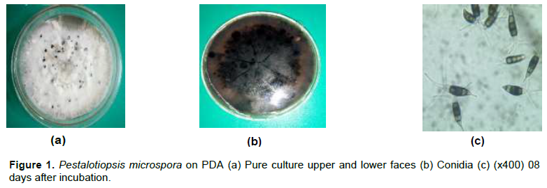
Morphocultural description of P. microspora on PDA culture medium
Macroscopic observation of P. microspora pure culture on PDA medium at room temperature of 25±2°C has presented an abundant and white mycelium after 08 days of incubation. The Petri dishes of 70 mm were totally filled after 08 days by the pathogen. The mycelial growth was radial. Microscopic observation of P. microspora isolates on PDA culture medium presented hyaline and cylindrical conidia. The lower end ends with a filamentous appendage while the more rounded upper end is extended by 03 filaments. Figure 1 shows macroscopic and microscopic observations of P. microspora conidia on PDA, 08 days after incubation.
Pathogenicity of P. microspora isolated from pineapple (smooth Cayenne variety)
Results of the pathogenicity test showed that P. microspora isolated from pineapple rotten fruits was pathogenic. The inoculated fruits were rotting compared to the fruits used as control. The rot developed by the inoculated fruit spread from the inoculation zone. This is demonstrated in Figure 2.

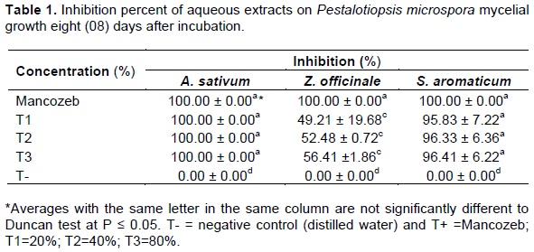
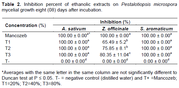
Effect of plant extracts on the mycelial growth of P. microspora
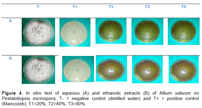
All extracts significantly inhibited the growth of the fungus tested, compared to the untreated control. Highest inhibition rates were obtained from both aqueous and ethanolic extracts of A. sativum and S. aromaticum. Aqueous and ethanolic extracts of Z. officinale had a dose-dependent inhibitory effect on the fungus, as higher doses were observed to inhibit the growth better than lower doses. Comparing the effect of the plant extracts to that of Mancozeb, the active ingredient of the synthetic fungicide Penncozeb 80WP, garlic extracts (aqueous and ethanolic) and clove extract (ethanolic) were found to produce a similar inhibitory effect as Mancozeb on the growth of P. microspora. However, there was a significant difference between the effect of Mancozeb and ginger (aqueous and ethanolic) at 20 and 40% concentrations and that of garlic (aqueous) at all concentrations. The inhibitory diameters are not statistically different from those of the positive control (Mancozeb). Extracts of Z. officinale revealed a lower inhibition percentage (70.35%) at the concentration of 80%, statistically different from the inhibition percentages of the positive control. This can be clearly demonstrated in Tables 1 and 2.
Figures 3 and 4 show the mycelial growth of P. microspora in the presence of plant extracts.

DISCUSSION
The results of pathogenicity test are in accordance with those of Tuanwei et al. (2018) who demonstrated that P. microspora is one of dominant pathogenic fungi causing rotten disease in harvested Chinese olive fruits. The in vitro trials showed that all the selected plants species, A. sativum, S aromaticum and Z. officinale displayed consistent antifungal activity against P. microsposra responsible for pineapple fruits rot in postharvest. The results of the antifungal tests indicated that for all tested plants extracts, garlic showed the highest antifungal activity against P. microsposra. The inhibition of growth of P. microsposra was total with both aqueous and ethanolic extracts. The results obtain with garlic extracts corroborate those of (Antu, 2013; Islam et al., 2004) who showed that garlic extracts were effective against Pestalotiopsis psidii affecting the guava plant (Psidium guajava). Similar results were obtained on other fungi by Chohan et al. (2011) who found that garlic extract was able to inhibit the in vitro growth of Fusarium oxysporum by 35%.
This can be explained by the fact that plants contain several phytochemicals known to play very important defensive roles against pathogens (Fawole et al., 2013). A. sativum possesses antifungal properties due to the presence of powerful antioxidants, sulphur compounds and numerous phenolic compounds such as allicine (Bourgoin et al., 2017).
These results were followed by clove antifungal activities showing significant effectiveness. This finding is in agreement with that of Abdulaziz et al. (2010), who obtained completed inhibition of Rhizotonia solani at the concentration of 1 2, 3 and 4% with clove extracts. This fungicidal effect of clove may result from eugenol which seems to be is the main component of clove oil and the strongest inhibitors of enzyme processes (Pepeljnjak, 2003). Antimicrobial activity of clove oil can also be attributed to the presence of an aromatic nucleus and a phenolic OH group that are known to be reactive and can form hydrogen bonds with –SH groups in the active sites of target enzymes, resulting in deactivation of enzymes in fungi (Alma, 2007).
Ethanolic extract of Z. officinale also presented a high antifungal activity against P. microspora. These results are in accordance with those of Tuanwei et al. (2018) who demonstrated that Ginger oleoresin effectively inhibited in vitro mycelial growth and spore germination of P. microspora responsible for Chinese olive fruits rot. Rawal and Adhikari (2016) ascertained that Z. officinale aqueous and ethanoilc extracts showed moderate to minimum antifungal in vitro activity on F. oxysporum f.sp lycopersici.
CONCLUSION
Hence, the objective of this study was to evaluate the effect of selected plants extracts on the P. microspora fungus responsible of pineapple fruits rot. Results showed extracts of A. sativum and S. aromaticum showed high antifungal activity gainst the pathogen which was not significantly different from the positive control with Mancozeb. Ethanolic extracts of A. sativum and S. aromaticum showed a total antifungal activity at all the concentrations tested against P. microspora. Considering the attribute of plants extracts, and broad-spectrum activities, successful development of such compounds as antifungal could promise success in multipurpose alternatives to conventional fungicides for management of plants diseases. Therefore, A. sativum and S. aromaticum extracts indeed possessed valuable products which could be exploited to control post-harvest diseases on pineapple fruits.
CONFLICT OF INTERESTS
The authors have not declared any conflict of interests.
REFERENCES
|
Abdulaziz A, Al-Ashkar, Younes M, Rashad (2010). Efficacy of some plants extracts against Rhizoctonia solani on pea. Journal of Plant Protection Research 50:3. |
|
|
Alma MH, Ertas M, Nitz S, Kollmannsberger H (2007). Chemical composition and content of essential from the bud of cultivated Turskish clove (Syzygium aromaticum L.). Bioresource Technology 2(2):265-269. |
|
|
Antu SK (2013). Studies on canker disease of guava (Psidium guajava) caused by Pestalotiopsis psidii Pat. Thesis presented for a Master of Science in Agriculture. |
|
|
Bag A, Chattopadhyay RR (2015). Evaluation of synergistic antibacterial and antioxidant efficacy of essential oils of spice and herbs in combination. PLoS One 10(7):e0131321. |
|
|
Baruwa OI (2013). Profitability and constraints of pineapple production in Osun State, Nigeria. 2013. Journal of Horticultural Research 21(2):59-64. |
|
|
Bourgoin MA, Garza Guajardo R, Philippe G, Souchet S (2017). Etude des propriétés antimicrobiennes de l'extrait d'ail (Allium sativum L.). Ecole Supérieure d'agricultures - F49000 Angers - France. |
|
|
Chaemsanit S, Matan N, Matan N (2018). Effect of peppermint oil on the shelf-life of dragon fruit during storage. Food Control 90:172-179. |
|
|
Chohan S, Atiq R, Mehmood MA, Naz S, Siddique B, and Yasmin G (2011). Efficacy of few plant extracts against Fusarium oxysporum f. sp. gladioli, the cause of corm rot of gladiolus. Journal of Medicinal Plants Research 5(16):3887-3890. |
|
|
Dhingra OD, Sinclair JB (1985). Basic Plant Pathology Methods. CRC Press, Florida P 325. |
|
|
Elliott ML, Broschat TK, Uchida JY, Simone GW (2004). Diseases and disorders of ornamental palms. American Phytopathological Society St. Paul. |
|
|
FAOSTAT (2017). Food and agriculture data. |
|
|
Fawole FJ, Sahu NP, Pal AK, Lakra WS (2013). Evaluation of antioxidant and antimicrobial properties of selected Indian medicinal plants. International Journal of Medicinal and Aromatic Plants 3(1):69-77. |
|
|
Islam MR, Hossain MH, Ali MR (2004). Identification of the causal agent of leaf spot of betelnut and in vitro evaluation of fungicides and plant extracts against it. Pakistan Journal of Biological Sciences 7(10):1758-1761. |
|
|
Li JY, Harper JK, Grant DM, Tombe BO, Bashyal B, Hess WM, Strobel GA (2001). Ambuic acid, a highly functionalized cyclohexenine with antifungal activity from Pestalotiopsis spp. and Monochaetia sp. Phytochemistry 6:463-168. |
|
|
Maharachchikumbura SSN, Guo LG, Chukeatirote E, Bahkali AH, Hyde KD (2011). Pestalotiopsis. Morphology, phylogeny, biochemistry and diversity. Fungal Diversity 50(1):167-187. |
|
|
Nyaka AIC, Owona NPA, Oumar D, Ntsomboh-Ntsefong G, Njonje SW, Ehabe EE (2017). Caracterization of Pestalotiopsis microspora, the causal agent of rubber leaf blight disease in Cameroon. Rubber Science 31(2):112:120. |
|
|
Okigbo RN, Emoghene AO (2009). Anti -fungal activity of leaf extract of some plant species on Mycospharella fijiensis merelet, the causal organism of black sigatoka disease of banana (Musa acuminata). Scientific Journal 4(4):20-31. |
|
|
Pepeljnjak S, Kosalec I, Kalodera Z, Kustrak D (2003). Natural Antimycotic from Croatian plants. pp. 49-79. In: "Plants Derieved Antymycotics, Current Trends and Future Prospects" (M. Rai, D.Mares, eds.). Haworth Press. Binghamtom, USA 88 p. |
|
|
Rawal P, Adhikari RS (2016). Evaluation of Antifungal Activity of Zingiber officinale against Fusarium oxysporum f.sp. lycopersici. Advances in Applied Science Research 7(2):59. |
|
|
Riaz H, Begum A, Raza SA, Khan ZM, Yousaf H, Tariq A (2015). Antimicrobial property and phytochemical study of ginger found in local area of Punjab, Pakistan. International Current Pharmaceutical Journal 4(7):405-409. |
|
|
Rongai D, Patrizio P, Barbara P and Filomena M (2015). Antifungal Activity of some Botanical Extracts on Fusarium oxysporum. Open Life Sciences 10(1):409-416. |
|
|
Santra HK, Banerjee D (2020). Natural Products as Fungicide and Their Role in Crop Protection. In: Singh J., Yadav A. (eds) Natural Bioactive Products in Sustainable Agriculture. Springer, Singapore. |
|
|
Tilasto (2019). Cameroon: Pineapples, production quantity (tons). |
|
|
Tuanwei C, Ju L, Binbin K, Mengshi L, Lijie D, Lingyan Z, Guoying C, Shaojun C, Hetong L (2018). Antifungal activity and action mechanism of Ginger oleoresin against Pestalotiopsis microspora isolated from Chinese olive fruits. Frontiers in Microbiology 9:2583. |
|
|
Ul-Haq S, Hasan SS, Dhar A, Mital V, Sahaf KA (2014). Antifungal properties of phytoextracts of certain medicinal plants against leaf spot disease of mulberry, Morus spp. Journal of Plant Pathology and Microbiology 5:224. |
|
|
Xu L, Kusakari S, Hosomi A, Toyoda H, Ouchi A (1999). Postharvest disease of grape caused by Pestalotiopsis species. Annals of the Phytopathological Society of Japan 65(3):305-311. |
|
|
Yusi MS (2016). The Analysis of Production Function and Farm Marketing efficiency of pineapple (Ananas comosus L. Merr) in South Sumatera Province, Indonesia. African Journal of Agriculture Research 11(23):1990-1998. |
|
Copyright © 2024 Author(s) retain the copyright of this article.
This article is published under the terms of the Creative Commons Attribution License 4.0


