Full Length Research Paper

ABSTRACT
Bacterial biofilms forming on indwelling urinary catheters continue to represent a public health problem because they are associated with urinary tract infections (UTIs). This study was undertaken to evaluate the ability of immobilized Lactobacillus acidophilus cells to inhibit biofilm formation on catheter surfaces. Urine bacteria and Lactobacillus species were isolated from urine and vaginal swabs (HVS), respectively. Immobilization of L. acidophilus on catheter samples was achieved using sodium alginate and the inhibition of urine bacteria by the immobilized Lactobacillus cells was evaluated by microscopy and viable cell count procedures following co-culture of the immobilized cells and urine bacteria. Results showed that pre-coating of catheter surfaces with L. acidophilus before exposure to urine bacteria significantly (p<0.05) reduced attachment of some urine bacteria to the catheter surfaces. Staphylococcus aureus, Klebsiella and Escherichia coli were significantly inhibited, while Pseudomonas aeruginosa was not inhibited. Furthermore, crude bacteriocin preparations from the Lactobacillus cells had antimicrobial activity against the urine bacteria. This study shows that pre-coating of catheter surfaces with L. acidophilus could be an effective strategy for controlling biofilm formation on urinary catheters.
Key words: Biofilm, catheter, Lactobacillus, immobilization, sodium alginate.
INTRODUCTION
Urinary tract infections (UTIs) account for about 25-40% of nosocomial infections, out of which about 90% are catheter-associated (Trautner and Darouiche, 2004; Ghanwate et al., 2012). The daily incidence of bacteriuria in catheterized patients is approximately 3–10% and among patients with bacteriuria, up to 25% will develop symptoms of local UTI, while about 3% will develop bacteremia (Saint et al., 2005). Studies have also shown that the duration of catheterization is the important factor
of bacteriuria (Stickler, 2008; Al-Mathkhury et al., 2011).
Several strategies have been attempted to control urinary catheter biofilms including application of antimicrobial ointments and lubricants, bladder instillation or irrigation, antimicrobial agents in the collection bags, impregnation of catheter with antimicrobial agent such as silver oxide or use of systemic antibiotics, but none of these strategies proved very effective (Donlan, 2001; Trautner and Darouiche, 2004). Currently, novel strategies are being attempted by researchers, including coating of catheters with enzymes, EDTA, biosurfactants, probiotics and other non-pathogenic organisms (Darouiche et al., 2001; Trautner et al., 2007; Borchert et al., 2008; Fracchia et al., 2010; Ghanwate et al., 2012; Sambanthamoorthy et al., 2014).
Lactobacilli are probiotic bacteria known to have a positive effect on the maintenance of human health and are considered to be generally safe (Merk et al., 2005; Borchert et al., 2008). These bacteria, which constitute an important part of the natural microbiota, are recognized as potential interfering bacteria. In particular, lactobacilli have long been known for their antimicrobial activity and capability to interfere with adhesion of pathogens to epithelial cells of the urogenital tract (Reid et al., 2001; Reid and Burton, 2002; Borchert et al., 2008; Fracchia et al., 2010; Ali 2012; Sambanthamoorthy et al., 2014), hence their application in vaginal suppositories for the treatment of vaginal and urinary tract infections (Reid and Burton, 2002; Uehara et al., 2006; Borchert et al., 2008). This property of bacterial interference of lactobacilli therefore presents a potential intervention strategy for the control of urinary catheter biofilms. Some investigators have reported attempts to inhibit biofilm-formation by applying Lactobacillus-derived bio-surfactants, acid supernatants, bacteriocin or whole cells to surfaces colonized by biofilm-forming bacteria (Maldonado et al., 2007; Fracchia et al., 2010; Al-Mathkhury et al., 2011; Sambanthamoorthy et al., 2014). In pre-coating and co-incubation assays, these researchers showed that more effective inhibition was achieved with higher concentrations of the Lactobacillus-derived substances.
Alginates are hydrocolloids and water-soluble biopolymers, which have been applied for immobilization and proliferation of cells (Andersen et al., 2012). This study evaluated the capability of Lactobacillus acidophilus cells, immobilized on catheter surfaces with sodium alginate, to inhibit the formation of biofilms by Escherichia coli, Klebsiella, Staphylococcus aureus and Pseudomonas aeruginosa in vitro.
MATERIALS AND METHODS
Collection of samples
Sterile Foley silicone catheters were purchased from the market; urine specimens were collected aseptically from outpatients attending a local hospital and from apparently healthy individuals for the isolation of urine bacteria; and high vaginal swab (HVS) specimens were collected from an apparently healthy female for isolation of Lactobacillus spp.
Isolation and identification of organisms
In order to isolate urine bacteria, loopfuls of the urine samples were cultured on MacConkey and Blood agar and incubated for 24 h at 37°C. Resultant colonies were sub-cultured onto fresh culture plates of the same media to obtain pure cultures. Then, isolates were characterized and identified, using standard microbiological procedures as described by Cheesebrough et al. (2000).
For Lactobacillus spp., the vaginal swab specimens were inoculated onto De Man Rogosa Sharpe (MRS) agar (Oxoid). The plates were incubated at 37°C in an anaerobic jar, for 24 to 48 h. Thereafter, the creamy-white colonies, suspected to be Lactobacillus, were sub-cultured on MRS medium to obtain pure cultures. The isolates were further characterized and identified by biochemical analyses as described by Cheesebrough et al. (2000).
Evaluation of growth of Lactobacillus in urine
Ten milliliters of urine was filter-sterilized to make the urine free from bacteria. The sterile urine was then used as a culture medium for Lactobacillus cells using an inoculum size of 106 cfu. The culture was incubated at 37°C in an anaerobic jar. Viable cell counts were taken from triplicate samples, each day, for seven days, to evaluate the survival and growth of the cells in urine.
Evaluation of anti-biofilm effect of Lactobacillus using cover-slip assay
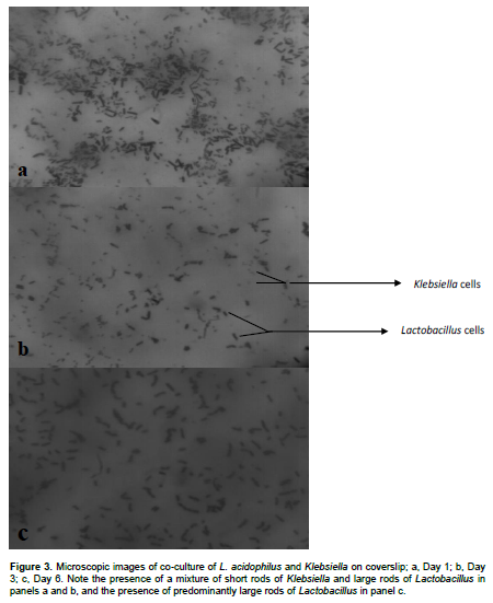
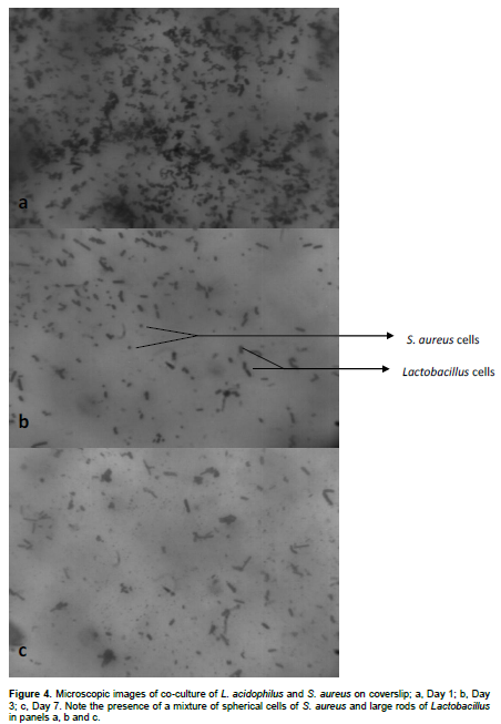
The ability of Lactobacillus cells to inhibit biofilm formation by urine bacteria was evaluated qualitatively by cover-slip assay by a modification of the method of Ghanwate et al. (2012). Sterile Petri dishes were filled each with 10 ml of MRS broth containing 1% sucrose. Then, sterile glass cover-slips were added to each Petri dish. Thereafter, each plate was inoculated with 0.1 ml of overnight culture of L. acidophilus and incubated anaerobically at 37°C for 24 h to coat the cover-slips with Lactobacillus. The next day, the Lactobacillus-coated cover-slips were extracted and introduced into culture dishes containing 10 ml broth-culture of different urine isolates (S. aureus, E. coli, Klebsiella sp. and P. aeruginosa) to produce co-cultures of the Lactobacillus and urine organisms. The dishes were then incubated aerobically at 37°C. On daily basis, a cover-slip from each culture set-up was removed, unattached cells were rinsed off with phosphate buffered saline and attached cells or biofilms were stained with 0.1% crystal violet for 5 min (for S. aureus) and Gram staining for E. coli, Klebsiella sp. and P. areuginosa. Finally, stained biofilms were observed microscopically and photographed using a 0.2 Mega pixel Motic camera. The co-cultures were monitored over a seven-day period.
Immobilization of Lactobacillus acidophilus on catheter pieces
MRS broth containing L. acidophilus cells was incubated for 48 h at 37°C. The broth was then centrifuged at 5000 rpm for 15 min. The supernatant was discarded while the pellet of cells was added into a beaker containing exactly 2% (w/v) sodium alginate solution. The catheter pieces were then introduced into the mixture and allowed to stand for 1 h. Thereafter, the catheter pieces were extracted and immersed in a beaker containing 2% CaCl2.2H2O to allow the formation of a gel. The set-up was incubated for 24 h for stability of the gel.
Evaluation of anti-biofilm capability of immobilized Lactobacillus cells
The catheter pieces containing immobilized L. acidophilus cells were immersed in broth cultures of the different urine isolates in separate Bijoux bottles. The bottles were allowed to stand for 7 days at 37°C. Untreated catheter sections also immersed in broth cultures of urine isolates served as control. Biofilm formation on the catheter sections was evaluated by viable cell count procedures. On each day of counting, two catheter pieces were picked from the various cultures and rinsed with sterile distilled water to remove unattached cells. Then, the attached cells were gently scraped off from both the outer and luminal surfaces of the catheters using a wire loop and introduced into sterile beakers containing 10 ml of phosphate buffered saline. The cells in the biofilm were dispersed using a magnetic stirrer. Serial dilutions were prepared and the cells inoculated onto MacConkey and blood agar plates for viable counts.
Preparation of crude bacteriocin of L. acidophilus
The isolated L. acidophilus was propagated in a 100 ml-conical flask containing 25 ml MRS broth and incubated anaerobically at 37°C for 24 h. Cells were separated by centrifugation at 5000 rpm for 20 min at room temperature. The supernatant was adjusted to pH 7 by using 1 N NaOH to remove the antimicrobial effect of organic acids. The clear solution was then filtered using 0.22 µ pore size filter.
Determination of antimicrobial activity of crude bacteriocin
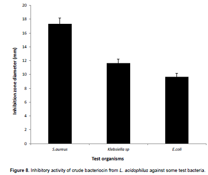
The inhibitory activity of the crude bacteriocin preparation against the urine isolates was determined using disc diffusion method. Paper discs of 6 mm diameter were prepared from Whatman No.1 filter paper. The discs were autoclaved for 15 min at 121°C and allowed to cool. Then, the sterile discs were impregnated with the bacteriocin preparation. The impregnated discs were placed on solidified Muller-Hinton agar seeded with 18 h cultures of the test organisms and incubated at 37°C for 24 h. The assay was performed in triplicates. Zones of inhibition were then measured and the values were recorded.
Statistical analysis
Analysis of variance (ANOVA) and LSD0.05 were employed to analyze the data on viable counts using Microsoft Excel 2007 application.
RESULTS
Isolation of bacteria from urine and HVS
Four bacteria were isolated from the urine specimens. These were: S. aureus, E. coli, Klebsiella sp. and P. aeruginosa. All four organisms were used in the biofilm studies. Two Lactobacillus species were identified from HVS, namely: L. acidophilus and Lactobacillus plantarum. The L. acidophilus was used for all the anti-biofilm studies.
Growth of Lactobacillus in urine
L. acidophilus cells were able to survive and multiply in urine, increasing in number from 6.5 x 10⁷ to 1.5 x 10⁸ cfu/ml by the fifth day of culture, after which there was a decrease in number (Figure 1).

Inhibition of urine bacteria by Lactobacillus cells on coverslips
Evaluation of the inhibitory activity of Lactobacillus cells against bacterial biofilms on coverslips showed that pre-coating of coverslip surfaces with Lactobacillus cells prior to exposure of the surfaces to urine bacteria reduced the number of urine bacterial cells attaching to the coverslips. For E. coli, cell numbers reduced relative to number of Lactobacillus cells from the third day of culture, while there was complete inhibition by the sixth day (Figure 2).

Similarly, numbers of attached cells of Klebsiella and S. aureus reduced from the third day and complete inhibition was achieved by the seventh day (Figures 3 and 4). For P. aeruginosa, on the other hand, reduction in number was observed only from the fifth day and attached cells were still observed by the end of the experiment on the seventh day (Figure 5).

Inhibition of catheter biofilms by immobilized Lactobacillus cells
Evaluation of attachment and growth of urine organisms on catheter pieces containing immobilized Lactobacillus cells was compared with growth on uncoated catheter pieces. The results showed significant (p<0.05) reduction in number of S. aureus and Klebsiella cells on the coated catheter pieces when compared with the uncoated catheters. E. coli and P. aeruginosa, on the other hand, showed no reduction. Representative graphs of the growth of S. aureus and P. aeruginosa are shown in Figures 6 and 7, respectively.


Inhibitory activity of crude bacteriocin of L. acidophilus against test isolates
The crude bacteriocin preparation from the Lactobacillus isolate was tested for its activity against the urine aisolates. The results showed that the highest activity was against S. aureus, followed by Klebsiella before E. coli (Figure 8).
DISCUSSION
The human normal bacterial flora is increasingly recog-nized as an important defense to infection. Lactobacilli in particular, are regarded to be generally safe and recognized as potent interfering bacteria. Lactobacilli and their derived substances have been used widely in both clinical and experimental trials to inhibit growth of other bacteria on various natural and artificial surfaces, including urinary catheters (Reid and Tieszer, 1994; Reid and Burton, 2002; Reid and Bruce, 2006; Maldonando et al., 2007; Ruiz et al., 2009; Barrons and Tassone, 2008; Fracchia et al., 2010; Ray, 2011; Al-Mathkhury et al., 2011; Sambanthamoorthy et al., 2014; Abd-Alkareem, 2014).
In this study, L. acidophilus was isolated from HVS of a healthy female. The Lactobacillus cells showed appreciable growth in urine for up to 5 days in culture, after which there was a decline in growth. The decline in growth after five days could be attributed to culture conditions, in which the cells had to survive in what could be described as stagnant urine.
Evaluation of the anti-biofilm potential of the Lactobacillus cells by cover-slip assay showed that pre-coating of the surfaces with Lactobacillus reduced the attachment and growth of urine bacteria. The effect was more pronounced against E. coli, Klebsiella and Staphylococcus, with near complete inhibition being achieved by the fifth day of co-culture. For P. aeruginosa, on the other hand, clearing required longer than seven days. The inhibitory effect observed in the cover-slip assay was confirmed quantitatively using viable cell count procedures following immobilization of Lactobacillus cells on catheter surfaces and exposure to urine bacteria. In the quantitative assay, however, more inhibition was recorded with S. aureus and Klebsiella than with E. coli and Pseudomonas.
The results in this study agree with those of Maldonado et al. (2007), in which whole cells and acid supernatant of Lactobacillus fermentum inhibited biofilm formation and growth of Klebsiella. Westbroek et al. (2010) reported that Streptococcus pyogenes was not inhibited by Lactobacillus crispatus and Lactobacillus jensenii. In Westbroek’s (2010) study, the Lactobacillus and Steptococcus cells were mixed before plating. It appears therefore that the inhibitory action requires pre-application of the Lactobacillus or its products to the surface before exposure to the biofilm-forming organism, as was demonstrated in the study by Fracchia et al. (2010). Similar results were also reported by Hall et al. (2000) and Darouiche et al. (2001), working with benign E. coli strains. In their studies, instillation of the benign E. coli strains into the bladders of patients with recurrent UTI and coating of urinary catheters with benign E. coli, respectively, significantly reduced the occurrence of UTI in the patients.
In the present study, cell-free supernatant from the L. acidophilus was also evaluated for its activity against the urine bacteria by disk-diffusion method and the results showed that S. aureus, Klebsiella and E. coli were inhibited, while Pseudomonas was resistant.
The results from this preliminary study showed that Lactobacillus has the potential to inhibit urinary catheter biofilms when applied to catheters and the inhibitory action could be both by removal of attachment surface for the urine bacteria and production of antibacterial substances.
The finding presents an interesting intervention strategy that should be explored further for actual use. Concerns that may come to mind in consideration of practical application are those relating to safety and stability of alginate, blockage of the catheter by the coating, as well as possibility of infection by the Lactobacillus. These questions would be answered by carrying out further trials using small animals.
CONFLICT OF INTEREST
There is no conflict of interest.
REFERENCES
|
Abd-Alkareem AY (2014). Lactobacillus acidophilus as antibiofilm formed by Staphylococcus aureus in vitro. Diyala. J. Med. 7:24-34. |
|
|
Ali OA (2012) Inhibition of uropathogenic Citrobacter adhesion by biosurfactants extracted from vaginal Lactobacillus acidophilus. J. Al-anbar Univers. Med. Sci. 10:11-19. |
|
|
Al-Mathkhury HJF, Ali AS, Ghafil JA (2011). Antagonistic effect of bacteriocin against urinary catheter associated Pseudomonas aeruginosa biofilm. N. Am. J. Med. Sci. 3:367-370. |
|
|
Andersen T, Strand BL, Formo K, Alsberg E, Chritensen BE (2012). Alginates as biomaterials in tissue engineering. Carbohydr. Chem. 37:227-258. |
|
|
Barrons R, Tassone D (2008). Use of Lactobacillus probiotics for bacterial genitourinary infections in women: a review. Clin. Therapeutics 30:453-468. |
|
|
Borchert D, Sheridan L, Papatsoris A, Faruquz Z, Barua JM, Junaid I, Pati Y, Chinegwundoh F, Buchholz N. (2008). Prevention and treatment of urinary tract infection with probiotics: Review and research perspective. Indian J. Urol. 24:139-144. |
|
|
Cheesebrough M (2000). District Laboratory Practice in Tropical Countries. Tropical Health Technology and Butterworth-Heinemann Ltd. Cambridge. 2:63-70. |
|
|
Darouiche RO, Donovan WH, Del Terzo M, Thornby JI, Rudy DC, Hull RA (2001). Pilot trial of bacterial interference for preventing urinary tract infection. Urology 58:339-344. |
|
|
Darouiche RO, ThornbyJI, Cerra-Stewart C, Donovan WH, Hull RA (2005). Bacterial interference for prevention of urinary tract infection:A prospective, randomized,placebo-controlled, double blind pilot trial. Clin. Infect. Dis. 41:1531-1534. |
|
|
Donlan RM (2001). Biofilms and device-associated infections. Emerg. Infect. Dis. 7:277-280. |
|
|
Fracchia L, Cavallo M, Allegrone G, Martinotti MG (2010). A Lactobacillus-derived biosurfactant inhibits biofilm formation of human pathogenic Candida albicans biofilm producers. Curr. Res. Technol. Educ. Top. Appl. Microbiol. Microb. Biotechnol. 2:827-837. |
|
|
Ghanwate NA, Thakare PV, Bhise PR, Dhanke A, Apotikar S (2012). Prevention of biofilm formation in urinary catheter by coating enzymes/gentamycin/EDTA. World Acad. Sci. Eng. Technol. 6:468-470. |
|
|
Hall R, Rudy D, Donovan W, Svanborg C, Wieser I, Stewart C, Darouiche R (2000). Urinary tract infection prophylaxis using Escherichia coli 83972 in spinal cord injured patients. J. Urol. 163:872-877. |
|
|
Maldonado NC, Silva de Ruiz C, Cecilia M, Nader-Macias ME (2007). A simple technique to detect Klebsiella biofilm-forming strains. Inhibitory potential of Lactobacillus fermentum CRL 1058 whole cells and products. Commun. Curr. Res. Educ. Top. Trends Appl. Microbiol. pp. 52-58. |
|
|
Merk K, Borelli C, Korting HC (2005). Lactobacillus-bacteria-host interactions with special regards to the urinary tract. Int. J. Med. Microbiol. 295:9-18. |
|
|
Ray K (2011). Infection: Lactobacillus probiotic could prevent recurrent UTI. Nature Rev. Urol. 8:292. |
|
|
Reid G, Tieszer C (1994). Use of lactobacilli to reduce the adhesion of Staphylococcus aureus to catheters. Int. Biodeterior. Biodegrad. 34:73-83. |
|
|
Reid G, Bruce AW (2002). Probiotics to prevent urinary tract infections: the rationale and evidence. World J. Urol. 24:28-32. |
|
|
Reid G, Bruce AW, Fraser N, Heinemann C, Owen J, Henning B (2001). Oral probiotics can resolve urogenital infections. FEMS Immunol. Med. Microbiol. 30:49-52. |
|
|
Reid G, Burton J (2002). Use of Lactobacillus to prevent infection by pathogenic bacteria. Microb. Infect. 4:319-324. |
|
|
Ruiz FO, Gerbaldo G, Asurmendi P, Pascual LM, Giordano W, Barberis IL (2009). Antimicrobial activity, inhibition of urogenital pathogens and synergistic interactions between Lactobacillus strains. Curr. Microbiol. 59:497-501. |
|
|
Saint S, Kaufman SR, Thompson M, Rogers MA, Chenowith CE (2005). A reminder reduces urinary catheterization in hospitalized patients. Joint Commission J. Qual. Patient Safety 31:455-462. |
|
|
Sambanthamoorthy K, Feng X, patel R, Patel S, Paranavitana C (2014). Antimicrobial and antibiofilm potential of biosurfactants isolated from lactobacilli against multi-drug-resistant pathogens. BMC Microbiol. 14:197. |
|
|
Stickler DJ (2008). Bacterial biofilms in patients with indwelling urinary catheters. Nature Clin. Practice Urol. 5:598-608. |
|
|
Trautner BW, Darouiche RO (2004). Role of biofilm in catheter-associated urinary tract infection. Am. J. Infect. Control 32:177-183. |
|
|
Trautner BW, Hull RA, THornby JI, Darouiche RO (2007). Coating urinary catheters with an avirulent strain of Escherichia coli as a means to establish asymptomatic colonization. Infect. Control Hosp. Epidemiol. 28:92-94. |
|
|
Uehara S, Monden K, Nomoto K, Seno Y, Kariyama R, Kumon H (2006). A pilot study evaluating the safety and effectiveness of Lactobacillus vaginal suppositories in patients with recurrent urinary tract infection. Int. J. Antimicrob. Agents 28:S30-34. |
|
|
Westbroek ML, Davis CL, Fawson LS (2010). Interactions of lactobacilli with pathogenic Streptococcus pyogenes. Infect. Dis. Obstet. Gynecol. pp. 289-743. |
|
Copyright © 2024 Author(s) retain the copyright of this article.
This article is published under the terms of the Creative Commons Attribution License 4.0


