Full Length Research Paper

ABSTRACT
Camel milk is a suitable substrate for the growth of protective bacterial flora. Detection of lactic acid bacteria producing antimicrobial substances from camel (Camelus dromedarius) milk in south Algeria against some food-borne pathogens is the subject of this work. Morphological, physiological and biochemical tests have identified four Lactobacillus isolates belonging to the following species: Lactobacillus fermentum, Lactobacillus helviticus, Lactobacillus plantarum and Lactobacillus acidophilus. In order to demonstrate the inhibitory effect of these bacteria in vitro, their antagonistic property was tested against six pathogenic strains often involved in food-borne illness: Staphylococcus aureus, Bacillus subtilis, Escherichia coli, Pseudomonas aeruginosa, Salmonella enteritidis and Shigella flexneri using the disc diffusion method. The antagonistic effect was manifested by the appearance of inhibition zones around the discs. The potential inhibitor was estimated by calculating diameter of inhibition zones which extend from 02 to 16 mm. All Lactobacillus isolates secreted into the culture medium inhibitory substances were able to inactivate the growth of pathogenic strains tested. L. plantarum has shown the largest inhibition zone against S. aureus (16 mm). These two strains were chosen to determine the nature of L. plantarum secreted substances responsible for the antagonistic effect. The obtained results have shown that L. plantarum inhibitory property against S. aureus resulted from the combined effect of several biological agents originating from their metabolic activities, especially organic acids and bacteriocins.
Key words: Antagonism, camel milk, Lactobacilli, food borne pathogens, mixed culture, pure culture.
INTRODUCTION
Food borne pathogenic bacteria such as Escherichia coli, Staphylococcus aureus and Salmonella are the cause of various pathologies and food borne illness. Pasteurization, fermentation and refrigeration are mostly used to preserve and prolong the shelf-life of food products. However, these methods do not constitute a sufficient guarantee to fight against microbial contamination. Excessive and uncontrolled use of chemical additives may create health risks for the consumer (Mami et al.,2010). Lactic acid bacteria have been used successfully, with few adverse effects to prevent antibiotic associated diarrhea, to treat severe infantile diarrhea and to treat various diarrheal illnesses (Tadesse et al., 2005). Several studies have exploited microbial interactions to protect food quality and to fight against undesirable microorganisms (Allouche et al., 2010). Hence search for new strains of lactic acid bacteria producing antimicrobial substances is a universal objective for creation of lactic leaven intended for a better food bio-preservation (Mami et al., 2010; Franz et al., 2010).
It has been shown that camel milk possesses a specific protective system, powerful against contamination flora. This system is composed of antagonistic substances such as protective proteins ((lysosomes, lactoperoxidases and lactoferrin), organic acids, hydrogen peroxide (H2O2), bacteriocins produced by lactic acid bacteria which limit the growth of certain pathogenic microbes (Barbour et al., 1984; Klaenhammer et al., 1994; Siboukeur, 2007; Jrad et al., 2013). However, only few studies reported the effect of lactic acid bacteria isolated from camel milk on pathogenic bacteria. Therefore, the aim of this study was to highlight lactic strains isolated from camel (Camelus dromedaries) milk belonging to nomads in the Wilaya of Ouargla (South Algeria) and to evaluate in vitro, their antimicrobial activities against various pathogenic strains encountered in dairy products and involved often in food-borne diseases.
MATERIALS AND METHODS
Biological material
Milk sample
The collection of milk was carried out on a camel (C. dromedaries), belonging to nomads in the wilaya of Ouargla (South Algeria), 6 years old, after her first pregnancy and one calving.
Pathogenic strains
The antimicrobial activity of lactic acid bacteria isolated from camel milk was tested on the growth of the following pathogenic strains: Salmonella enteritidis (ATCC 25928), Staphylococcus aureus (ATCC 6538), Escherichia coli (ATCC 4157), Pseudomonas aeruginosa (ATCC 25853), Bacillus subtilis (ATCC 6633) and Shigella flexneri (ATCC29903) obtained from Microbiology Laboratory of Saidal Group and Microbiology Laboratory of Pasteur Institute, Algiers.
Isolation and identification of lactic acid bacteria from camel milk
The isolation was performed on Man-Rogosa and Sharp (MRS) agar, medium suitable for specific research of Lactobacilli. The cultures were incubated for 48 to 72h at 30°C under anaerobic conditions. The purification was done by carrying successive streaking of individual colonies on MRS agar and incubation at 30°C for 48 h until colonies with same size, shape, color, were obtained, indicating therefore the purity of the strain. The identification was based on the determination of macroscopic characters (colonies aspect on MRS agar), microscopic characters (shape, Gram reaction, mobility and sporulation), physiological characters (catalase test, the growth at different temperatures, salt tolerance and production of CO2) and biochemical characters (fermentation of carbohydrates using the API 50 CH gallery). The fermentation results of the 49 sugars in the API50CH gallery were treated by ApiWeb software (BioMérieux) to identify lactic acid bacteria with a similarity rate of 100% (Alaoui et al., 2016). The conservation of the pure strains was performed on inclined solid medium (MRS agar). Then, the cultures were maintained at 4°C and every four weeks, the strains were inoculated in new medium (MRS agar).
The antagonism test
Preparation of bacterial pre-cultures
A young lactic culture of 18 h of each isolate was inoculated into a test tube containing 10 mL of MRS broth. The tube was then incubated at 37°C for 18 h under anaerobic conditions. The pathogenic strains were each inoculated in a test tube containing 10 mL of nutrient broth and incubated at 37°C for 18 h.
Standardization of bacterial pre-cultures
The inoculum size of each pathogen and lactic acid bacteria was standardized against the McFarland turbidity standard No. 0.5 using a spectrophotometer at a wave length of either 600 or 625 nm. The McFarland 0.5 standard was used in the preparation of standardized bacterial inoculums for the susceptibility test to antimicrobial agents. It corresponds approximately to a homogenous bacterial suspension of 1.5 x 10⁸ cells/mL (McFarland, 1907).
Detection of antagonism by the disc diffusion method
This method consists of pouring 20 mL of Trypic Soja Agar (TSA) medium into sterile Petri dishes. After solidification, the dishes were flooded with 1 mL of pathogenic strains pre-cultures. After drying, sterile filter discs (9 mm) were dipped in the lactic acid bacteria pre-cultures incubated for 18 h and placed on solidified TSA agar seeded with test microorganisms pre-cultures. The diffusion of substances responsible for bacterial interactions was improved by incubation at 37°C for 24 h (Tadesse et al., 2005).
Estimation of inhibitory potential of lactic acid bacteria
The antimicrobial activity is revealed by the appearance of inhibition zones around the discs. Inhibition zones (Zi) were measured according to the Equation 1.

According to Guessass (2007), a positive result is when the inhibition zone diameter is greater than 2 mm. Antibacterial activity tests were done in triplicates and the mean values were recorded.
Confirmation of the inhibition and determination of the inhibitor nature
The antibacterial activity was confirmed in vitro by a test on reconstituted bovine milk in interaction conditions between the two types of microorganisms (lactic acid bacteria of camel milk and pathogenic bacteria). The experiment was carried out with lactic strains that showed the greatest inhibitor potential (high value of ). For that, a sterile sample of 150 mL reconstituted bovine milk was sterilized at 120°C for 20 s then contaminated with 1 mL of S. aureus pre-culture. After, the contaminated sample was divided into three series of tubes. In tube 1, the sample was directly incubated at 37°C for 8 h (Pure culture). In tube 2, the contaminated bovine milk, was inoculated with 1 mL of L. plantarum pre-culture (Lb3) and incubated at 37°C for 8 h (mixed culture); whereas, the contaminated sample in tube 3 was inoculated with 1 mL of neutralized supernatant of L. plantarum pre-culture resulting from centrifugation at 8000 rpm for 10 min at 4°C (Siboukeur and Siboukeur, 2013). The resulting supernatant was adjusted to pH 6.5 with NaOH (1 N) to remove the antibacterial activity which could be exerted by organic acids and incubated in the same conditions (Jrad et al., 2013). Growth and variation in cell number of S. aureus in pure and mixed cultures were estimated by counting the colonies of S. aureus on Chapman agar every 2 h of the incubation period for the 3 series. For that, decimal dilutions (10-1 to 10-5) of each sample were made. Only the dishes containing between 30 and 300 colonies were taken into consideration (Guessas et al., 2006). A volume of 0.1 mL from the dilution 10-1 for each sample was plated on the surface of the Chapman agar in order to count S. aureus colonies (Kaban et al., 2006). The colonies were counted to determine the number of CFU/mL using the following equation:

V: The inoculated volume, D: concerned dilution.
The final result of colonies count was converted into logarithm (Log10 cfu / mL) according to the established recommendations of Alomar et al. (2008). In this experiment, the authors aimed to confirm the inhibition of S. aureus by L. plantarum by comparing the variation in S. aureus cells number during 8 h of incubation in pure culture (series 1) and mixed culture (series 2 and 3).
Then, in order to show the nature of the agents responsible for the inhibition of S. aureus by L. plantarum, the authors tried to show the effect of organic acids produced by L. plantaum in first stage, and to eliminate the effect of these organic acids by neutralizing the supernatant of L. plantarum pre-culture with NaOH (1N) to prove the existence of the inhibiting agents other than the organic acids. The enumeration of viable S. aureus cells and pH measurement were done in triplicate every 2 h from the dilution 10-1 for the three series and the mean values were recorded.
Statistical analysis
Microsoft Excel software (Microsoft Excel 2010) was used to plot the curve of inactivation. Experiments were done in triplicates and the means of the three data sets are presented. Analysis of variance was performed using statistical software SPSS. In all cases, significant difference was based on the 5% level (P ≤ 0.05).
RESULTS
Isolation and identification of lactic acid bacteria from camel milk
Macroscopic appearance
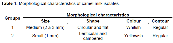
In the present study, a total of 32 isolates were recovered on MRS agar medium from camel milk sample. They were divided into two groups with different morphological characteristics as shown in the Table 1.
Microscopic appearance
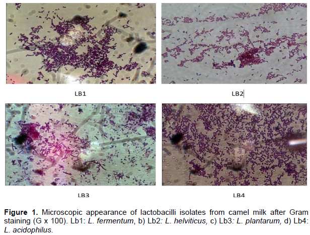
Gram reaction revealed that among the 32 isolates, 14 were retained as Gram (+), rod shaped, individual, in pairs and in chains. Therefore, Lactobacilli was potentially isolated as shown in Figure 1.
Biochemical characterization
Among the 14 Gram (+) isolates, four of them were catalase (-) and were retained. The results of morphological, physiological and biochemical tests of isolated Lactobacilli from camel milk are shown in Table 2.
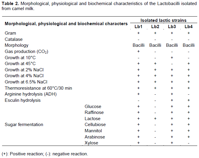
The macroscopic, microscopic and physiological characteristics showed that all camel milk isolates belong to the genus Lactobacillus. Fermentation of carbohydrates using API 50 CH Gallery system showed that Lactobacillus isolates belong to the following species: L. fermentum (Lb1), L. helveticus (Lb2), L. plantarum (Lb3) and L. acidophilus (Lb4). This biochemical identification was based on the comparison with fermentation profiles of Biomerieux database for these species with 100% similarity.
Results of antagonism
The antagonistic effect was manifested by the appearance of clear and translucent inhibition zone around the discs. Diameter of inhibition zones produced by Lactobacillus isolates on the test pathogenic strains are shown in Table 3.
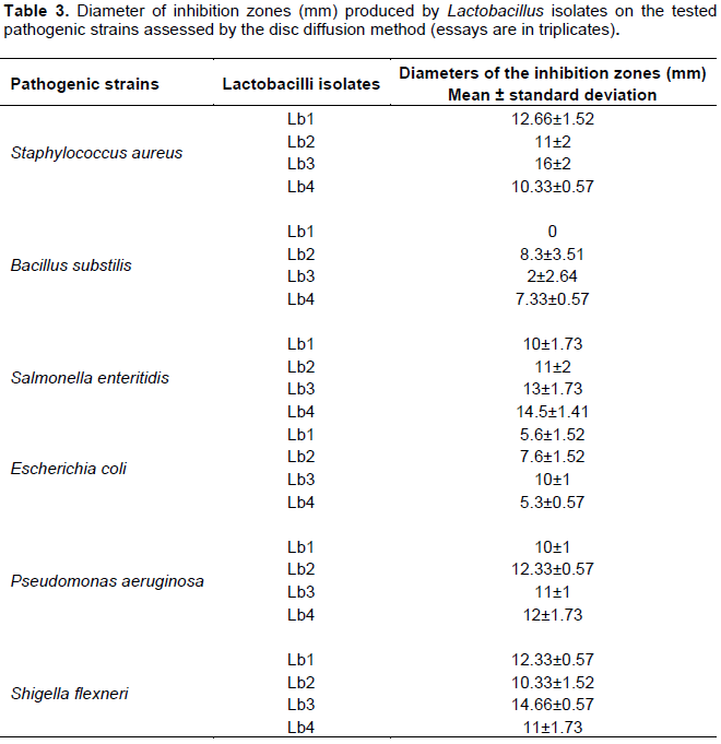
All Lactobacillus isolated from camel milk inhibited the growth of all pathogenic strains tested to varying degrees, except Lb1 which was inactive against B. subtilis. This concluded that Lb. fermentum, L. helveticus, L. plantarum and L. acidophilus are characterized as producers of inhibitory substances against pathogenic strains. The inhibition zones are clear with distinct borders; diameters of inhibition zones varied from 02 (lower activity observed for L. plantarum against Bacillus subtilis) to 16 mm (higher activity observed for L. plantarum against S. aureus). Therefore, L. plantarum was retained as the strongest inhibitor against the tested pathogenic strains.
Confirmation of the inhibition and determination of the inhibitor nature
Confirmation of the inhibition
The growth kinetics of S. aureus in the presence and absence of L. plantarum is shown in Figure 2. The bacterial count was carried out every 2 h during 8 h of treatment at 37°C. In the sample of tube 1 (reconstituted milk + S. aureus), the survival fraction of S. aureus did not significantly increase or decrease since the contaminated milk was not treated by Lb3.
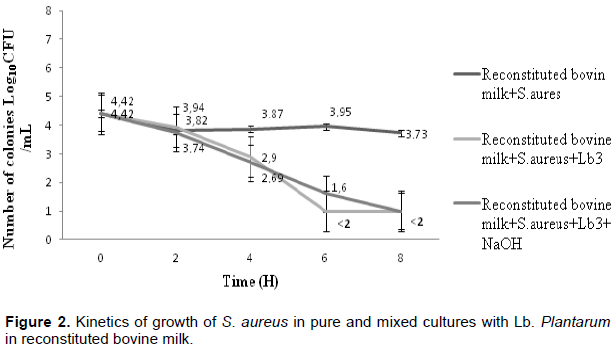
However, when comparing the bacterial count of tube 2 (reconstituted milk + S. aureus + L. plantarum) and 3 (reconstituted bovine milk + S. aureus + L. plantarum + NaOH 1N) to tube 1, a drastic S. aureus reduction was noticed just after 2 h of treatment. The number of viable cells per milliliter declined from 4.42 to 2.9 log10 cfu/mL for the tube 2 and 2.69 log10 cfu/mL for tube 3 after 4 h of treatment. And S. aureus cells number became less than 2 log10 cfu/mL within 6 and 8 h for the tubes 2 and 3, respectively (Figure 2). These differences were not statistically significant on the 5% level (p≥0.05), (0.77 >0.05) and (0.87>0.05).
Nature of inhibitor
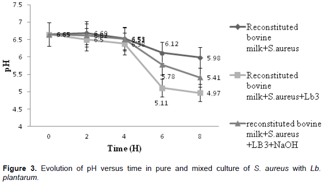
The milk pH measurement was done every 2 h of treatment. A pH decrease was shown in Figure 3 for the three milk samples (tubes 1, 2 and 3). In tube 1 (untreated sample), pH was changed insignificantly during 8 h of incubation. In contrast, for tubes 2 and 3 (treated samples), pH decreases considerably from the 4th h to reach a value of 4, 97 and 5, 41 respectively. These differences were not statistically significant on the 5% level (p≥0.05), (0.81>0.05 and 0.56>0.05).
DISCUSSION
In the present study, a total of 32 lactic acid bacteria were isolated from raw camel milk sample. Morphological, physiological and biochemical tests identified four Lactobacillus isolates belonging to the following species: L. fermentum, L. helviticus, L. plantarum and L. acidophilus. Carbohydrate degradation tests by the API 50 CH galleries allowed us to differentiate these Lactobacillus species with fermentation profiles of 100% similar to those given by ApiWeb software (BioMérieux) for these same species. The positive reading is given by a change of the color under the effect of pH variation in the API 50 CH galleries wells. These classic galleries API 50 CH were used to differentiate 15 lactobacillus isolates from the 112 lactic isolates of camel milk that belonged to the species: L. brevis, L. delbrueckii and L. fermentum in the work of Alaoui et al. (2016). Similar to our findings, L. fermentum (Lb1) isolated in this study were among the lactobacilli isolated from camel milk in the work of Khandelwal et al. (2015) and Alaoui et al. (2016). For L. helveticus (Lb2) and L. acidophilus (Lb4), they were isolated from goat milk in the work of Badis et al. (2005), and isolation of L. plantarum (Lb3) from raw goat milk and raw camel milk was described in several studies (Guessass and Kihal, 2004; Badis et al., 2004 a, b; Mami et al., 2008). All Lactobacillus isolated from camel milk inhibited the growth of majority of the tested pathogenic strains in varying degrees. The inhibition zones of diameter of L. fermentum (Lb1) against the tested pathogenic strains varied from 5.6 to 12.66 mm. The greatest inhibition recorded was against S. aureus without any inhibition against B. subtilis. Foster and Hall (1991) reported that some microorganisms produce an acid-tolerance response system that protects them against severe acid stress for longer periods. According to Cogan et al. (1997), the good acidifying lactic acid bacteria are those which are able to reduce the pH of the milk from its initial value of approximately 6.6 to 5.3 over a period of 6 h. However, the pH variation technique (ΔpH) allows classifying the lactobacilli into rapidly acidifying strains, with medium acidification rate and low acidification rate. On this basis and from the data shown in Figure 3, L. fermentum was able to reduce the pH of bovine milk from 6.65 to 5.11 after 6 h of incubation (severe acid stress). Therefore, L. fermentum is a rapidly acidifying strain. This may be the reason for resistance of B. subtilis to L. fermentum as compared to the other tested strains which probably belong to the other less acidifying categories. For L. helveticus (Lb2), inhibition zones diameters varied from 7.6 to 12.33 mm. In comparison with the work of other investigators that reported the weak antibacterial activity against P. aeroginosa by L. casei and L. bulgaricus isolated from various foods (Nigatu et al., 2015), the current results showed good inhibitory effect against P. aeruginosa by L. helveticus. Whereas, L. helveticus (Lb2) was more active against E. coli as compared to those isolated from cow's milk in the work of Allouche et al. (2010). For L. acidophilus (Lb 4), inhibition zones diameters varied from 5.3 to 14.5 mm. The greatest inhibition was against Salmonella enteritidis. Itoh et al. (1995) and Tahara and Kanatani (1996), showed that it is also possible that L. acidophilus exerts inhibitory activity only against bacteria taxonomically closest to the producing strain. L. plantarum (Lb3) showed the larger zone of inhibition against S. aureus (16 mm). The inhibition of S. aureus and E. coli by L. plantarum isolated from goat milk have already been described by Mami et al. (2008), Todorov et al. (2004) and Karthikeyan and Santosh (2009). According to Trias et al. (2008), the diameter of the inhibition zones varies with the type of culture medium used and the species used as a target strain or indicator strain.
In series 1 (untreated sample), the survival fraction of S. aureus did not significantly increase or decrease during the incubation period. The low growth of S. aureus in untreated sample of reconstituted bovine milk medium can be explained by the existence of an intrinsic antimicrobial activity of the lactic acid bacteria found in this milk against S. aureus. In contrast, the important reduction in S. aureus cells number (Log 10cfu/mL<2) in mixed culture with L. plantarum (series 2 and 3) after 8 ho of treatment confirmed the bactericidal effect of L. plantarum on S. aureus. This same event was observed after 72 h in the work of Mami et al. (2008). According to Lairini et al. (2014), Lactobacillus are rapidly acidifying strains. The observed decrease in pH for the series 2 and 3 resulted from the production of organic acids (lactic and acetic acid) by L. plantarum isolated from camel milk. However, for series 1 (untreated sample), pH reduction was due to acid production by S. aureus. The inhibition of S. aureus in parallel with the pH decrease in the medium means that the acidity caused by organic acids produced by L. plantarum is an inhibitory agent for the growth of S. aureus. This decrease in pH has as consequence, a significant inhibition of S. aureus (less than 2 Log 10cfu /mL) from the 6th h of treatment for series 2. McLean and McGroarty (1996) showed that about 60% of the antimicrobial activity of lactic acid bacteria was removed when the filtrates were neutralized to pH 6.5 with NaOH. However, addition of the neutralized L. plantarum pre-culture supernatant to the S. aureus culture has only delayed this event (charge less than 2 Log 10cfu /mL) in series 3. It can be concluded that organic acids are probably not the unique agent responsible for this phenomenon, other metabolites could be implicated. Inhibition of S. aureus by the production of hydrogen peroxide is excluded because this one has a catalase (Alomar et al., 2008). The H2O2 released by lactic acid bacteria strains inhibit bacteria which do not possess defenses against oxidative stress (Ouwehand and Vesterlund, 2004). The production of bacteriocins by L. plantarum is widely accepted (Ouwehand and Vesterlund, 2004; Todorov et al., 2004; Karthikeyan and Santosh, 2009). Therefore, inhibitory properties of L. plantarum against S. aureus are also owed to the production of bacteriocins. Lactobacillus strains isolated from camel (Camelus dromedaries) milk in this study showed different inhibitory activities against the tested pathogenic strains, this can also be explained by the quantity and the structural variability of bacteriocins produced (Richard, 1996).
The results obtained showed that the antibacterial property of L. plantarum isolated from camel milk against S. aureus result from the synergistic (combined) effect of several biological factors originating from their metabolic activities, especially the organic acids and unknown bacteriocins.
CONCLUSION
Lactic acid bacteria isolated from camel milk in this study are able to produce antibacterial substances to eliminate the presence of tested pathogenic bacteria. Camel milk differs from milk of other species by the presence of a powerful protector system; it constitutes a source of new antimicrobial strains. It appeared that the synergistic effect of several biological factors originating from their metabolic activities (organic acid, bacteriocins, H2O2) produced by lactic acid bacteria derived from camel milk was responsible for the self-purification effect of stored camel milk which is kept for many hours in relatively high temperatures (about 28°C). This study showed also the inhibitory activity of lactic acid bacteria derived from camel milk observed in vitro against pathogenic species that may accidentally contaminate milk, indicating the possibility of exploiting this activity for use as a means of food bio-preservation and to preserve the health of the consumer against pathogenic bacteria involved in food poisoning deemed in the summer season.
CONFLICT OF INTERESTS
The authors have not declared any conflict of interests.
REFERENCES
|
Alaoui Ismaili M, Guilal J, Hamama A, Saidi B, Zahar M (2016). Identification de bactéries lactiques du lait cru de chamelle du sud du Maroc. Int. J. Multi-disciplinary Sci. 1(1):81-94. |
|
|
Allouche FN, Hellal A, Laraba A (2010). Etude de l'activité antimicrobienne des souches de lactobacilles thermophiles utilisées dans l'industrie laitière. Rev. Nat. Technol. 3:13-20. |
|
|
Alomar J, Loubiere P, Delbes C, Nouaille S, Montel MC (2008). Effect of Lactococcus garvieae, Lactococcus lactis and Enterococcus faecalis on the behaviour of Staphylococcus aureus in microfiltred milk. Food Microbiol. 25(3):502-508. |
|
|
Badis A, Guetarni D, Moussa-Boudjemaa B, Henni DE, Tornadijo ME, Kihal M (2004). Identification of cultivable lactic acid bacteria isolated from Algerian raw goat's milk and evaluation of their technological properties. Food Microbiol. 21(3):343-349. |
|
|
Badis A, Guetarnib D, Moussa-Boudjemaa B, Henni DE, Kihal M (2004). Identification and technological properties of lactic acid bacteria isolated from raw goat milk of four Algerian races. Food Microbiol. 21(5):579-588. |
|
|
Badis A, Laouabdia-Sellami N, Guetarni D, Kihal M, Ouzrout MR (2005). Caractérisation phénotypique des bacteries lactiques isolées à partir de lait cru de chèvre de deux populations caprines locales « arabia et kabyle ». Sci. Technol. 23:30-37. |
|
|
Barbour EK, Nabbut NH, Frerichs WN, Al Nakhli HM (1984). Inhibition of pathogenic bacteria by camel's milk: relation to whey lysosome and stage of lactation. J. Food. Prot. 47(11):838-840. |
|
|
Cogan TM, Barbosa M, Beuvier E, Bianchi-salvadori B, Cocconcelli PS, Fernandes I, Gomez J, Gomez R, Kalanzopoulos G, Ledda A, Medina M, Rea MC, Rodriguez (1997). Characterization of lactic acid bacteria in artisanal dairy products. J. Dairy Res. 64(3):409-421. |
|
|
Foster JW, Hall HK (1991). Inducible pH homeostasis and the acid tolerance response of Salmonella typhimurium. J. Bacteriol. 173(16):5129-5135. |
|
|
Franz CMAP, Cho GS, Holzapfel WH, Galvez A (2010). Safety of lactic acid bacteria. Biotechnology of lactic acid bacteria: Novel applications. pp. 341-360. |
|
|
Guessass B (2007). Les potentialités métaboliques des bactéries lactiques isolées du lait cru de chèvre dans le bio-contrôle de Staphylococcus aureus. Thèse de Doctorat. Université d'Oran Es-Senia. Algérie. |
|
|
Guessass B, Kihal M (2004). Characterization of lactic acid bacteria from Algerian arid zone raw gaot's milk. Afr. J. Biotechnol. 3(6):339-342. |
|
|
Itoh T, Fujimoto Y, Kawai Y, Satto T (1995). Inhibition of food-borne pathogenic bacteria by bacteriocin from Lactobacillus gasseri. Lett. App. Microbiol. 21(3):137-141. |
|
|
Jrad Z, El Hatmi H, Fguiri I, Arroum S, Assadi M, Khorchani T (2013). Antibacterial activity of Lactic acid bacteria isolated from Tunisian camel milk. Afr. J. Microbiol. Res. 7(12):1002-1008. |
|
|
Kaban G, Kaya M (2006). Effect of starter culture on growth of Staphylococcus aureus in sucuk. Food Control 17(10):797-801. |
|
|
Karthikeyan V, Santosh SW (2009). Isolation and partial characterization of bacteriocin produced from Lactobacillus plantarum. Afr. J. Microbiol. Res. 3(5):233-239. |
|
|
Khandelwal D, Joshi H, Chaudhary BL (2015). Antagonistic effect of Lactobacilli of Camel Milk against Aeromonas veronii isolated from Pichola lake, Udaipur, Rajasthan, India. Res. J. Rec. Sci. 4:170-172. |
|
|
Klaenhammer TR, Fremaux C, Hechard Y (1994). Activités antimicrobiennes des bactéries lactiques. In. De Roissard and Luquet (Eds), Bactéries Lactiques I. Tech. Doc, Lavoisier, Paris. Pp. 353-365. |
|
|
Lairini S, Beqqali N, Bouslamti R, Belkhou R, Zerrouq F (2014). Isolement des bactéries lactiques à partir des produits laitiers traditionnels Marocains et formulation d'un lait fermenté proche du Kéfir. Afr. Sci. 10(4):267-277. |
|
|
Mami A, Hamedi AR, Henni JE, Kerfouf A, Kihal M (2010). Anti-bacterial activity of Lactobacillus plantarum isolated from Algerian raw gaot's milk against Staphylococcus aureus. Les technologies de laboratoire. 5:26-33. |
|
|
Mami A, Henni JE, Kihal M (2008). Anti-bacterial activity of Lactobacillus Species isolated from Algerian raw gaot's milk against Staphylococcus aureus. World. J. Dairy Food. Sci. 3(2):39-49. |
|
|
McFarland J (1907). The nephelometer: an instrument for estimating the number of bacteria in suspensions used for calculating the opsonic index and for vaccines. J. Am. Med. Assoc. 49(14):1176-1178. |
|
|
McLean NW, McGroarty JA (1996) .Growth inhibition of metronidazole-susceptible and metronidazole-resistant strains of Gardnerella vaginalis by lactobacilli in vitro. Appl. Environ. Microbiol. 62(3):1089-1092. |
|
|
Nigatu JM, Tuji FA, Tefera AT (2015). Evaluation of the antagonistic effect of six mixed cultures of lactic acid bacteria, isolated from the Ethiopian fermented milk ergo, against some food-borne pathogens inoculated into the Ethiopian cottage cheese ayib. Afr. J. Microbol. Res. 9(29):1789-1797. |
|
|
Ouwehand AC, Vesterlund S (2004). Antibacterial components from lactic acid bacteria. Food Science and Technology, New York. Marcel Dekker. 139:375-396. |
|
|
Richard J (1996). Utilisation de bactériocines pour la production d'aliments plus sûrs : mythe ou réalité ? Le lait. 76(1-2):179-189. |
|
|
Siboukeur A, Siboukeur O (2013). Effect of Cameline Nisin isolated from Lactococcus lactis sub sp. lactis on Staphylococcus aureus sp. Emir. J. Food Agric. 25(5):398-402. |
|
|
Siboukeur O (2007). Etude du lait camelin collecté localement : Caractéristiques physico-chimiques et microbiologiques ; aptitude à la coagulation. Thèse de Doctorat. INA. |
|
|
Tadesse G, Ephraim E, Ashenafi M (2005). Assessment of antimicrobial activity of lactic acid bacteria isolated from borde of Shamita, traditional Ethiopian fermented beverage, on some food borne pathogens and effect of growth medium in the inhibitory activity. Internet J. Food Saf. 5:13-20. |
|
|
Tahara T, Kanatani K (1996). Isolation, partial characterization and mode of action of acidocin J1229, a bacteriocin produced by lactobacillus acidophilus JCM 1229. J. Appl. Bacteriol. 81(6):669-677. |
|
|
Todorov SD, Van Reenen CA, Dicks LMT (2004). Optimization of bacteriocin production by Lactobacillus plantarum ST13BR, a strain isolated from barely beer. J. Gen. Appl. Microbiol. 50(3):149-157. |
|
|
Trias R, Ba-eras L, Badosa E, Montesinos E (2008). Bioprotection of Golden Delicious apples and Iceberg lettuce against food-borne bacterial pathogens by lactic acid bacteria. Int. J. Food Microbiol. 123(1):50- 60. |
|
Copyright © 2024 Author(s) retain the copyright of this article.
This article is published under the terms of the Creative Commons Attribution License 4.0


