African Journal of
Microbiology Research

  • Abbreviation: Afr. J. Microbiol. Res.
  • Language: English
  • ISSN: 1996-0808
  • DOI: 10.5897/AJMR
  • Start Year: 2007
  • Published Articles: 5233

Full Length Research Paper

Screening of bioactive compound, antimicrobial activity producing halophilic isolates from the saltpans of Thoothukudi district

Deepalaxmi R. K.
  • Deepalaxmi R. K.
  • Department of Microbiology, St.Mary’s College (Autonomous), TamilNadu, India.
  • Google Scholar
Gayathri C.
  • Gayathri C.
  • Department of Microbiology, St.Mary’s College (Autonomous), TamilNadu, India.
  • Google Scholar


  •  Received: 23 February 2018
  •  Accepted: 29 March 2018
  •  Published: 21 April 2018

 ABSTRACT

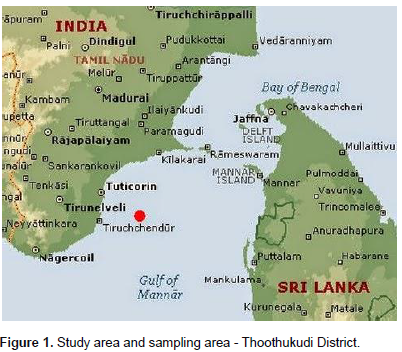
Extreme environments harbor a number of microbes producing novel bioactive compounds. The aim of our study is to isolate and identify bioactive compound producing halophiles. Marine soil sediments were collected from the solar saltpans of Thoothukudi District, Tamil Nadu, India. Based on colony morphology, two species were isolated and identification was done by using morphological and biochemical tests. The extracts of cell-free supernatant of the two halophilic isolates were screened for bioactive compound and tested for antimicrobial activity against human pathogenic bacteria such as Staphylococcus aureus, Pseudomonas sp, Klebsiella sp, Vibrio sp, Escherichia coli and fungi Aspergillus niger and Penicillium chrysogenum by the agar cup diffusion method. The results were then compared to standard antibiotics which showed 80% of similar activity in 50 μL/g concentration. In addition, the arbitrary unit of two isolates was calculated against S. aureus which produced enhanced inhibitory results. Hence our finding illustrated that Thoothukudi saltpan might be considered as a resource for novel bioactive compounds.

Key words: Halophilic bacteria, bioactive compound, anti-microbial activity, arbitrary unit, Thoothukudi saltpan.


 INTRODUCTION

The Earth’s surface consists of 70% water, which is inhabited by 80% of all life forms with greater diversity and consequently, marine environment can be described or characterized as a number of different scales, ranging from ocean-level processes which occur at species and genetic level (Bruckner, 2002; Connor et al., 2002). Marine soil has been widely explored as the source of microorganisms, possessing a large number of bioactive molecules. Halophiles are a group of microorganisms that live in saline environments and are economically important because it produces several bioactive compounds which are useful for many pharmaceutical industries (Ghosh et al., 2010). Among the halophilic microbial forms, the halophilic bacterial forms are mostly known for its secondary metabolites such as proteins, amino acids, etc. Notwithstanding, there is an enormous difficulty in isolation and culturing marine bacteria. Significant progress has been achieved in this field, and investigations of bioactive compounds produced by the marine species are rapidly increasing (Dennis and Shimmin, 1997; Wagner et al., 2002). In recent years, marine microorganisms have become important in the study of novel microbial products exhibiting antibacterial, antiviral, antitumor as well as anti-coagulant and cardioactive properties. Thousands of marine bacilli are known to contain antibiotic substance and less than 1% has been examined for their pharmaceutical activity. Although it was proved that Halophilic bacteria have a potent activity for the production of antimicrobial compounds and their antimicrobial spectrum against pathogenic microorganisms differ. Thoothukudi district in India possesses considerable theoretical, biological and conservation importance, but the biodiversity of this area is poorly characterized due to a lack of experts and inaccessibility. The present study has investigated the halophilic bacteria for their bioactive secondary metabolites. Therefore, these bioactive compounds might be consider as therapeutics directly or used as lead structures for drug innovation (Proksch et al., 2002; Kamat and Kekar, 2004). Based on the foregoing evidence the present study has been aimed to isolate and identify halophilic bacteria from the saltpan environment of Thoothukudi district and evaluate their potential for the production of bioactive compounds which serves as a source of anti-microbial agents against pathogenic bacteria and fungi.


 MATERIALS AND METHODS

Study area
 
Thoothukudi district is located on the South-East of Tamil Nadu state. The district covers an area of 4621km2 bounded by the districts of Virudhunagar and Ramanathapuram on the East and Gulf of Mannar on the South East and by Tirunelveli district on the West and South West. Its geographical co-ordinates are 8°470’’ North, 78°80’’ East (Figure 1).


Sampling area
 
In this study, the saline soil sample was collected from solar salt pans and coastal areas of Thoothukudi district, Tamil Nadu, India. The collected soil samples were placed in sterile polythene bags and containers and immediately transported to laboratory for further analysis.
 
Physico – chemical parameter analysis
 
Physical parameters such as atmospheric temperature, pH and temperature of the brine were analyzed using the standard methods (Strickland and Parson, 1972). Determinations of chemical parameter like sodium, chloride, sulphate, calcium, magnesium, potassium a analysed in soil using standard methods (Vogel, 1978).
 
 Isolation of halophilic bacteria from the saline soil sample
 
The soil samples were serially diluted in the range of 10-1, 10-2, 10-3, 10-4, 10-5, 10-6, and 10-7 in a series of test tubes. Mineral salt (MM63) medium was prepared with an increased concentration of NaCl (6 g / 100 ml). The pH of the medium was adjusted to 7 to 7.4. The samples were spread plated and are incubated at 37°C for 48 h.
 
Preparation of pure culture of halophilic bacterium species
 
Mineral salt (MM63) medium slant was prepared in test tubes and the isolated colony obtained in the Petri plates were taken (leaving out mixed culture colony) and streaked in the medium in a zigzag manner. Then test tubes were screwcapped and incubated at 37°C for 48 h.
 
Identification of the halophilic isolates
 
Morphological and biochemical studies
 
Gram staining: A thin smear of the bacterial colony was prepared on a clean slide. The slide was fixed by passing through the flame. The smear was covered with crystal violet for 30 s, and then rinsed with water and air dried. The smear was covered with iodine for 30 s, rinsed with water, decolorized with 95% ethanol and washed with water then, counterstained for about 30 s with safranin and washed with water. The slide was examined under oil immersion (1,000x). Colonial appearances were examined after incubated for 3 to 7 days.
 
Biochemical tests: The halophilic isolates were screened for many biochemical tests (Indole, methyl-red, voges–proskauer, citrate utilization, motility catalase, oxidase, D-Galactose, D-Fructose, D-glucose, Sucrose and Lactose) using standard procedures of Bergeys Manual.
 
Solvent extraction method
 
Two bacterial samples were cultured in nutrient broth containing 6 g/ml of NaCl. The cultures were then centrifuged separately and the supernatant was collected. The cell free supernatants were mixed in three different solvents; Dimethyl sulfoxide (DMSO), ethyl alcohol and phenol separately and the mixture were kept at 4°C overnight. The organic and aqueous phases were tested for antimicrobial activity through well diffusion method (Birbir et al., 2004; Birgul et al., 2002; Gesheva and Vasileva-Tonkova, 2012; Sawale et al., 2014).
 
Screening of antimicrobial activity producing halophilic bacteria
 
Anti-bacterial assay
 
The supernatants of each suspension were assessed for antibacterial property against bioassay strains of bacteria viz., E.coli, Staphylococcus aureus, Vibrio sp, Pseudomonas sp, and Klebsiella sp (Pathogens were isolated from the wound samples of infected patients and identified by morphological and biochemical test). To perform this test, bioassay strain was cultivated on Mueller Hinton agar and wells were made in plate agar using sterile cork borer. 50 and 100 μl of each supernatant were added to each well and plates were incubated at 35°C for 24 h. The exhibition of a clear zone of growth inhibition was observed, measured and quantified which were considered as antimicrobial activity.
 
Quantification of antibacterial activity
 
For accurate quantification of inhibition area, the anti bacterial activity score was calculated. Zones of inhibition against various test organisms were measured from the above antibacterial activity in mms and data was computed using the reported quantification procedure (Velho- Pereira and Kamat, 2011) to obtain:
 
 (i) Percent area specific differential antibiotic activity score
 
 
Where AWG is area on the plate without growth of test pathogen [area of the zone of inhibition-area of the plug (28.26 mm 2)] and TSA is the total swabbed area of the pathogen on the plate (Circumference of a Petri plate=6358.5 mm 2).
 
(ii) Percent overall inhibition efficiency score (POIES), was calculated using the following equation:
 
 
Where, TNIS is total number of inhibited species and TNTS is total number of test species. The ideal score for multispecific inhibition would be 100.
 
Anti-fungal assay
 
Different fungal cultures (Aspergillus niger and Penicillium chrysogenum) were swabbed on sterile petri plates containing sterile potato dextrose agar media. The supernatant of cell-free extracts of 50 and 100 µl were then inoculated into the well and tested against the above fungal cultures through well diffusion method. Zone of inhibition around the well was observed, measured and recorded.
 
Antimicrobial susceptibility assay
 
The susceptibility of antibiotics test was carried out by the following standard procedure against test organisms onto Mueller Hinton agar plates using antibiotics Streptomycin, Erythromycin and Ampicillin (Dubey and Maheshwari, 2002). The zones of inhibition by the antibiotics were recorded and compared with the antimicrobial activity of the two bacterial isolates.
 
Arbitrary unit (au) of bioactive compound
 
 
To determine an arbitrary unit of the bioactive compound produced by halophilic bacteria isolates, the bacterial culture was serially diluted (10-2, 10-4, 10-8, 10-16, 10-32, 10-36, 10-40 and 10-52) then, 100 μl of each dilution was added into wells of seeded Muller Hinton agar by S. aureus. The plates were incubated at 35°C for 24 h and the arbitrary unit of each bioactive compound was determined by the reciprocal of the highest dilution, exhibiting the antimicrobial effect (Hashemi et al., 2014).


 RESULTS

Saline soil samples were collected from the coastal areas of Thoothukudi district. Analysis of physicochemical parameters of the marine soil sample is indicated as follows; atmospheric temperature 34°C, temperature of the brine 40°C, pH 7.5, moisture content 30%, ash content 6.4%, Chloride 12.3%, Sulfate 1.63%, Calcium 0.08%, Magnesium 0.54%, Sodium 1.27% and Potassium 0.061%. Thirty four colonies with two different morphology in mineral salt medium (MM63) were chosen for bacterial isolation. The selected white colored colony was named as GD3007 and the Reddish brown colony was named as DM0207. Different sizes and shapes of colonies were observed after incubation period of 96 h (Table 1). The isolated pure cultures were subjected to various biochemical characterization and the results were indicated in Table 2. The cell free supernatant extracts were obtained by the above mentioned solvent extraction method which is used in determining anti-microbial activity. The phenolic extract of halophilic isolate GD3007 showed potent activity against all test organisms whereas the halophilic isolate DM0207 inhibited the growth of all test organisms except S. aureus. The overall inhibition efficiency score for halophilic isolates (GD3007 and DM0207) was calculated against the test organisms in both 50 and 100 µl (Table 3). Among the three solvent extracts, phenol showed potent activity against the test organisms A. niger and P. chrysogenum when compared with other two solvents DMSO and Ethyl alcohol in 100 µl (Table 4, Figure 2). The phenol solvent extract showed equal activity as bacterial antibiotics streptomycin, erythromycin and ampicillin and fungal antibiotic ketoconazole in 50 µl. Arbitrary unit of the halophilic isolates (GD3007 and DM0207) was determined against S. aureus and the inhibition effect of the test organism was found till dilution factor 10-48 for isolate GD3007 and 10 -52 for isolate DM0207. Phenol solvent extract shows better zone of inhibition activity which indicates that the antimicrobial component may have maximum partition coefficient in phenol.


 DISCUSSION

The use of halophilic microorganisms in the industrial application has been increased. It produces a wide range of bioactive compounds such as enzymes (protease, amylase, cellulase etc) (Ganesan et al., 2010), extracellular polysaccharides (EPS), proteins, etc. Marine bacteria were screened for antagonistic activity against terrestrial microbes including Salmonella typhi, S. aureus, Escherichia coli, Enterobacter aerogenes and Streptococcus mutants (Agricultural Culture Collection of TamilNadu) as test microorganisms. Antimicrobial activity was determined by using the purified extract which was eluted with ethyl acetate by agar diffusion method using 3 h broth culture which was then compared with MacFarland standard 0.5. The maximum antibacterial activity was noted in Bacillus subtilis (21.5 mm) against E. coli, B. subtilis (19 mm) against S. aureus, Bacillus licheniformis (18.5 mm) against S. aureus, Bacillus cereus (16.5 mm) against E. aerogenes, B. licheniformis (16.5 mm) against E. aerogenes .The minimum antibacterial activity were observed in B. cereus (14.5 mm) against S. typhi, Bacillus pumilus (13.75 mm) against S. typhi and B. pumilus (13 mm) against E. coli (Kannahi and Eshwari, 2016). Same appreciable result was recorded in the present study against different test pathogens. Antimicrobial activity was assayed in duplicate using a standard paper disc assay (Mearns- Spragg et al., 2012). Anti fungal test was performed by agar well diffusion method against A. niger and Penicillium notatum by Hashemi et al. (2014). The zone of inhibition was observed after incubating at 27°C for 36 h. In addition, a study demonstrated the inhibitory effect performed till 10-128 dilution by B. licheniformis against S. aureus.  Similarly, the results of the study by Nezami et al. (2016) revealed that the halophilic isolates had the ability to produce secondary metabolites which have bioactivity against human pathogen. Thus halophilic strains finds application in developing pharmacological lead compounds against disease causing human pathogens..


 CONCLUSION

In this present study, two isolates GD3007 and DM0207 showed a wide range of inhibition zone in the primary screening against pathogenic bacteria such as E.coli, S. aureus, Vibrio, Pseudomonas and Klebsiella and fungi such as A. niger and P. chrysogenum. The crude extracts of antimicrobial compound were tested for antibacterial activity by well diffusion method. More yield of crude extract was produced with phenol solvents. The reason for the increased yield was due to the lack of water and complete miscibility in organic solvents (Phenol) of the supernatant growth. Thus, the results of this investigation revealed that the marine bacteria collected from the sediments of saltpan might be a potent source of novel antibiotics. Upon conducting tests for bioactive compound production, it was found that they produced bioactive compound against certain bacteria and fungi. The bacteria produced bioactive compounds which are extracellular in nature. Further analysis is needed in future to explore the type of bioactive compounds by isolated microbes and the knowledge which can lead to the discovery of various products that may be of medicinal as well as industrial use. 


 CONFLICT OF INTERESTS

The authors have not declared any conflict of interests.


 ACKNOWLEDGEMENTS

Authors are thankful to the principal of this Institution for providing the requisite facility in carrying out this study.


 REFERENCES

Holt JG, Krieg NR, Sneath PA, Staley JT, Williams ST (1994). Bergey's Manual of determinate bacteriology. 9th edn (Lipponcott Williams & Wilkins), London.


Birbir M, Ogan A, Calli B, Mertoglu B. (2004). Enzyme characteristics of extreme halophilic archaeal community in Tuzkoy Salt Mine, Turkey. World J. Microbiol. Biotechnol. 20(6):613-621.
Crossref


Bruckner AW (2002). Life-saving products from coral reefs. Issues Sci. Technol. 18(3):39-44.


Connor DW, Breen J, Champion A, Gilliland PM., Huggett D, Johnston C, Shardlow M (2002). Rationale and criteria for the identification of nationally important marine nature conservation features and areas in the UK. JNCC, Peterborough Google Scholar.


Dennis PP, Shimmin LC (1997). Evolutionary divergence and salinity-mediated selection in halophilic archaea. Microbiol. Mol. Biol. Rev. 61(1):90-104.


Dubey RC, Maheshwari DK (2002). Practical microbiology. S. Chand Pvt. Limited.


Ganesan S, Manoharan N, Naveenkumar S, Velsamy G, Manivannan SP (2010). Study on proteolytic treatment of textile fabric softness and smoothening using halophilic bacterial biopolymers. Int. J. Environ. Sci. 1(4):567.


Gesheva V, Vasileva-Tonkova E (2012). Production of enzymes and antimicrobial compounds by halophilic Antarctic Nocardioides sp. grown on different carbon sources. World journal of microbiology and biotechnology. 28(5):2069-2076.
Crossref


Ghosh R, Chattopadhyay PK, Chattopadhyay B, Pal D (2010). Antibiotic resistance profile of halophilic microorganisms isolated from tannery effluent. 9(1):80-86.


Hashemi T, Baseri SM, Bahador N (2014). Isolation of Halophilic Bacteria from Maharlu salt Lake-Iran and their evaluation for the production of bioactive compounds. Int. J. Mol. Clin. Microbiol. 1:365-370.


Kamat T, Kerkar S (2004). Studies on a bioactive compound produced by a halotolerant salt pan isolate. In Conference on Microbiology of the Tropical Seas (COMITS), National Institute of Oceanography, Goa. MB 10.


Kannahi M, Eshwari NT (2016). Extraction, Purification and Antibacterial Activity of Bioactive Compounds from Marine Bacillus Species. Int. J. Pure Appl. Biosci. 4(4):244-254.
Crossref


Mearns‐Spragg A, Bregu M, Boyd KG, Burgess JG (1998). Cross‐species induction and enhancement of antimicrobial activity produced by epibiotic bacteria from marine algae and invertebrates, after exposure to terrestrial bacteria. Letters Appl. Microbiol. 27(3):142-146.
Crossref


Nezami S, Joseph K, Ahsan A, Aparna LVS, Sharma SR, Jha SK (2016). Screening Of commercially important halophilic microbial community for bioactive compounds. Int. J. Pharm. Chem. Biol. Sci. 6(3):309-321.


Proksch P, Edrada R, Ebel R (2002). Drugs from the seas–current status and microbiological implications. Appl. Microbiol. Biotechnol. 59(2-3):125-134.
Crossref


Sawale A, Kadam TA, Karale MA, Kadam OA (2014). Antimicrobial activity of secondary metabolites from halophilic Bacillus pumilus sp. Int. J. Curr. Microbiol. Appl. Sci. 3(6):506-512.


Strickland JD, Parsons TR (1972). A practical handbook of seawater analysis (2nd ed.). Bull. Fish. Res. Ed. Canada. Ottawa, Canada. pp. 167-310.


Velho-Pereira S, Kamat NM (2011). Antimicrobial screening of actinobacteria using a modified cross-streak method. Indian J. Pharm. Sci. 73(2):223.
Crossref


Vogel AI (1978). A Text Book of Quantitative Inorganic Analysis, ELBS and Longman, London. Google Scholar. P 609.


Wagner-Döbler I, Beil W, Lang S, Meiners M, Laatsch H (2002). Integrated approach to explore the potential of marine microorganisms for the production of bioactive metabolites. In Tools Appl. Biochem. Eng. Sci. pp. 207-238.
Crossref


          */?>