Full Length Research Paper
ABSTRACT
The study aimed to evaluate the in vivo antiplasmodial activity of an aqueous extract of aerial parts of Ipomoea pes-caprea in mice infected with Plasmodium berghei. Extracts of Ipomoea pes-caprea were obtained from the decoction of the dried leafy stem of the plant. The extracts obtained were used for the evaluation of the antiplasmodial activity which was done according to two methods: the Rane test was used to evaluate the curative effect of the aqueous extracts of Ipomoea pes-caprea while the Peters test was used for the suppressive effect. Four concentrations of the aqueous extract (25, 50, 100 and 200 mg/kg) were administered orally during the tests. The results showed a dose-dependent decrease in parasitaemia. The dose of 200 mg/kg bwt inhibited parasitaemia with a percentage suppression equal to 50.89% without resulting in significant body weight loss of the animals (p?0.05), but showing a survival rate identical to that of chloroquine. This study showed that the aqueous extract of I. pes-caprea has antiplasmodial properties at the dose of 200 mg/kg bwt against P. berghei. It would be a good candidate to be tested in human parasite, that is, Plasmodium falciparum, for development of other antimalarial drugs.
Key words: Antiplasmodial activity, Ipomoea pes-caprea, Plasmodium berghei.
INTRODUCTION
409,000 people die from it, with the majority of reported deaths occurring in sub-Saharan African countries, where most victims are children under 5 years old and pregnant women (Ong’echa et al. 2006). Furthermore, the World Health Organization (WHO) African region bears a disproportionate share of the global malaria disease burden. In 2019, more than half of the world's cases were recorded in six African countries, including Ivory Coast (WHO, 2007). Also, malaria is one of the three most deadly infectious diseases in humans (Vitoria et al., 2009).
According to WHO malaria treatment procedures, several available drug substances are used as first-line treatment. Unfortunately, strains resistant to these drugs have been reported in some areas, such as Thailand-Cambodia in 2008 (Noedl, 2008). Indeed, the valorization of plants of therapeutic interest is a millennium goal and should be included in primary health care. To this end, according to WHO resolution AFR/RC 50/R3 of 31 August 2000 (Regional Committee for Africa, 2000), African countries should develop regional strategies on traditional medicine to undertake scientific work on the efficacy, safety and toxicity of medicinal plants and promote their optimal use in primary health care delivery systems. Several plants are used in traditional medicine for the treatment of malaria and fever in malaria endemic areas. However, few studies have been done on these plants to scientifically confirm their effectiveness. The plant studied in this study is Ipomoea pes-caprea, a member of the Convolvulaceae family that is found on all tropical and subtropical beaches. Several pharmacological properties have been demonstrated in I. pes-caprea, such as anti-inflammatory, cytotoxic, analgesic, and antimicrobial properties (Meira et al., 2012). An in vitro study demonstrated that methanolic extracts of I. pes-caprea had an antiplasmodial effect on chloroquine-resistant strains of Plasmodium (3D7 strain), with an IC50 estimated at 15 µg/mL (Pothula and Kanikaram, 2015). This antiplasmodial effect is corroborated with the traditional uses of I. pes-caprea for the treatment of malaria in the Comoros Islands (Kaou et al., 2008). The objective of the present study was to evaluate the in vivo antiplasmodial activity of an aqueous extract of leafy stems of I. pes-caprae on Swiss mice with the aim to contribute to the development of an antimalarial phytomedicine from aqueous extract of leafy stems of I. pes-caprea harvested in Ivory Coast.
MATERIALS AND METHODS
Plant
Fresh samples of leaves and stems of I. pes-caprea (IPC) were collected from the seashore of Jacqueville, a town located at 60 km from Abidjan (Ivory Coast). A specimen of the leafy stem was identified at the National Floristic Center (NFC) under the number UCJ004223.
Animal
Thirty-five adults male and female Swiss mice weighing 20±3 g, aged 9 weeks and bred in the animal house of the laboratory of the Department of Pharmacology of the Pharmaceutical and Biological Sciences of Training and Research Unit of the Felix Houphouët-Boigny University (Ivory Coast) were used to perform the experiments. All animals were maintained under laboratory environmental conditions of temperature (20±2°C) and humidity (40%), with a cycle of 12 h light and 12 h darkness. The mice were fed with a diet (IVOGRAIN®) consisting of proteins, vitamins, and minerals with free access to water. The experiments were conducted in accordance with the animal guidelines: Institute of Laboratory Animal Resources (ILAR) (Wolfle, 1996).
Microbiological material
Chloroquino-sensitive NTA strains of Plasmodium berghei (ATCC MRA-159, MR4, ATCC® Manassas, Virginia) from USA stored in liquid nitrogen at 180°C at Pasteur Institute in Cocody-Abidjan (Ivory Coast) were used as microbiological material for the evaluation of the antiplasmodial activity.
Chemical reagents
The chemical reagents are artemether vial (Artemether Ubi 20 mg injectable®, UBIPHARMA), chloroquine powder (SIGMA), diethyl ether (Ether Gifrer®, GIFRER BARBEZAT), pure solution of Giemsa (LABKEM), and physiological saline 0.9% NaCl (Sodium chloride 0.9% PHN, PHARMIVOIRE).
Preparation of the aqueous extract of I. pes-caprea
The plant part was washed under running water. The clean fresh plant material was dried for 5 weeks before being coarsely pulverized with a grinder and boiled for 30 min (Visht and Chaturvedi, 2012). The decoctate obtained was filtered on blotting paper using a filtration device consisting of a gallows and two glass funnels. Following filtration, the decoctate was placed in a 45°C oven in porcelain containers for 48 h to produce an extract powder to aid in the preparation of the various doses to be administered to the animals during the experiment. The dry extract powder obtained was stored in the refrigerator until the beginning of the experiment.
Anti-plasmodial test
4-Day suppressive test
The anti-plasmodial test of I. pes-caprea was based on the method of Peters et al. (1975). Five donor mice were infected with P. berghei and when the parasitaemia reached 20%, blood was collected and the pellet dissolved in PBS. 200 µl of 107 parasitized red blood cells/µl were inoculated intraperitoneally into all mice in the experiment.
A total of 15 mice divided into 5 groups (3 mice per group) were inoculated with P. berghei on the first day of the experiment (Day 0). Group 1 was used as a control and received distilled water (1 mL/100 g/day) orally. Mice in group 2 received 10 mg/kg chloroquine orally and mice in groups 3 to 5 received 50, 100 and 200 mg/kg of leafy stem extracts of I. pes-caprea orally, respectively. The treatment was carried out for 4 consecutive days as previously described.
On the fifth day of the experiment (Day 4), blood smears from each mouse were prepared on slides and mixed with methanol and stained with 10% Giemsa at pH 7.2 for 15 min.
Rane’s curative test
It is a six day curative test performed by inoculating healthy mice with P. berghei strains (Ryley and Peters, 1970). A total number of twenty mice were randomly selected and fasted the day before the experiment. An inoculum was prepared from the blood of mice infected with the P. berghei strain. The parasitemia of the animals is determined then, with a scalpel blade, the carotid artery of each infected mouse was transected and the blood was collected using an EDTA tube placed below the section. A portion of the parasitized blood was collected for blood count (CBC) at the hematology laboratory of the Pasteur Institute to determine the number of red blood cells. The other part is transferred to a 15 ml Falcon tube, then centrifuged at 3000 rpm for 5 min to obtain a red blood cell pellet. From the CBC data and the parasitemia of the animals, a volume of parasitized pellet is diluted with a volume of saline so that 200 µL of the mixture contains 107 parasitized red blood cells. All mice were infected with 0.2 mL of a parasite inoculum containing 107 P. berghei infected red blood cells.
After 7 days, the blood of the animals is taken by caudal scission; a thin blood smear stained with Giemsa is made in order to determine the parasitemia of the animals. The infected mice whose parasitaemia is above 20% were used to formulate 5 groups of 4 mice. Group 1 was used as a control and received distilled water (1 mL/100 g/day) orally. Mice in group 2 received 10 mg/kg chloroquine orally and mice in group 3 received artemether injection intraperitoneal (12 mg/kg/day). Groups 4 and 5 received 25 and 200 mg/kg of dry leafy stem extract of the plant orally, respectively. The treatment was carried out for 5 consecutive days as described earlier.
Blood smears from each mouse were prepared on slides and fixed with methanol pure over a period of six days from the first day of administration of the substances. The slides were stained with 10% Giemsa solution at pH 7.2 for 15 min (WHO, 2016).
Parasitaemia evaluation
The number of parasitized red blood cells (PRCs) was counted under a light microscope at grossing 100 to determine the percentages of parasitaemia and percentages of parasitaemia suppression of the animals according to the following formulae (Kalra et al., 2006):
% Parasitaemia = Number of parasitized red blood cells* / Total number of red blood cells*
where *Number of parasitized red blood cells is the total number of parasitized red blood cells read on several fields of the smear slide and *Total number of red blood cells is the total number of red blood cells read on several fields of the smear slide.
%Suppression = (Average parasitaemia of negative control* - Average parasitaemia of treated batch*) / Average parasitaemia of negative control where *Average parasitaemia of negative control is the average parasitaemia of animals in the batch treated with salineand *Average parasitaemia of treated batch is the average parasitaemia of animals in the batch treated with I. pes-caprea extracts.
Measurement of weight variation of mice
The body weight of the animals was measured throughout the experiment using a weighing scale for mice. A comparison of the mean body weights was made with the control group and percentage changes in mean body weights were determined for each group according to the following formula:
% Variation in weight = (Mean body weight at D0* - Mean body weight at D4* / Mean body weight at D0) × 100
where *D0 is the first day of the suppressive test experiment and *D4 is the fourth day of the suppressive test experiment.
The mortality of each group was monitored every day of the experiment by considering the survival rate as the number of living mice in a group at the end of the experiment compared to the total number of mice at the beginning of the experiment in the same group. The survival rate for each group was calculated as follows (Chandel and Bagai, 2010):
Survival rate = (Number of living mice in a group at the end of the experiment / Number of mice in that group at the beginning of the experiment) × 100
Statistical analysis
All values were expressed as mean plus or minus the mean standard error (Mean±MSE) with n = 4. Data were statistically analyzed using a one-way ANOVA followed by a Kruskal-Wallis’s test for multiple comparison of the means of different batches of animals. Descriptive statistics tests were also performed. Values were considered significant for p ≤ 0.05.
RESULTS
4-Day suppressive test
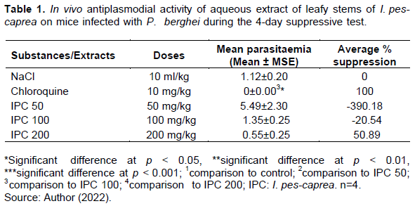
Table 1 shows the results obtained after the suppressive test in this study. It is noted that the aqueous extract of I. pes-caprea at 200 mg/kg resulted in a suppression of parasitaemia estimated at 50.89% while the other doses of extracts did not cause a decrease in parasitaemia. Chloroquine showed a 100% suppression of parasitaemia of mice.
Rane’s curative test
Following the administration of extracts of I. pes-caprea at 25 mg/kg, a significant increase in parasitaemia was noted from the 2nd to 3rd day of the experiment before being maintained in a slight decrease from the 4th day until the end of the experiment. With the extracts of I. pes-caprea at 200 mg/kg, a slight increase in parasitaemia was also noted from day 2 which lasted until day 3, then the parasitaemia decreased on day 4 and remained constant until the last day of the experiment. The parasitaemia of the batch that received chloroquine showed a clear and constant decrease from the 2nd day of treatment to reach zero values (parasitaemia = 0%) from the 4th day. As for the animals treated with artemether, the parasitaemia decreased sharply from the second day of the experiment, then the parasitaemia remained constant from the 3rd day with values close to 0.
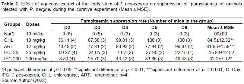
The percentages of suppression derived from the average parasitaemia values indicate that the treatment with plant extracts inhibited the advance of parasitaemia by -15.83 and 32.2% at the doses of 25 and 200 mg/kg, respectively (Table 2). Chloroquine treatment (10 mg/kg) resulted in 84.5% suppression and 81.95% for the lot treated with artemether (12 mg/kg).
Effect of extracts on body weight of mice
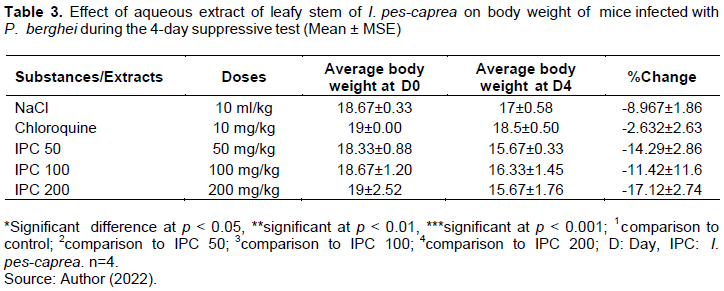
Percentage changes from D0 to D4 show that the 50, 100, and 200 mg/kg dose result in greater weight less than the control lot (p?0.05) (Table 3). The reference lot (chloroquine) showed less body weight change than the control (p?0.05).
Effect of extracts on mouse survival
Death was observed in all batches (Table 4). Death occurred more quickly (at the second day of the experiment) in batches treated with control and artemether. A high number of death was also observed with the batches that received artemether, the control, and the 25 mg/kg extracts. The 25 mg/kg dose had a lower survival rate than the control. While the survival rates of the batches that received the 200 mg/kg extracts and chloroquine were identical to each other and lower than the control and artemether.

DISCUSSION
The aim of this study was to evaluate the in vivo antiplasmodial activity of aerial parts of I. pes-caprea, a plant with multiple effects. This work is a preliminary study on I. pes-caprea and its originality lies in the fact that no experimental study has evaluated the in vivo anti-plasmodial activity of I. pes-caprea. Previous studies on the plant were limited to in vitro antiplasmodial activity and other activities such as antinociceptive, anti-inflammatory, antioxidant, antihistaminic, antibacterial, antifungal, anticancer, immunostimulant, hepatoprotective and antidiabetic.
The antimalarial effect of an aqueous extract of I. pes-caprea was evaluated on Swiss mice according to the 4-day suppressive test method and the curative test. In the three in vivo models used for the antiplasmodial chemotherapy study, the determination of the percentage of parasitaemia of suppression is the most reliable parameter. An antimalarial test compound is considered an active compound (possesses antiplasmodial activity) when it shows an average suppression of parasitemia greater than or equal to 30% in both suppressive and curative tests (Bantie et al., 2014). The results revealed an average inhibition of 50.89% of the parasitaemia of animals treated with the aqueous extract of I. pes-caprea at the dose of 200 mg/kg while the dose of 25 mg/kg did not result in a decrease in parasitaemia (Table 1). The 200 mg/kg dose showed a higher antiplasmodial effect to that of several medicinal plants known in Ivorian, Nigerian and Ethiopian traditional medicine.
Among other medicinal plants, there are the hydroethanol extract of Anthocleista djalonensis (35.91 to 200 mg/kg) from Ivory Coast (Attemene et al., 2018), the butanolic (36% at 240 mg/kg) and chloroformic (31% at 240 mg/kg) extracts of Alchornea cordifolia harvested in Nigeria (Nnamdi et al., 2017), the methanolic (37% at 200 mg/kg) and aqueous (11% at 200 mg/kg) extracts of Croton macrostachys (Bantie et al., 2014), the aqueous (29% at 200 mg/kg) and hydroethanolic (36% at 200 mg/kg) extracts of Strychnos mitis (Fentahun et al., 2017) and the methanolic extracts of Acanthus polystachyus (34% at 200 mg/kg) all collected in Ethiopia (Derebe and Wubetu, 2019).
The antiplasmodial effect of I. pes-caprea at 200 mg/kg is close to that of an ethanolic extract of Ajuga bracteosa (42.4% suppression at 250 mg/kg) harvested in India (Chandel and Bagai, 2010). The anti-plasmodial effect found in this study is however lower than that of the ethylacetic and N-hexane fractions of A. cordifolia (76 and 60.5% at 240 mg/kg) harvested in Nigeria (Nnamdi et al., 2017) and the hydroethanolic extract of Ziziphus mauritiana (58.68% at 200 mg/kg) harvested in Ivory Coast (Attemene et al., 2018).
The current antiplasmodial effect is corroborated by the results of an in vitro study conducted in India on different types of aqueous extracts of I. pes-caprea which showed an IC50 of 15 µg/mL of the methanolic root extracts (Pothula and Kanikaram, 2015).
The suppression of parasitaemia with the 200 mg/kg dose during the curative test was gradual before being maintained over time. From 8% at D2, it increased to 25% at D3 and then parasitaemia increased to 49% at D6 (Table 2). Since the rate of suppression of parasitaemia does not reach that of the reference substances (artemether and chloroquine), it could be assumed that I. pes-caprea at 200 mg/kg developed a parasitostatic or weakly schizontocidal effect (Fentahun et al., 2017).
With regard to the evolution of the weight of the mice during the suppressive test, there was no significant decrease in body weight of the mice given saline solution and those of all groups. Studies with other medicinal plants had shown that their administration in anti-plasmodial models significantly reduced the body weight of mice in control batches compared to chloroquine-treated batches (Chandel and Bagai, 2010; Kalra et al., 2006).
Animals given 25 mg/kg of the aqueous extract of I. pes-caprea showed a survival rate close to that of the control (Table 4); while at 200 mg/kg, the survival rate of the animals was higher than that of the control and even artemether group. This could reflect a decrease in the virulence of P. berghei in the presence of I. pes-caprea. The effect of the 200 mg/kg dose on animal survival is similar to that of other medicinal plants used in traditional medicine against malaria; that is, were methanolic extracts of A. cordifolia at 360 mg/kg and aqueous extracts of Tinospora baenzigeri at 500 mg/kg, which showed survival times similar to those of chloroquine (Nnamdi et al., 2017; Ounjaijean et al., 2019).
The parasitostatic mechanism reduces the adverse effects of parasite destruction on animal morbidity and mortality. Several studies conducted on medicinal plants had revealed that plant extracts had a weak schizontocidal effect as found with the 200 mg/kg dose of I. pes-caprea.
Doxycycline also has a parasitostatic effect on Plasmodium which is used in the management of malaria. Doxycycline is combined with quinine, artesunate or artemether to improve cure rates (Holmes and Charles, 2009). Indeed, drug combinations are generally made when a germ is left stationary by a first substance as is the case with doxycycline (Raoult et al., 1990). In future studies, I. pes-caprea could be evaluated in combination therapies.
The parasitostatic effect of doxycycline occurs at 100 mg. Also, the literature reports that its oral LD50 is 1870 mg/kg. However, the LD50 of I. pes-caprea (above 2000 mg/kg) is far from the parasitostatic dose (200 mg/kg). This makes I. pes-caprea safer to use than doxycycline.
These preliminary findings with 200 mg/kg support the continuation of the study with higher doses (400 and 600 mg/kg). The presence of active secondary metabolites such as saponins, polyphenols, flavonoids, and terpenoids may explain the antimalarial activity of the 200 mg/kg dose of plant extracts. According to Derebe and Wubetu, (2019), alkaloids, anthraquinones, saponins, phenols, and flavonoids would play an important role in the appearance of the antimalarial effect.
CONCLUSION
This study showed that the aqueous extract of I. pes-caprea has antimalarial properties at a dose of 200 mg/kg. This dose had no effect on the animals' survival and did not cause a decrease in body weight.
CONFLICT OF INTERESTS
The authors have not declared any conflict of interests.
ACKNOWLEDGEMENT
The authors thank the Institut Pasteur de Cocody (Abidjan) for making the Plasmodium berghei strain available for the study.
REFERENCES
|
Attemene SDD, Beourou S, Tuo K, Gnondjui AA, Konate A, Toure AO, Kati-coulibaly S, Djaman JA (2018). Antiplasmodial activity of two medicinal plants against clinical isolates of Plasmodium falciparum and Plasmodium berghei infected mice. Journal of Parasitic Diseases 42(1):68-76. |
|
|
Bantie L, Assefa S, Teklehaimanot T, Engidawork E (2014). In vivo antimalarial activity of the crude leaf extract and solvent fractions of Croton macrostachyus Hocsht. (Euphorbiaceae) against Plasmodium berghei in mice. BMC Complementary and Alternative Medicine 14(1):1-10. |
|
|
Chandel S, Bagai U (2010). Antiplasmodial activity of Ajuga bracteosa against Plasmodium berghei infected BALB/c mice. Indian Journal of Medical Research 131(3):440-444. |
|
|
Derebe D, Wubetu M (2019). Antimalarial Activity of Hydroalcoholic Root Extract of Acanthus polystachyus Delile (Acanthaceae) Against Plasmodium berghei-Infected Mice. Journal of Evidence-Based Integrative Medicine 24:1-6. |
|
|
Fentahun S, Makonnen E, Awas T, Giday M (2017). In vivo antimalarial activity of crude extracts and solvent fractions of leaves of Strychnos mitis in Plasmodium berghei infected mice. BMC Complementary and Alternative Medicine 17(13):1-12. |
|
|
Holmes NE, Charles PGP (2009). Safety and efficacy review of doxycycline. Clinical Medicine: Therapeutics 1:471-82. |
|
|
Kalra BS, Chawl S, Gupta P, Valecha N (2006). Screening of antimalarial drugs: an overview. Indian Journal and Pharmacology 38:5-12. |
|
|
Kaou AM, Mahiou-Leddet V, Hutter S, Aïnouddine S, Hassani S, Yahaya I, Azas N, Ollivier S (2008). Antimalarial activity of crude extracts from nine African medicinal plants. Journal of Ethnopharmacology 116(1):74-83. |
|
|
Meira M, Silva EP, David JM, David JP (2012). Review of the genus Ipomoea: traditional uses, chemistry and biological activities. Brazilian Journal of Pharmacognosy 22(3):682-713. |
|
|
Nnamdi A, Ettebong E, Davis K (2017). Antiplasmodial and antioxidant activities of methanolic leaf extract and fractions of Alchornea cordifolia 6(4):171-177. |
|
|
Noedl H, Se Y, Schaecher K, Smith BL, Socheat D, Fukuda MM (2008). Evidence of artemisinin-resistant malaria in western cambodia. New England Journal of Medicine 359(24):2619-2620. |
|
|
Ong'echa JM, Keller CC, Were T, Ouma C, Otieno RO, Landis-Lewis Z, Ochiel D, Slingluff JL, Mogere S, Ogonji GA, Orago AS, Vulule JM, Kaplan SS, Day RD, Perkins DJ (2006). Parasitaemia, anemia, and malarial anemia in infants and young children in a rural holoendemic Plasmodium falciparum transmission area. American Journal of Tropical Medicine and Hygiene 74(3):376-385. |
|
|
Ounjaijean S, Kotepui M, Somsak V (2019). Antimalarial Activity of Tinospora baenzigeri against Plasmodium berghei- Infected Mice. Journal of Tropical Medicine 2019:1-6. |
|
|
Peters W, Portus JH, Robinson BL (1975). The chemotherapy of rodent malaria, XXII. Annals of Tropical Medicine & Parasitology 69(2):155-171. |
|
|
Pothula VVS, Kanikaram S (2015). In vitro antiplasmodial efficacy of mangrove plant, Ipomoea pes-caprae against Plasmodium falciparum (3D7 strain). Asian Pacific Journal of Tropical Disease 5(12):947-956. |
|
|
Raoult D, Drancourt M, Vestris G (1990). Bactericidal Effect of Doxycycline Associated with Lysosomotropic Agents on Coxiella burnetii in P388D1 Cells. Antimicrobial Agents aand Chemotherapy 34(8):1512-1514. |
|
|
Regional Committee for Africa (2000). Promoting the role of traditional medicine in health systems. Strategy for the African Region 2. |
|
|
Ryley JF, Peters W (1970). The antimalarial activity of some quinolone esters. Annals of Tropical Medicine & Parasitolog, 64(2):209-222. |
|
|
Visht, S., Chaturvedi S (2012). Isolation of natural products. Journal of Current Pharma Research 2(3):584-599. |
|
|
Vitoria M, Granich R, Gilks CF, Gunneberg C, Hosseini M, Were W, Raviglione M, De Cock KM (2009). The global fight against HIV / AIDS, tuberculosis, and malaria current status and future perspectives. American Journal of Clinical Pathology 131(6):844-848. |
|
|
Wolfle TL (1996). Institute of Laboratory Animal Resources (ILAR). Guide for the Care and Use of Laboratory Animals. National Academy Press Washington 7:287. |
|
|
World Health Organization (WHO) (2007). Report of the WHO Interregional Workshop on the Use of Traditional Medicine in Primary Health Care. WHO96 p. |
|
|
World Health Organization (WHO) (2016). Giemsa staining of blood smears for plasmodium. WHO: 6 p. |
|
Copyright © 2024 Author(s) retain the copyright of this article.
This article is published under the terms of the Creative Commons Attribution License 4.0


