Full Length Research Paper
ABSTRACT
Zanthoxylum zanthoxyloides has been used for decades in traditional medicine across West African countries to treat several ailments such as malaria and pain. However, sometimes it is used without information on its toxicity level. This present work aimed to explore the toxicity level of Zanthoxylum zanthoxyloides bark aqueous extract (AZZ) on albino Wistar rats after single and repeated administrations. For the acute toxicity evaluation, a single dose of the extract at 3, 5 and 7.5 g/kg of the extract was administered orally, and the animals were kept under observation for mortality and clinical signs for the first four hours, and then once every day for 14 days. For the sub-acute toxicity evaluation, the extract was administered to the animals orally at doses of 0.5, 0.75 and 1 g/kg daily for 28 days. On the 15th day (acute evaluation) or 29th day (sub-acute evaluation), the animals were weighed and anaesthetized and then blood was collected for haematological and biochemical analysis. Vital organs (liver, kidneys and lungs) were removed and weighed. No mortality or toxicological behavior was observed, and there was no statistically significant difference between the treated and control groups for whole body weight and organ weights. Also, the results showed no statistically significant effects on haematological and biochemical parameters. Thus, AZZ may be considered as nontoxic both after single and repeated administrations.
Key words: Zanthoxylum zanthoxyloides, bark extract, toxicity level, clinical signs, Wistar rats, hematology, biochemical parameters.
INTRODUCTION
Zanthoxylum zanthoxyloides (Lam.) is distributed in the Savannah of West Africa and the Coastal areas from Senegal to Cameroon. It is known all over Africa for various traditional medicinal uses. In Togo, the stem and stem bark are traditionally used for malaria treatment (Denou et al., 2016; Koudouvo et al., 2016), and the root bark macerate is used to treat central nervous system disorders such as epilepsy and paralysis (Kantati et al., 2016). Many studies have been done on some parts of this plant to elucidate its biological and physiological activities. Studies were conducted for its antibacterial (Kamdem et al., 2015; Wouatsa et al., 2013; Gardini et al., 2009; Ngassoum et al., 2003), antimalarial (Gansané et al., 2010; Kassim et al., 2005; Nyangulu et al., 2005), antioxidant and anti-inflammatory (Larsen et al., 2015; Diatta et al., 2014; Chaaib et al., 2003), analgesic and anthelmintic (Azando et al., 2017; Olounladé et al., 2012; Prempeh and Mensah-Attipoé, 2008) activities. Ogwal-Okeng et al. (2003) evaluated the acute toxicity of Z. zanthoxyloides root bark methanolic extract and found that the extract was safe up to a dose of 5 g/kg.
Since earliest time, plants have been used throughout populations worldwide as a source of natural products in traditional medicine, also called “non-conventional medicine” (Dosseh et al., 2015). Medicinal plant use in therapeutics has increased significantly in the last few decades due to an increased interest in natural substances (Lee et al., 2012; Castro et al., 2009). According to the World Health Organisation (WHO) in developing countries, medicinal plants occupy an important and significant place in primary health care for more than 80% of the population living in those countries (Nath et al., 2011). Although traditional herbal medicine may be considered safe, some products might cause side effects at high doses after single administration, and others may have potential side effects after repeat administration. Many reports about the safety of the herbs used in traditional medicine are often related to hepatotoxicity and nephrotoxicity (Dosseh et al., 2015; Debelle et al., 2008; Tédong et al., 2007). This suggests the need for traditional practitioners to keep abreast of the reported incidence of renal and hepatic toxicity and other possible side effects caused by the ingestion of traditional medicinal products. Hence, even if the medicinal plants have been in use for centuries, toxicological evaluation of those medicinal plants is required.
This present study aimed to investigate the toxicity level of Z. zanthoxyloides aqueous bark extract after single and repeated (28 days) oral administrations on albino Wistar rats.
MATERIALS AND METHODS
Plant material
Z. zanthoxyloides bark was collected in the northern part of Togo (Pya) in February 2019. A botanist from the Botany and Vegetal Ecology Department authenticated the plant, and a voucher specimen has been deposited in the national herbarium of the department (reference number Togo15491).
Experimental animals
University of Jos Animal House Unit provided male Wistar rats weighing between 150 and 200 g. The animals were kept under ambient temperature, with 12 h light and 12 h dark cycle and had free access to food and water. All animal procedures were performed in accordance with the recommendations of proper care and use of laboratory animals after approval (No. UJ/FPS/F17-00379) from University of Jos (Nigeria) Ethics Committee. Before each experiment, the animals were fasted overnight with liberal access to water.
Preparation of crude extract
The plant material was air-dried and put into powder in Biochemistry Department using a mortar. The powder (500 g) was macerated with 1.5 L of distilled water for 24 h at room temperature, and the mixture was filtered and kept for concentration in the oven. The same procedure was repeated twice with the substrate. The whole filtrate from the three days maceration was concentrated to dryness in an oven at 70°C, which yielded a residue of 6.6%. The dry extract (brown, crystallized) was stored at -4ºC.
Determination of the lethal dose 50 (LD50)
The LD50 determination process was two phases of 24 h each interconnected as described by Lorke (1983).
Phase one
This phase required nine animals divided into three groups of three animals each. Each group of animals were administered the extract at varying doses - 10, 100 and 1000 mg/kg. The animals were placed under observation for 24 h to monitor their behaviour as well as mortality.
Phase two
This phase involves the use of three animals, which were distributed into three groups of one animal each. The animals were administered higher doses - 1600, 2900 and 5000 mg/kg of the extract and then observed for 24 h for behaviour as well as mortality.
Acute toxicity evaluation
Oral acute toxicity evaluation was carried out using single test dose method (Dosseh et al., 2015; Bakoma et al., 2013; Tédong et al., 2007). Five groups of five rats each were used in this evaluation. The study was performed by administering the extract at varying doses- 3, 5 and 7.5 g/kg body weight, orally. The fourth and fifth groups were designated as positive and negative controls and given 5 mL/kg of diluted dimethyl sulfoxide (DMSO) 5% solution and distilled water, respectively. The animals were individually observed for immediate signs of toxicity and mortality for a period of 4 h, and at least once daily for 14 days for delayed mortality and toxicity symptoms such as changes in the skin and fur, eyes, mucous membranes, salivation, diarrhoea, convulsion, lethargy and coma. On the 15th day after administration, animals were weighed and blood samples were collected under ethyl ether anaesthesia by retro-orbital bleeding into tubes with and without EDTA for haematological and biochemical analysis, respectively. White blood cell count (WBC), red blood cell count (RBC), platelet count (PLT), haemoglobin (HGB), haematocrit (HCT), mean corpuscular volume (MCV), and mean corpuscular haemoglobin concentration (MCHC) were determined using an automated haematology analyser (BC-5300, Mindray, China). Biochemical analysis was performed using the serum obtained after centrifugation of total blood without anticoagulant for 15 min at 2,500 rpm. An automated spectrophotometer (Cobas c 111, India) was used to determine the following parameters: Alkaline phosphatase (ALP), aspartate aminotransferase (AST), alanine aminotransferase (ALT), creatinine (CRE), and urea (URE). Vital organs such as lungs, liver and kidneys were carefully removed, grossly examined and weighed to determine their relative weights as [(organ weight/total body weight) ×100].
Sub-acute toxicity evaluation
A repeated dose oral toxicity evaluation was carried out according to Bakoma et al. (2013) and Dosseh et al. (2015). Twenty male rats were randomly divided into four groups of five animals each and treated orally. The first group was taken as control and received normal saline (0.9% NaCl). The three other groups were the treatment groups and received varying doses of the extract - 500, 750, and 1000 mg/kg body weight, respectively. The administration was done each day for 28 days. The rats were observed at least once a day for mortality or morbidity, changes in posture, skin, eyes, fur, mucous membranes and behaviour. The body weights of the rats were evaluated every 7 days. On the 29th day, blood samples were collected from overnight-fasted rats under ether anaesthesia by retro-orbital bleeding into tubes with and without EDTA for haematological and biochemical analysis, respectively. The same parameters above in acute evaluation were determined. Animals were then sacrificed by cervical dislocation and internal organs, including liver kidneys, and lungs, were carefully removed. These organs were then weighed to determine their relative weights. The liver and kidneys were conserved in 10% formalin for histopathological analysis using the method described by Choji et al. (2015) and Avwioro (2011).
Organs were harvested and fixed in 10% formalin for three days, cut into thin slices of 5 mm × 2 mm × 1 mm thick, and then placed in running tap water for five minutes. The tissues were then dehydrated by passing through ascending grades of ethanol as follows: 70, 80, 90, 95%, three changes of absolute ethanol, cleared in two changes of xylene, and infiltrated in two changes of paraffin wax oven placed at 4?C. The SPIN tissue processor, STP 120 (Thermo Scientific), was used and the tissues were subjected to each stage for two hours, making a total of twenty-two hours. The tissues were embedded using embedding cassettes on a tissue Tek Embedding Centre (SLEE MPS/P2) and cooled rapidly on the cooling component. Tissues were then sectioned using a rotary microtome (MICROM HM340E Thermo Scientific) set at 4 micromes, picked on slides, and ready for staining. Sections were dewaxed and hydrated by passing through two changes of xylene and descending grades of alcohol (100, 80 and 70%) for three minutes each and then into water, stained in Harris’ haematoxylin solution for 5 min, and washed in running water. Sections were differentiated in 1% acid alcohol and then washed well in water, blued in Scott’s tap water substitute for 5 min and rinsed briefly in distilled water, counterstained in 1% aqueous eosin for two min, washed well in water, dehydrated in descending grades of alcohol, cleared in xylene and mounted in DPX (Destrene, Plasticiser and Xylene). The sections were placed in slide carriers and placed in a 40?C oven to dry overnight, then read microscopically.
Statistical analysis
The results are presented as mean ± standard error of the mean (SEM). Statistical analysis was performed by one-way analysis of variance (ANOVA) followed by Tukey’s multiple comparison test to evaluate significant differences between groups. Mean differences with p values less than 0.05 w considered statistically significant. All statistical analyses were carried out using Graph Pad prism 8.0.1 Software.
RESULTS
Lethal dose 50 (LD50):
The calculation of the LD50 was carried out in two phases successively. No rat died during the first and second phases. This suggests that the LD50 is above 5,000 mg/kg body weight.
Acute oral toxicity study
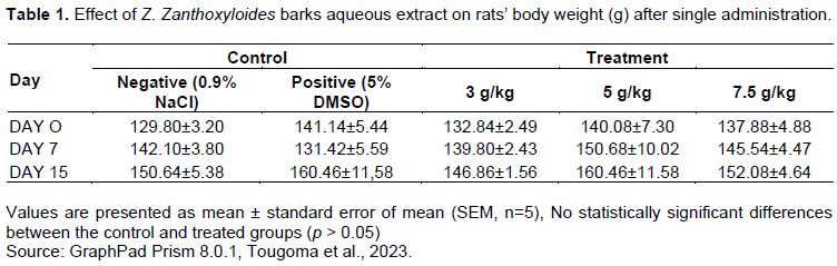
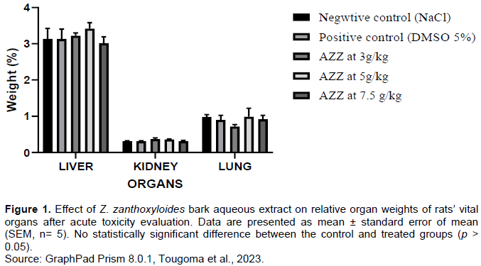
The single administration of the extract at different doses did not cause any mortality or clinical signs of toxicity. The mean body and relevant organ weights are recorded in Table 1 and Figure 1 respectively. No statistically significant differences were observed between control and extract treated groups either for the body weight or relative organ weight (p > 0.05).
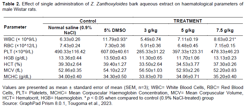
Haematological and biochemical analysis results are recorded in Tables 2 and 3. There were no statistically significant differences (p > 0.05) between control and extract-treated groups for all biochemical parameters analysed. For the haematological parameters, there was no statistically significant difference between control and treated groups except for white blood cell (WBC) count at 7.5 g/kg, where the result showed a statistically significant augmentation (p < 0.05) in the number of white blood cells when compared to the control.

Sub-acute toxicity
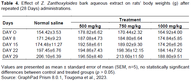
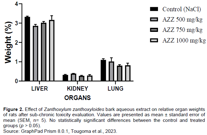
Animals were treated orally and repeatedly once daily for 28 days with AZZ (500, 750 and 1000 mg/kg) and normal saline (as control). No mortality or signs of toxicity were recorded after extract administration. The mean body and relative organ weights are recorded in Table 4 and Figure 2, respectively. There was no statistically significant difference (p > 0.05) between control and treated groups either in the body weight or relative organs weights.
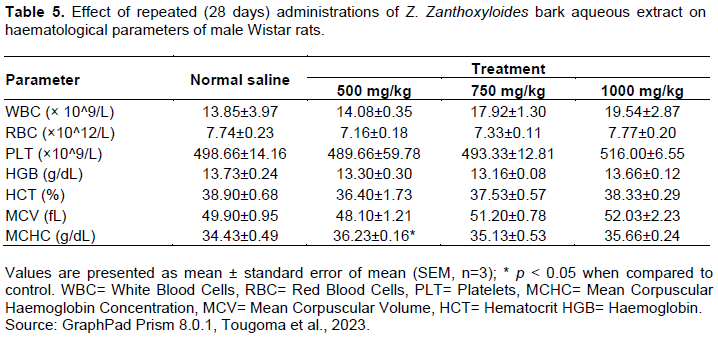
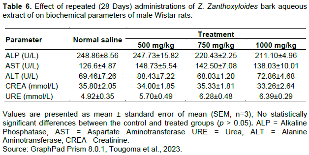
The haematological and biochemical parameters results are recorded in Tables 5 and 6 respectively. When MCHC values were compared, there was statistically significant difference between 500 mg/kg-treated group and control (p < 0.05). No statistically significant differences in other haematological parameters between the other treated groups and control (p > 0.05). All biochemical parameters analysed showed no statistically significant differences between control and treated groups (p > 0.05).
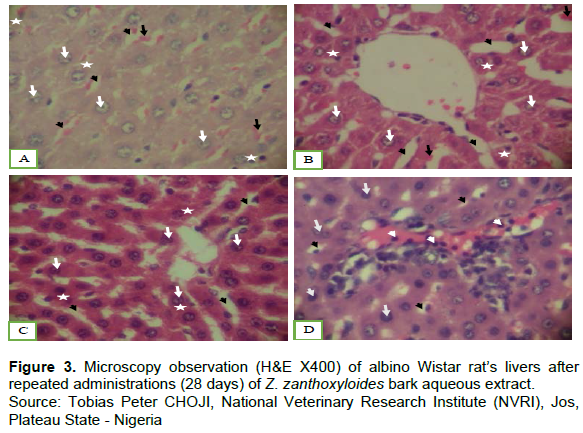
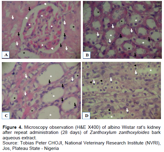
Microscopy observation of the liver in all groups showed in general normal morphology of the hepatocytes. For all groups, the nuclei and the cytoplasm were intact with normal aspect. There was presence of hepatic sinusoids containing in some places red blood cells. In the fourth group (AZZ at 1000 mg/kg), there was presence of inflammatory cells within hepatocytes (Figure 3). Microscopy observation of the kidney in all groups showed normal morphology of renal cells with intact nuclei surrounded by intact cytoplasmic components. The glomeruli had normal appearance and surrounded by clear zone of capsular space. However, in the fourth group (AZZ at 1000 mg/kg) there was degeneration of the tubule and nuclear hypertrophy (Figure 4). The figure showed normal morphology as demonstrated by intact nuclei (white arrows) surrounded by intact cytoplasmic components (white stars). The hepatocytes are interspersed by hepatic sinusoids (black arrowheads) some of which contain red blood cells (black arrows) and inflammation of the hepatocytes evident by the presence of inflammatory cells (mononuclear cells) (white arrowheads) (A: Normal saline group; B: AZZ at 500 mg/kg; C: AZZ at 750 mg/kg; D: AZZ at 1000 mg/kg). The figure showed normal morphology as demonstrated by cells presented with intact nuclei (white arrows) surrounded by intact cytoplasmic components. The glomeruli (white stars) appear normal and were surrounded by a clear zone of capsular space (white arrowheads). The glomerular capsules (black arrows) present their characteristic simple squamous epithelial cells. Black star = urinary pole. D is showing tubular degeneration evident by perinuclear vasculation and nuclear hypertrophy as shown by vacuoles (Black arrowheads) surrounding the nuclei (white arrows) (A: Normal saline group; B: AZZ at 500 mg/kg; C: AZZ at 750 mg/kg; D: AZZ at 1000 mg/kg).
DISCUSSION
Natural product from medicinal plants has gained an important interest in recent times and the population in developing countries uses many of these plant products for their primary health care. Such remedies are often believed to be harmless, since these treatments are natural and commonly used for self-medication without supervision. However, the use of these plant products may expose the population to toxicological risks. There is little scientific data on the safety of Z. zanthoxyloides bark. In fact, several investigations on Z. zanthoxyloides root and leaves have reported that the plant has various pharmacological proprieties (Diatta et al., 2014; Wouatsa et al., 2013; Zahoui et al., 2010; Ouattara et al., 2009; Chaaib et al., 2003) but few works have been done on its bark, especially on the toxicity level. It was necessary to evaluate the toxicity of the bark of the plant for human use. So, this present work aimed to investigate the toxicity levels in single and repeated administration of Z. zanthoxyloides bark aqueous extract.
The LD50 toxicity categories modified from the probable lethal dose for humans using oral treatment in the rat for comparison were defined as follows: Less than 5 mg/kg, extremely toxic; between 5-50 mg/kg, highly toxic; between 50-500 mg/kg, moderately toxic; between 500-5000 mg/kg, slightly toxic; greater than 5000 mg/kg, practically non-toxic (Ghost, 1984). After administration of the high dose (5000 mg/kg), there was no mortality. The extract is considered as practically non-toxic with the LD50 greater than 5000 mg/kg. This finding is in agreement with that of Ogwal-Okeng et al. (2003), who found that the methanolic extract of the root-bark of Z. zanthoxyloides was slightly toxic with the LD50 = 5000 mg/kg body weight. Also, Yadav and Tangpu (2009) in their work on Zanthoxylum rhesta (DC), another subspecies of Zanthoxylum used in traditional medicine in India, found that the LD50 of the leaf methanolic extract when given orally was 2737,34 g/kg body weight and considered as slightly toxic.
Generally, reduction in body weight and internal organ weights are sensitive indices of toxicity after exposure to toxic substances (Dosseh et al., 2015; Ouedraogo et al., 2013; Teo et al., 2002). Index to diagnose whether an organ has been affected after exposure to injury is to calculate the organ to body weight ratio (Bakoma et al., 2013; Rosidah et al., 2009). Hence, if rats are exposed to toxic substances, the weights of the damaged organ will either increase or decrease, as will the organ to body weight ratio. During this study, single and repeated administrations of AZZ did not affect either whole body weight or the relative organ weights of specific vital organs (liver, kidneys and lungs).
The liver and kidneys are vital organs in the body and their functions are very important for the organism. Any dysfunction of these two organs will affect the functioning of the entire body. Haematological parameters can be influenced by toxicity of extracts administered, as these extracts have ability to initiate the acute phase response (Veerappan et al., 2007). Alkaline phosphatase (ALP), aspartate aminotransferase (ASP), alanine aminotransferase (ALT), creatinine (CREA) and Urea (URE) levels in the blood are markers for liver and kidney function (Liaqat et al., 2018; Costa-Silva et al., 2008; Rebecca et al., 2002). According to Abarikwu et al. (2018), anytime the creatinine level reduced and urea level increases, there is no kidney damage but Nisha et al. (2017) reported that renal toxicity should be considered only when creatinine and urea level increased parallel to each other. Single administration at doses of 3, 5 and 7.5 g/kg body weight and repeated administrations (28 days) at doses of 500, 750 and 1000 mg/kg body weight of the extract did not significantly affect the haematological and biochemical parameters except the significant increase of MCHC at 500 mg/kg. These data show that Z. zanthoxyloides bark aqueous extract did not practically induce damage to the liver or kidneys. The effect on MCHC could be due to the agglutination of red blood cell (RBC) or to hyperchromic anaemia (abnormal shaped RBC). The difference between the results of works using the same plant can be explained by the part and the type of extracts used. In fact, to confirm the toxic nature of any plant product, it is good to consider several factors that can alter its toxicity profile, including the growth stage, the maturity and the specific part (s) used of the plant; the storage conditions, and the seasonal variation in the relative abundance of phytochemicals (El Halaly et al., 2004). Gbate et al. (2021) showed that there were no significant changes in AST, ALT, bilirubin, creatinine and urea level when Z. zanthoxyloides root bark was used to formulate albino rats feed. Liaqat et al. (2018) during their work using leaves and stem essential oils of Zanthoxylum armatum (Rutaceae) did not find any changes in total erythrocyte count (TEC), total leukocyte count (TLC), packet cell volume (PCV), mean corpuscular volume (MCV) and mean corpuscular haemoglobin concentration (MCHC) levels when compared to the control. No significant depletion of all liver and kidney injury markers level was observed. Histopathology analyses of our study show inflammation of the liver and kidneys at the dose of 1000 mg/kg body weight, while at doses of 500 and 750 mg/kg body weight the slides show normal cells (Figures 3 and 4). Excessive administration of xenobiotics induces immunologic reactions including cell dysfunction, cells degeneration, and natural and adaptive immune response (Alayunt et al., 2019). Inflammation of cells may be due to production of proinflammatory cytokines, especially TNF-α, IL-1β, IL-6 mainly produced by macrophages and monocytes. This is the primary mediator in systemic toxicity of liver and kidney damage (Bonzo et al., 2015, Ganey and Roth, 2001). According to Hammam et al. (2013) and Amdur et al. (2016), the increase of TNF-α levels play an important role in liver and kidney damage. The repeated administration of the extract at the dose of 1000 mg/kg body weight may lead to cytokines production and cause liver and kidney inflammation. This inflammation at 1000 mg/kg did not significantly affect liver and kidney haematological and biochemical parameters as no changes were observed (Tables 5 and 6).
CONCLUSION
It can be concluded from the above results that acute (3000, 5000 and 7500 mg/kg BW) and sub-chronic (500, 750 mg/kg BW) administration of Z. zanthoxyloides aqueous stem bark extract can be considered safe due to its ability in not causing death nor producing any toxicity signs or evident symptoms after single and repeated administrations. Hence, Z. zanthoxyloides bark may be exploited as a medicinal product in treatment of diseases in humans at reasonable doses. However, more investigations are needed before its use for therapeutic purposes.
CONFLICT OF INTERESTS
Authors declare no conflict of interest.
REFERENCES
|
Abarikwu SO, Njoku RC, Onuah CL (2018). Aged coconut oil with a high peroxide value induces oxidative stress and tissue damage in mercury-treated rats. Journal Basic and Clinical Physiology and Pharmacology 24(4):1-12. |
|
|
Alayunt ÖN, Aksoy L, Karafakioglu YS, Sevimli S (2019). Assessment of anti-inflammatory and antioxidant properties of Safranal on CCl4-induced oxidative stress and inflammation in rats. The Annals of the Brazilian Academy of Sciences 91(2):1-11. |
|
|
Amdur RL, Feldman HI, Gupta J, Yang W, Kanetsky P, Shlipak M, Rahman M, Lash JP, Townsend RR, Ojo A, Roy-Chaudhury A, Go AS, Joffe K, He J, Balakrishnan VS, Kimmel PL, Kusek JW, Raj DS (2016). Inflammation and progression of CKD: The CRIC Study. Clinical Journal of the American Society of Nephrology 11(9):1546-1556. |
|
|
Avwioro G (2011). Histochemical uses of haematoxylin - a review. Journal of Physics Conference Series 1(5):24-34. |
|
|
Azando EVB, Olounlade AP, Hounzangbe-Adote MS, Tam Ha TB, Fabre N, Valentin A (2017). Control of gastro-intestinal parasites with essential oil from Zanthoxylum zanthoxyloides (Fagara zanthoxyloides). Revue de Medecine Veterinaire 168(10-12):205-212. |
|
|
Bakoma B, Berké B, Eklu-Gadegbeku K, Agbonon A, Aklikokou K, Gbeassor M, Creppy EE, Moore N (2013). Acute and sub-chronic (28days) oral toxicity evaluation of hydroethanolic extract of Bridelia ferruginea Benth root bark in male rodent animals. Food and Chemical Toxicology 52(1):176-179. |
|
|
Bonzo JA, Kelly R, Freeman K, Deibert E, Amaral KB, Ferguson SS, Andersen ME, Witek RP, Lecluyse EL (2015). Differential effects of Trovafloxacin on TNF-α and IL-6 profiles in a rat hepatocyte-Kupffer Cell Coculture System. Applied In Vitro Toxicology 1(1):45-54. |
|
|
Castro LS, Perazzo FF, Maistro EL (2009). Genotoxicity testing of Ambelania Occidentalis (Apocynaceae) leaf extract in vivo. Genetics and Molecular Research 8(2):440-447. |
|
|
Chaaib F, Queiroz EF, Ndjoko K, Diallo D, Hostettmann K (2003). Antifungal and Antioxidant Compounds from the Root Bark of Fagara zanthoxyloides. Planta Medica 69(4):316-320. |
|
|
Choji TP, Ogenyi SI, Ngokere AA, Chollom SC, Jugu, KP, Lokason SD (2015). Evaluation of the conventional versus two rapid microwave processing techniques using the masson trichrome histochemical methtod. Journal of Advances in Biology and Biotechnology 4(3):1-15. |
|
|
Costa-Silva JH, Lima CR, Silva EJR., Araujo AV, Fraga MCCA, Ribeiro e Ribeiro A, Arruda AC, Lafayette SSL, Wanderley AG (2008). Acute and subacute toxicity of the Carapa guianensis Aublet (Meliaceae) seed oil. Journal of Ethnopharmacology 116(3):495-500. |
|
|
Debelle FD, Vanherweghem J, Nortier JL (2008). Aristolochic acid nephropathy?: A worldwide problem. Kidney International 74(2):158-169. |
|
|
Denou A, Koudouvo K, Togola A, Aziati KY, Esseh J, Ajavon CA, Essien K, Aklikokou K, Sanogo R, Diallo D, Gbeassor M (2016). Traditional knowledge on antimalarial plants having analgesic properties, used in Togo Maritime Region. Journal of Ethnobiology and Traditional Medecine 126(1):1160-1170. |
|
|
Diatta W, Sy G, Manga C, Diatta K, Fall A, Bassene E (2014). Research of anti-inflammatory and analgesic activities of extracts of leaves of Zanthoxylum zanthoxyloides (Lam) zepernick and timber (Rutaceae). International Journal of Biological and Chemical Sciences 8(1):128-133. |
|
|
Dosseh K, Amegnona A, Messanvi G (2015). Antioxidant and toxicological studies of ethanolic root extract of Byrsocarpus coccineus. Journal of Medicinal Plants Research 9(36):940-949. |
|
|
El Halaly J, Israili ZH, Lyoussi B (2004). Acute and chronic toxicological studies of Ajuga iva in experimental animals. Journal of Ethnopharmacology 91(1):43-50. |
|
|
Ganey PE, Roth RA (2001). Concurrent inflammation as a determinant of susceptibility to toxicity from xenobiotic agents. Toxicology 169(3):195-208. |
|
|
Gansané A, Sanon S, Ouattara LP, Traoré A, Hutter S, Ollivier E, Azas N, Traore AS, Guissou IP, Sirima SB, Nebié I (2010). Antiplasmodial activity and Cytotoxicity of semi purified fractions from Zanthoxylum zanthoxyloides Lam. Bark of Trunk. International Journal of Pharmacology 6(6):921-925. |
|
|
Gardini F, Belletti N, Ndagijimana M, Guerzoni ME, Tchoumbougnang F, Zollo PHA, Micci C, Lanciotti R, Kamdem SLS (2009). Composition of four essential oils obtained from plants from Cameroon, and their bactericidal and bacteriostatic activity against Listeria monocytogenes, Salmonella enteritidis and Staphylococcus aureus. African Journal of Microbiology Research 3(5):264-271. |
|
|
Gbate M, Ashamo OM, Kayode AL (2021). Toxicological evaluation of Zanthoxylum zanthoxyloides (Lam) Zepernick & Timler root bark used as biopesticide and medicine. Journal of Medicinal Plants Research 15(2):10-117. |
|
|
Ghost MN (1984). Fundamentals of Experimental Pharmacology. 2nd ed. Kolkata (India): Calcutta, Scientific Book Agency. |
|
|
Hammam O, Mahmoud O, Zahran M, Sayed A, Salama R, Hosny K, Farghly A (2013). A possible role for TNF-α in coordinating inflammation and angiogenesis in chronic liver disease and hepatocellular carcinoma. Gastrointestinal Cancer Research 6(4):107-114. |
|
|
Kamdem SLS, Belletti N, Tchoumbougnang F, Essia-Ngang JJ, Montanari C, Tabanelli G, Lanciotti R, Gardini F (2015). Effect of mild heat treatments on the antimicrobial activity of essential oils of Curcuma longa, Xylopia aethiopica, Zanthoxylum xanthoxyloides and Zanthoxylum leprieurii against Salmonella enteritidis. Journal of Essential Oil Research 27(1):52-60. |
|
|
Kantati YT, Kodjo KM, Dogbeavou KS, Vaudry D, Leprince J, Gbeassor M (2016). Ethnopharmacological survey of plant species used in folk medicine against central nervous system disorders in Togo. Journal of Ethnopharmacology 181(1):214-220 |
|
|
Kassim OO, Loyevsky M, Elliott B, Geall A, Amonoo H, Gordeuk VR (2005). Effects of root extracts of Fagara zanthoxyloides on the in vitro growth and stage distribution of Plasmodium falciparum. Antimicrobial Agents and Chemotherapy 49(1):264-268. |
|
|
Koudouvo K, Esseh K, Denou A, Aziati K, Ajavon C, Afanyibo YG, Agbonon A, Sanogo R, Dougnon J, Aklikokou K, Aguiyi JC, Diallo D, Mensah GA, Gbeassor M (2016). Ethno-pharmacological study of antimalarial medicinal recipes from Togo for the formulation of a phytomedicine for the management of malaria. Bulletin de La Recherche Agronomique Du Benin 79:54-70. |
|
|
Larsen BHV, Soelberg J, Jäger AK (2015). COX-1 inhibitory effect of medicinal plants of Ghana. South African Journal of Botany 99(1):129-131. |
|
|
Lee M, Shin I, Seo C, Kim J, Han S, Shin H (2012). Subchronic oral toxicity studies of the traditional herbal formula Bangpungtongseong-san in Crl?: CD ( SD ) rats. Journal of Ethnopharmacology 144(3):720-725. |
|
|
Liaqat I, Riaz N, Saleem QA, Tahir HM, Arshad M, Tahir HM, Arshad N (2018). Toxicological Evaluation of Essential Oils from Some Plants of Rutaceae Family. Evidence-Based Complementary and Alternative Medicine 2018:4394687. |
|
|
Lorke D (1983). A New Approach to Practical Acute Toxicity Testing. Archives Toxicology 54(1):275-287. |
|
|
Nath S, Choudhury MD, Roychoudhury S, Talukdar AD, Sirotkin AV, Baková Z, Kadasi A, Maruniakova N, Kolesárová A (2011). Restorative aspect of castor plant on mammalian physiology. Journal of Microbiology Biotechnology and Food Sciences 1(2):236-246. |
|
|
Ngassoum MB, Essia-Ngang JJ, Tatsadjieu LN, Jirovetz L, Buchbauer G, Adjoudji O (2003). Antimicrobial study of essential oils of Ocimum gratissimum leaves and Zanthoxylum xanthoxyloides fruits from Cameroon. Fitoterapia 74(3):284-287. |
|
|
Nisha R, Srinivasa Kannan SR, Thanga Mariappan K, & Jagatha P (2017). Biochemical evaluation of creatinine and urea in patients with renal failure undergoing hemodialysis. Journal Clinical Pathology and Laboratory Medicine 1(2):1-5. |
|
|
Nyangulu JM, Hargreaves SL, Sharples SL, Mackay SP, Waigh RD, Duval O, Mberu EK, Watkins WM (2005). Antimalarial benzo[c] phenanthridines. Bioorganic and Medicinal Chemestry 15(8):2007-2010. |
|
|
Ogwal-okeng JW, Obua C, Anokbonggo WW (2003). Acute toxicity effects of the methanolic extract of Fagara zanthoxyloides (Lam.) root-bark. African Heath Sciences 3(3):124-126. |
|
|
Olounladé PA, Azando EVB, Hounzangbé-Adoté MS, Ha TBT, Leroy E, Moulis C, Fabre N, Magnaval JF, Hoste H, Valentin A (2012). In vitro anthelmintic activity of the essential oils of Zanthoxylum zanthoxyloides and Newbouldia laevis against Strongyloides ratti. Parasitology Research 110(4):1427-1433. |
|
|
Ouattara B, Jansen O, Angenot L, Guissou I P, Frederich M, Fondu P, Tits M (2009). Antisickling properties of divanilloylquinic acids isolated from Fagara zanthoxyloides Lam. (Rutaceae). Phytomedicine 16(2-3):125-129. |
|
|
Ouedraogo GG, Ouedraogo M, Lamien-sanou A, Lompo M, Olga M, Guissou PI (2013). Acute and Subchronic Toxicity Studies of Roots Barks Extracts of Calotropis procera ( Ait .) R . Br Used in the Treatment of Sickle Cell Disease in Burkina Faso. British Journal of Pharmacology and Toxicology 4(5):194-200. |
|
|
Prempeh ABA, Mensah-Attipoe J (2008). Analgesic Activity of Crude Aqueous Extract of the root bark of Zanthoxylum zanthoxyloides. Ghana Medical Journal 42(2):79-83. |
|
|
Rebecca MA, Ishii-Iwamoto E L, Grespan R, Cuman RKN, Caparroz-Assef SM, de Mello JCP, Bersani-Amado CA (2002). Toxicological studies on Stryphnodendron adstringens. Journal of Ethnopharmacology 83(1-2):101-104. |
|
|
Rosidah MFY, Sadikun A, Ahmad M, Akowuah AG, Asmawi ZM (2009). Toxicology evaluation of standardized methanol extract of Gynura procumbens. Journal of Ethnopharmacology 123(2):244-249. |
|
|
Tédong L, Dzeufiet PDD, Dimo T, Asongalem EA, Sokeng SN, Flejou JF, Callard P, Kamtchouing P (2007). Acute and subchronic toxicity of Anacardium occidentale Linn (Anacardiaceae) leaves hexane extract in mice. African Journal of Traditional, Complementary and Alternative Medicines 4(2):140-147. |
|
|
Teo S, Stirling D, Thomas S, Hoberman A, Kiorpes A, Khetani V (2002). A 90-day oral gavage toxicity study of D -methylphenidate and D , L -methylphenidate in Sprague - Dawley rats. Toxicology 179:183-196. |
|
|
Veerappan A, Miyazaki S, Kadarkaraisamy M, Ranganathan D (2007). Acute and subacute toxicity studies of Aegle marmelos Corr., an Indian medicinal plant. Phytomedicine 14:209-215. |
|
|
Wouatsa VNA, Misra L, Kumar S, Prakash O, Khan F, Tchoumbougnang F (2013). Aromatase and glycosyl transferase inhibiting acridone alkaloids from fruits of Cameroonian Zanthoxylum species. Chemistry Central Journal 7(1):125-140. |
|
|
Yadav AK, Tangpu V (2009). Therapeutic efficacy of Zanthoxylum rhetsa DC extract against experimental hymenolepis diminuta (Cestoda) infections in rats. Journal of Parasitic Diseases 33(1-2):42-47. |
|
|
Zahoui SO, Zirihi NG, Soro YT, Traore F (2010). Hypotensive effect of an aqueous extract of Zanthoxylum zanthoxyloides (Lam.) Waterman (Rutaceae). Phytotherapie 8(6):359-369. |
|
Copyright © 2024 Author(s) retain the copyright of this article.
This article is published under the terms of the Creative Commons Attribution License 4.0


