Full Length Research Paper
ABSTRACT
The present investigation was to examine the in vivo antioxidant activity and lipid peroxidation activities of different extracts of aerial parts of Chomelia asiatica (Linn). High fat diet rats demonstrated fundamentally decreased the levels of tissues enzymatic antioxidant and non-enzymatic antioxidant (Glutathione). The level of thiobarbuturic acid reactive substance (TBARS) is reduced in high-fat diet (HFD) rats when compared and control group. Administration of ethyl acetate extract of Chomelia asiatica in high fat diet rats were indicated altogether (p<0.001) increased the levels of antioxidant enzymes, for example, superoxide dismutase (SOD), catalase (CAT), glutathione peroxidase (GPx), glutathione reductase (GR) and level of non enzymatic antioxidant glutathione (GSH) when contrasted and HFD rats (Group II). The ethyl acetate extract of C. asiatica in high fat diet rats were discovered lowered the concentration of TBARS when contrasted and HFD rats. In comparison of all the three extracts treated group with standard group, the ethyl acetate extract of C. asiatica showed significant (p<0.001) result than that of other groups. Taking into account the outcomes, we concluded that the ethyl acetate extract of C. asiatica is a significant source of antioxidant, which may be useful in keeping the advancement of different oxidative stresses.
Key words: Chomelia asiatica, in vivo antioxidant, lipid peroxidation, rats.
INTRODUCTION
MATERIALS AND METHODS
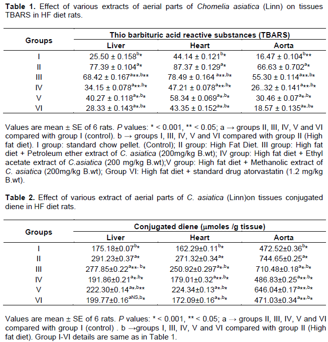
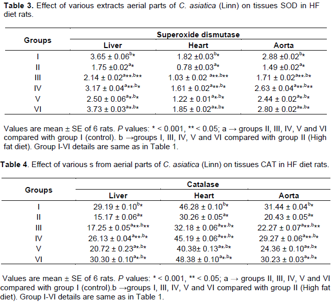
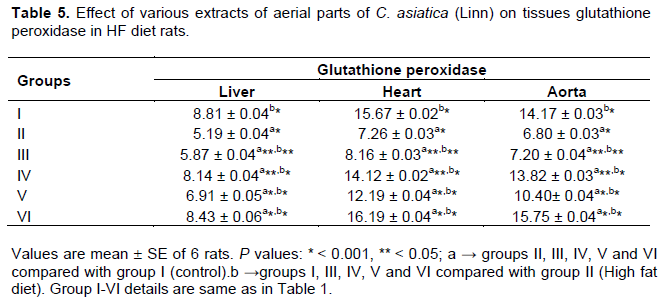
RESULTS
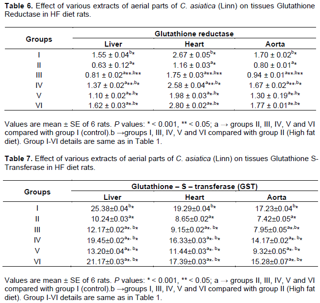
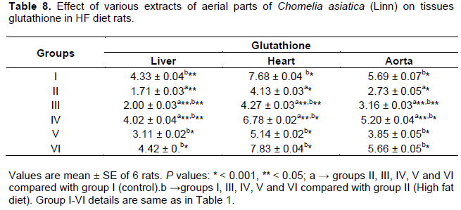
DISCUSSION
CONCLUSION
CONFLICT OF INTERESTS
The authors have not declared any conflict of interests.
ACKNOWLEDGMENT
REFERENCES
|
Amutha D, Shanthi S, Mariappan V (2012). Antiinflammatory effect of Tarenna asiatica in carrageenan induced lung inflammation. Int. J. Pharm. Pharmaceut. Sci. 4:344-347. |
|
|
Anjanadevi N, Menaga S (2013). Wound healing potential of Tarenna asiatica leaves. J. Theor. Exp. Biol. 10:75-80. |
|
|
Anonymous (1976). The wealth of India: Raw materials. New Delhi, Council of Scientific and Industrial Research. pp. 130-131. |
|
|
Badami S, Gupta MK, Suresh B (2003). Antioxidant activity of ethanolic extract of Striga orobanchioides, J. Ethnopharmacol. 85:227-230. |
|
|
Batra S, Singh SP, Srivasta VML (1989). Xanthine oxidase, Superoxide dismutase, Catalase and lipid peroxidation in mastomys nataensis effect of dipentalonema viteae infection, Indian J. Exp. Biol. 27:1067. |
|
|
Boccio GD, Lapenna D, Porreca E, Pennelli A, Savini F, Feliciani P, Ricci G, Cuccurullo F (1990). Aoertic antioxidant defense mechanisms: time related changes in cholesterol fed rabbits. Atherosclerosis pp. 81-127. |
|
|
Chance B, Greenstein DS (1992). The mechanism of Catalase actions-steady state analysis. Arch. Biochem. Biophys. 37:301-339. |
|
|
De La Cruz JP, Quintero L, Villalobos MA, Sanchez de.la Cuesta F (2000). Lipid peroxidation and glutathione system in hyperlipedemic rabbits influence of olive oil administration. Biochem. Biophys. Acta.1485:36. |
|
|
Ellman GL (1959). Tissue sulfhydroyl groups. Arch. Biochem. Biophy. 82:70. |
|
|
Folch J, Lees M, Sloane GH (1957). A simple method for the isolation and purification of total lipids from animals tissues. J. Biol. Chem. 226:497. |
|
|
Habig WH, Pabst MJ, Jakoby WB (1974). Glutathione – S – transferase, the first enzymatic step in mercaptouric acid formation. J. Biol. Chem. 249:7130- 7139. |
|
|
Halliwell B, Gutteridge JMC (1984). Lipid peroxidation, oxygen radicals, cell damage, and antioxidant therapy, The Lancet, 323:1396-1397. |
|
|
Halliwell B (1997). Advances in pharmacology, Academic Press 38:3-17. |
|
|
Harborne JB (1984). Phytochemical methods 11 Edn.In Chapman &, Hall. New York. pp. 4-5. |
|
|
Izawa S, Inoue Y, Kimura A (1996). Importance of Catalase in the adaptive response to hydrogen peroxide analysis of a catalasaemic Saccharomyces Cerevisae. Biochem. J. 320: 61-67. |
|
|
Jayaprakasha GK, Selvi T, Sakariah KK (2003). Antibacterial and antioxidant activities of grape (Vitis vinifera) seed extract. Food Res Int., 36: 117–122. |
|
|
Jayasinghe ULB, Jayasooriya CP, Bandara BMR, Ekanayake SP, Merlini Assante LG (2002). Antimicrobial activity of Sri Lankan Rubiaceae and Meliaceae. Fitoterapia. 73:424-427. |
|
|
Kakkar P, Das B, Visvanathan PN (1984). A modified spectrophotometric assay of SOD, Indian J. Biochem. Biophys. 21:130-132. |
|
|
Khan SA, Lee K, Minhas KM, Gonzalez DR, Raju SV, Tejani AD (2004). Neuronal nitric oxide synthase negatively regulates xanthine oxidoreductase inhibition of cardiac excitation-contraction coupling. Proc. Natl. Acad. Sci. USA. 101:15944. |
|
|
Kottai Muthu A, Sethupathy S, Manavalan R, Karar PK (2005). Hypolipidemic effect of methanolic extract of Dolichos biflorus Linn in high fat diet fed rats. Ind. J. Exp. Biol. 43:522-525. |
|
|
Mau JL, Lin HC, Song SF (2002). Antioxidant properties of several specialty mushrooms. Food Res. Int, 35: 519-526. |
|
|
Mavis RD, Stellwagen E (1968). Purification and Subunit Structure of Glutathione Reductase from Bakers' Yeast. J. Biol. Chem. 243:809-814. |
|
|
Meister A (1984). New aspects of glutathione biochemistry and transport selective alterations of glutathione metabolism. Nutr. Rev. 42:397. |
|
|
Nichans WH, Samulelson B (1968). Formation of malondialdehyde from phospholipid arachidonate during microsomal lipid peroxidation. Euro. J. Biochem. 6: 126-130. |
|
|
Rajakaruna N, Harris CS, Towers GHN (2002). Antimicrobial activity of plants collected from serpentine outcrops in Sri Lanka. Pharmaceut. Biol. 40:235-244. |
|
|
Rajashree GR, Rajmohan J, Augusti KT(1998). Antiperoxide effect of garlic Protein in alcohol fed rats. Ind. J. Exp. Biol. 36:60. |
|
|
Ramabharathi V, Apparao AVN, Rajith G (2014). Phytochemical investigation and evaluation of antibacterial and antioxidant activities of leaf-bud exudate of Tarenna asiatica (L.) Kuntze ex K. Schum. Indian J. Nat. Prod. Resour. 5:48-51. |
|
|
Ramarao N, Henry AN (1996). The ethnobotany of Eastern Ghats in Andhra Pradesh, India. Calcutta. Botanical Survey of India. |
|
|
Rao DM, Rao UVUB, Sudharshanam D (2006). Ethno-medico-botanical studies from Rayalaseema region of southern Eastern Ghats, Andhra Pradesh, India. Ethnobotanical Leaflets. 10:198-207. |
|
|
Rotruck JT, Pope AL, Ganther HE, Swanson AB, Hatman DG, Hoekstra WG (1973). Selenium; Biochemical roles as a component of glutathione peroxidise. Science 179:588. |
|
|
Sethupathy S, Elanchezhiyan C, Vasudevan K, Rajagopal G (2002). Antiatherogenic effect of taurine in high fat fed rats. Ind. J. Exp. Biol. 40: 1169. |
|
|
Sinha AK (1972). Colorimetric assay of catalase. Anal. Biochem. 47:389. |
|
|
Thampi HBS, Manoj G, Leelamma S, Menon VG (1991). Dietary fibre and lipid peroxidation: Effects of dietary fibre on levels of lipids and lipid peroxides in high fat diet, Ind. J. Exp. Biol. 29:563. |
|
|
Vinothkumar D, Murugavelh S, Prabhavathy AK (2011). Phytosociological and ethnobotanical studies of sacred groves in Pudukottai district, Tamil Nadu, India. Asian J. Exp. Biol. Sci. 2: 306-315. |
|
|
Waynforth BH (1980). Injection techniques. Experimental and surgical techniques in the rats, Academic Press, London. 3 p. |
|
Copyright © 2024 Author(s) retain the copyright of this article.
This article is published under the terms of the Creative Commons Attribution License 4.0


