Full Length Research Paper
ABSTRACT
The genus Osmundaria (Rhodophyta, Ceramilaes, Rhodomelaceae) comprises tropical and temperate regions red marine seaweeds species. Osmundaria obtusiloba is distributed from the northeastern coast of Brazil to the state of Rio de Janeiro. Studies with ethanol extract of red seaweed O. obtusiloba showed the antiviral potential of this alga. Hence, this study examined in BALB/c mice the acute toxicity after oral administration of O. obtusiloba crude extract. Then, female BALB/c mice received a single dose of O. obtusiloba extract by gavage at 550 mg/kg and their behaviors were monitored for a 14 day period. The biochemical and histological changes in the liver, kidney, stomach and spleen were analyzed. O. obtusiloba extract did not significantly change behavior, body weight, hematological or biochemical profiles. The organs of the animals did not show significant alterations when submitted to treatment with O. obtusiloba extract. In conclusion, the in vivo results revealed that O. obtusiloba has low toxicity and it can be and may be the target of further studies of biological activity.
Key words: Osmundaria obtusiloba; acute toxicity; seaweeds; preclinical tests.
INTRODUCTION
A variety of natural substances have been isolated from extracts of the genus Osmundaria J.V. Lamouroux (Order Ceramiales, family Rhodomelaceae), however, there is a predominance of bromophenols (Popplewell and Northcote, 2009), which are molecules that have one or more rings benzene, with varying degrees of halogenation and hydroxyl groups (Liu et al., 2011). These molecules present interesting biological activities described in the literature, for example, antimicrobial (Barreto and Meyer, 2006), antiviral (de Souza et al., 2012) and cytotoxic (Popplewell and Northcote, 2009).
The Osmundaria obtusiloba (C. Agardh) R.E. Norris species is characterized as a robust plant (Rhodophyta, Ceramiales, Rhodomelaceae), which can measure from 10 to 15 cm in height, with flat apices of 3 to 4 mm wide, showing dark red coloration (Carvalho et al., 2006). Several biological activities have been described from O. obtusiloba, mainly for their extracts, such as the fraction of O. obtusiloba rich in lectin that inhibited the trypsin and α-amylase enzymes, indicating its potential use in the production of drugs against diabetes (de Oliveira et al., 2009) or the ethanol extract that was able to inhibit the replication of Zika virus while maintaining low cytotoxicity (CC50=525 µg/ml) (Cirne-Santos et al., 2017). Antiviral activity against HSV-1 and HSV-2 has also been described for glycolipids extracted from O. obtusiloba (de Souza et al., 2012). In the work of de Alencar et al. (2016) the 70% EtOH was most effective solvent for extracting phenolic compounds from red seaweeds when compared to hexane, also O. obtusiloba EtOH extract presented high antioxidant activity. Already, the O. obtusiloba methanolic extract showed to present bromophenols (Carvalho et al., 2006).
Due to the discovery of several biological activities previously described, in particular the activity against the Zika virus, it is necessary to make the first preclinical tests to investigate the degree of toxicity of the ethanol extract of this seaweed, aiming the development of new drugs. Toxicity studies provides information on toxic doses and therapeutic indices of drugs and this type of studies in animals is vitally needed to determine the safety of medicinal plants for a future clinical study (Al-Afifi et al., 2018). There are still few toxicity studies of seaweed extracts in animals and the present work aims to evaluate the acute toxicity of the O. obtusiloba ethanol extract.
MATERIALS AND METHODS
Algae and extraction
Specimens of O. obtusiloba were collected at Rasa Beach, Armação de Búzios, Rio de Janeiro State, Brazil (lat. 22° 45’40”, long. 41° 54’ 32”). The seaweeds were washed with local water and separated from sediments, epiphytes, and other associated organisms. The material was dried at room temperature for about seven days, triturated using an industrial blender and weighed (140 g) on a semi-analytical scale. The crushed seaweed was exhaustively extracted with ethanol at room temperature. The extract was evaporated under reduced pressure, yielding a brownish residue (5 g). For oral administration the extract was diluted in 1% dimethyl sulphoxide (DMSO).
Biological studies
Animal model
Three groups of six female BALB/c mice, three months old, weighing 19 to 25 g were used for acute toxicity tests (Garrido, 2016). The animals were observed into our bioterium in Virology laboratory kept in polypropylene cages at 25 ± 2°C, under a 12/12 h light/dark cycle, with food and water ad libitum. All the tests were performed according to the protocols already approved by the Ethics Committee on Animal Use of the Fluminense Federal University (CEUA-UFF) with certificate number 798.
Experimental protocol: Acute toxicity (14 days)
To evaluate the acute toxicity over 14 days, the animals were divided into three groups: I) O. obtusiloba extract – 550 mg/kg (n = 6); II) 1% DMSO - vehicle (n = 6); III) Saline – negative control (n=6). These were administered in single oral dose of 200 μl. The animal behavior was observed throughout both experiments. At the end of the experimental period (14 days), the animals were euthanized by anesthetic overdose (ketamine + xylazine). Body weights were measured on D0 (first day), before extract administration, D7 (7th day) and D14 (14th day) of the experimental period. The protocol and concentrations used in this study were based on the OECD 423 guidelines (OECD, 2008). The concentration of 550 mg/kg of the extract was chosen based in low in vitro cytotoxicity of this extract and the dose-response curve in the OECD 425 guidelines (OECD, 2001). Blood samples were collected at the end of the experiment (14th day). Biochemical parameters tests used samples collected in BD-Microtainer® (Clot Activator/SSTTM Gel–Amber) vials to analyze: alanine aminotransferase (ALT), aspartate aminotransferase (AST) and blood urea nitrogen (BUN) (Garrido, 2016). The results were obtained with an automatic biochemistry meter (BS-210-Bioclin).
Histological analysis
At necropsy, the liver, kidney, heart, spleen and stomach were removed for histological processing. Thus, the organs were fixed in 10% Carson formalin and after tissue slices of all organs were routinely processed for paraffin embedding. After the processing of the organs, the pieces were cut into 5 mM microtome according to the literature (Musumeci, 2014)and were prepared and stained by Mayer hematoxylin/eosin (Sigma–Aldrich). Then, the slides were analyzed by conventional microscopy.
Statistical analysis
The data were analyzed by one-way analysis of variance (ANOVA) followed by Tukey test using GraphPad Prism version 5 program. A P value of <0.05 was considered statistically significant.
RESULTS AND DISCUSSION
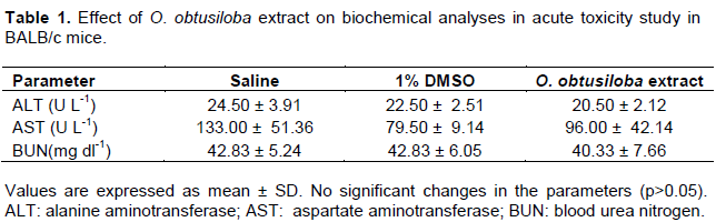
To evaluate the acute toxicity of O. obtusiloba extract (550 mg/kg) three groups were formed, a group receiving the ethanol extract, another group receiving only the vehicle and a group receiving saline by gavage. All animals treated with O. obtusiloba extract, 1% DMSO and saline survived for 14 days. There were no significant adverse clinical signs or changes in body weights (p > 0.05) (Figure 1). From the serum were determined the transaminases ALT and AST, parameters used for the evaluation of liver function and BUN, which can be used to estimate the renal function (Roy et al., 2015). The results showed that the group that received the oral administration of O. obtusiloba extract had no significant difference compared to the control group (saline) and vehicle (p>0.05) in all the biochemical parameters analyzed (Table 1) and remained within normal range (Kifayatullah et al., 2015).

Histological findings corroborate with those clinical observed during the experiment and biochemical analyses. We analyzed organs extracted from the experiment mice and they did not show significant changes in morphology in the 550 mg/kg O. obtusiloba extract group when compared with control groups (vehicle and saline) with spleen exception. Therefore, the liver was histologically normal, and structures of the hepatic wire and centrilobular veins may be evident corroborating with the AST and ALT analyzes (Figure 2A, B and C). Sasidharan et al. (2010) by testing another red seaweed observed no significant signs of toxicity, nor did a single administration of 2000 mg/kg methanol extract of Gracilaria changii cause death during the 14-day observation period. In another acute toxicity work of our group (period of 10 days) with dolabelladienotriol, a natural product derived from brown algae, Dictyota friabilis (as Dictyota pfaffii), all of the animals that received dolabelladienotriol presented a moderate increase in mitosis of hepatocytes and focal areas of hydropic cells in the medulla of the kidneys, even though this study indicates that dolabelladienotriol has low toxicity in administered dose range (Garrido et al., 2011).
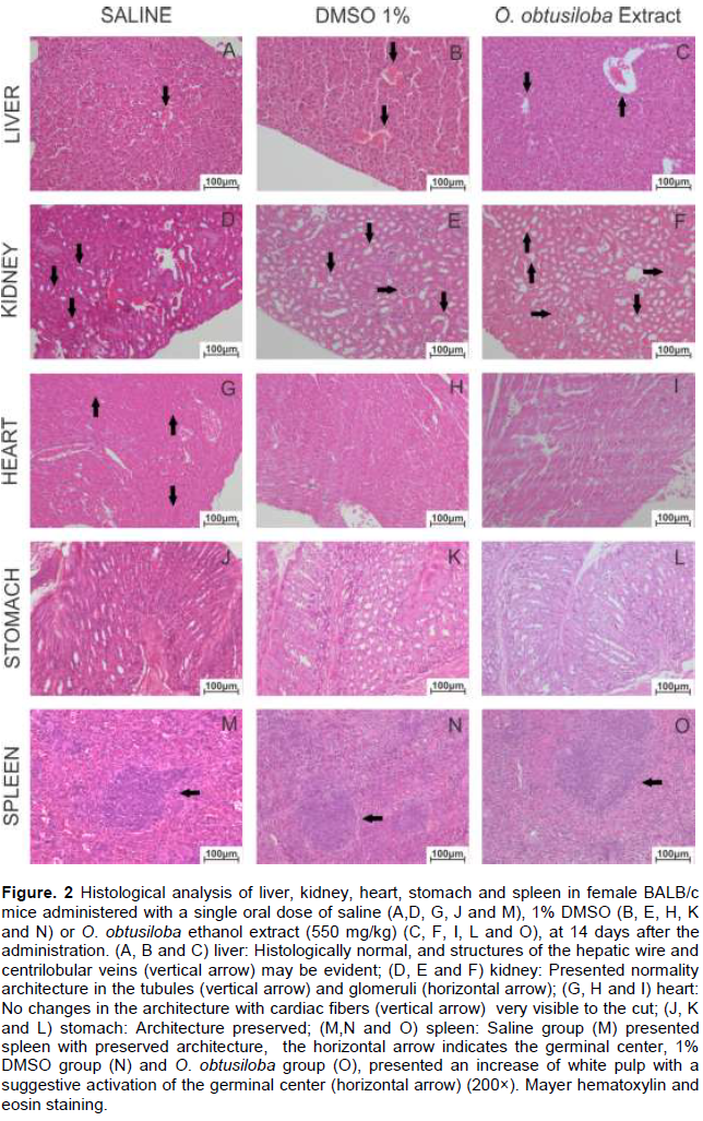
The architecture of the kidneys presented normality in the tubules and glomeruli (Figure 2D, E and F) and urea parameters (Table 1). The low toxicity was later confirmed by the subchronic toxicity study of this product (Garrido et al., 2017). Already for the extract of O. obtusiloba has been reported the presence of bromophenols (Osako and Teixeira, 2013). The heart showed no changes in the architecture with cardiac fibers very visible to the cut (Figure 2G, H and I). Finally, we analyze the histological structure of stomach and observed no changes (Figure 2J, K and L), just as Garrido et al. (2011) found in the acute toxicity study of dolabelladienotriol. However in the spleen, as noted in the 1% DMSO group and O. obtusiloba group, was presented an increase of white pulp with a suggestive activation of the germinal center, possibly due to the DMSO solvent used for solubilization of the extract (Figure 2M, N and O). These results, together with the biochemical parameters analyzed, demonstrate the low acute toxicity of the extract of this alga in tested concentration.
CONCLUSIONS
The result indicates that the single oral dose of administration of O. obtusiloba ethanol extract (550mg/Kg) in our acute toxicity study after 14 days did not produce any significant toxic effect in BALB/c mice. Hence, further studies should be carried out to confirm the low toxicity of this extract with its continuous and prolonged use.
CONFLICT OF INTERESTS
The authors have not declared any conflict of interests.
REFERENCES
|
Al-Afifi NA, Alabsi AM, Bakri MM, Ramanathan A (2018). Acute and sub-acute oral toxicity of dracaena cinnabari resin methanol extract in rats. BMC Complementary and Alternative Medicine 18(1):50. |
|
|
Barreto M, Meyer J (2006). Isolation and antimicrobial activity of a lanosol derivative from osmundaria serrata (rhodophyta) and a visual exploration of its biofilm covering. South African Journal of Botany 72(4):521-528. |
|
|
Carvalho LRd, Guimarães SM, Roque NF (2006). Sulfated bromophenols from Osmundaria obtusiloba (c. Agardh) re norris (rhodophyta, ceramiales). Brazilian Journal of Botany, 29(3):453-459. |
|
|
Cirne-Santos C, Barros CDS, Nogueira CCR, Campos RM, Teixeira V, Ferreira D,Paixão ICNDP (2017). Inhibition of zika virus by marine algae. |
|
|
de Alencar DB, de Carvalho FCT, Rebouças RH, dos Santos DR, dos Santos Pires-Cavalcante KM, de Lima RL, Baracho BM, Bezerra RM, Viana FA,dos Fernandes Vieira RHS (2016). Bioactive extracts of red seaweeds pterocladiella capillacea and osmundaria obtusiloba (floridophyceae: Rhodophyta) with antioxidant and bacterial agglutination potential. Asian Pacific Journal of Tropical Medicine 9(4):372-379. |
|
|
de Oliveira MN, Freitas ALP, Carvalho AFU, Sampaio TMT, Farias DF, Teixeira DIA, Gouveia ST, Pereira JG (2009). Nutritive and non-nutritive attributes of washed-up seaweeds from the coast of ceará, brazil. Food Chemistry 115(1): 254-259. |
|
|
de Souza LM, Sassaki GL, Romanos MTV, Barreto-Bergter E (2012). Structural characterization and anti-HSV-1 and HSV-2 activity of glycolipids from the marine algae Osmundaria obtusiloba isolated from southeastern brazilian coast. Marine Drugs 10(4):918-931. |
|
|
Garrido V, Barros C, Tonelli M, Teixeira G, Ocampo P, Silva GB, Giongo V, Paixão ICNDP (2016). Acute toxicity evaluation of aminomethylnaphthoquinone (amnq 1) in balb/c mice. IJPR 6(06):217. |
|
|
Garrido V, Barros C, Melchiades VA, Fonseca RR, Pinheiro S, Ocampo P, Teixeira VL, Cavalcanti DN, Giongo V, Ratcliffe NA (2017). Subchronic toxicity and anti-HSV-1 activity in experimental animal of dolabelladienetriol from the seaweed, Dictyota pfaffii. Regulatory Toxicology and Pharmacology 86:193-198. |
|
|
Garrido V, Teixeira GA, Teixeira VL, Ocampo P, Ferreira WJ, Cavalcanti DN, Campos S, Pedruzzi Md, Olaya P,dos Santos CC (2011). Evaluation of the acute toxicity of dolabelladienotriol, a potential antiviral from the brown alga Dictyota pfaffii, in balb/c mice. Revista Brasileira de Farmacognosia 21(2):209-215. |
|
|
Kifayatullah M, Mustafa MS, Sengupta P, Sarker MMR, Das A, Das SK (2015). Evaluation of the acute and sub-acute toxicity of the ethanolic extract of pericampylus glaucus (lam.) merr. In balb/c mice. Journal of Acute Disease 4(4):309-315. |
|
|
Liu M, Hansen PE,Lin X (2011). Bromophenols in marine algae and their bioactivities. Marine Drugs 9(7):1273-1292. |
|
|
Musumeci G (2014). Past, present and future: Overview on histology and histopathology. Herbert Publications. |
|
|
Organisation for Economic Co-operation and Development (OECD) (2001). 423: Acute oral toxicity-up-and-down procedure. OECD Guidelines for the Testing of Chemicals, Section 4. |
|
|
Organisation for Economic Cooperation and Development (OECD) (2008). 425: Acute oral toxicity-up-and-down procedure. OECD Guidelines for the Testing of Chemicals. |
|
|
Osako K, Teixeira VL (2013). Natural products from marine algae of the genus Osmundaria (rhodophyceae, ceramiales). Natural Product Communications 8(4):533-538. |
|
|
Popplewell WL,Northcote PT (2009). Colensolide a: A new nitrogenous bromophenol from the New Zealand marine red alga Osmundaria colensoi. Tetrahedron Letters 50(49):6814-6817. |
|
|
Roy S, Majumdar S, Singh AK, Ghosh B, Ghosh N, Manna S, Chakraborty T,Mallick S (2015). Synthesis, characterization, antioxidant status, and toxicity study of vanadium–rutin complex in balb/c mice. Biological Trace Element Research 1-18. |
|
|
Sasidharan S, Darah I, Noordin MKMJ (2010). In vitro antimicrobial activity against Pseudomonas aeruginosa and acute oral toxicity of marine algae Gracilaria changii. New Biotechnology 27(4):390-396. |
|
Copyright © 2024 Author(s) retain the copyright of this article.
This article is published under the terms of the Creative Commons Attribution License 4.0


