Full Length Research Paper
ABSTRACT
The effect of extracellular calcium on isolated uterine muscle contractions stimulated by acetone leaf extract of Anogeissus leiocarpa was investigated in a rat. About 12 m segment of the uterine muscle strip was mounted initially in a thermostatically regulated organ bath (37°C) containing normal Physiological Salt Solution (PSS; De Jalon) and later inphysiological salt solution devoid of CaCl2. The extract contracted the uterine muscle preparation in a concentration-related manner in normal PSS, with 0.53 mg/ml as the lowest active concentration. This contractile response was abolished by the calcium channel blocker, verapamil HCl (2 µg/ml). The extract however, did not evoke any contractile activity on the isolated tissue in a calcium-free media (PSS), even when caffeine (2 mmol) known to release calcium via the calcium induced-calcium release (CICR) mechanism was added to the perfusate. The results demonstrated the requirement for extracellular calcium for the extract mediated contractions and the inability of the extract to access calcium from intracellular storage sites such as the sarcoplasmic reticulum and the mitochondria. Therefore, the influx of Ca2+ into the cell cytosol is a pre-requisite for the extract-mediated uterine muscle contraction.
Key word: Anogeissus leiocarpa, thermostatically organ bath, verapamil, caffeine, uterus.
INTRODUCTION
Uterine muscle contraction is driven by the development of action potentials across the plasma membrane, resulting from a transient increase in the Intracellular Ca2+ concentration (Rasola and Bernardi, 2011), by the presence of contractile elements, and a conducting system between uterine cells (Wray et al., 2001). Intracellular calcium is normally stored in a special intracellular organelle known as sarcoplasmic reticulum (SR) that is either membrane bound or is located towards the center of the cell (Bradley et al., 2002). A prerequisite for myometrial contraction is an increase in intracellular free Ca2+ concentration which can occur through release from this intracellular storage or by influx from the extracellular compartment or both (Kuo and Ehrlich, 2015).The contraction can be elicited by different agonists or by electrical depolarization resulting in a rapid increase in cytosolic calcium ion concentration (Kuo and Ehrlich, 2015), and its binding to a messenger protein, calmodulin that becomes activated (Zhang et al., 2012). Consequently, there is a conformational change that allows the calmodulin-calcium complex to interact with the contractile protein myosin light chain (MLC) by activating its hitherto inactive enzyme, myosin light chain kinase (MLCK; Stull et al., 2011) that phosphorylates MLC, resulting in MLC cross-bridging with actin to bring about contraction. Uterine muscle relaxation, on the other hand, can be accomplished by dephosphorylating the myosin light chain protein by myosin light chain phosphatase (MLCP), dissociation of the calcium-calmodulin complex (Martín et al., 2014)or when some other mechanisms come into play such as, reduced release of calcium by the SR or reduced calcium entry into the cell or calcium efflux into the extracellular space via Ca2+-Adenosine triphosphatase (Ca2+-ATPase) and Na+-Ca2+ exchanger systems resulting in fall in cytoplasmic Ca2+ concentration (Wray et al., 2001, 2003; Pehlivanoglu et al., 2013) or through inhibition of myosin light chain kinase by increased intracellular concentration of cyclic adenosine monophosphate (cAMP) (Martín et al., 2014).
Verapamil is a voltage-dependent calcium channel blocker (Lacinová, 2005).Calcium channel blockers are considered as class - IV antiarrhythmic agents (Hondeghem and Katzung, 1984). Calcium channels blockers are specially concentrated in the sinoatrial and atrioventricular nodes where they are used to decrease impulse conduction through AV node, thus protecting the ventricles from atrial tachyarrhythmias(Li et al., 2010). Calcium channels are also present in the smooth muscles lining blood vessels, and by relaxing the tone of this smooth muscles calcium channel blockers dilate the blood vessels (Hondeghem and Katzung, 1984). Propranolol is a non-cardioselective sympatholytic beta blocker that is lipid soluble and also a sodium channel blocker. It is a non-selective beta blocker that blocks the action of epinephrine (adrenaline) and norepinephrine (noradrenaline) at both β1- and β2-adrenergic receptors (Steenen et al., 2015; Motiejunaite et al., 2020). Caffeine is a receptor antagonist at all adenosine receptor sub-types [A1, A2A, A2B, and A3 receptors] (Cao et al., 2015; Ferré, 2016). Antagonism at these receptors stimulates the medullary vagal, vasomotor, and respiratory centers, which increases respiratory rate, reduces heart rate, and constricts blood vessels (Ferré, 2008). Adenosine receptor antagonism also promotes neurotransmitter release (such as, monoamines and acetylcholine) which endows caffeine with its stimulant effects (Glade, 2010).
In addition to the numerous medicinal uses of A. leiocarpus in folk medicine, local livestock farmers in North Eastern Nigeria claim that ingestion of leaves of A. leiocarpus induces abortion in small ruminants (Chiroma et al., 2019).This study was therefore designed to examine the contractile effects of acetone leaf extract of this medicinal plant on isolated uterine muscle strips of the rat, and the possible role of calcium in this contractile process as a prelude to its in vivo effects on female reproduction. The research will also shed light on the veracity of its claimed abortifacient effects in small ruminants (sheep and goat) by local livestock farmers in the study area.
MATERIALS AND METHODS
Sample collection, identification and extraction
The leaves of A. leiocarpus were collected from a rural community in Lassa, Askira/Uba Local Government Area, Borno State. It was identified by a Botanist with the Department of Biological Sciences, University of Maiduguri. It was deposited in the herbarium of Veterinary Physiology and Biochemistry with herbarium number Vet. Species 208 A. The fresh leaves were air-dried under shade and grounded into fine powder using pestle and mortar. Two hundred and twenty grams of the plant material was extracted with 2Ltres of acetone using soxhlet method (Jensen, 2007). The product was filtered using Whatman filter paper (18cm) and was concentrated in hot air oven at 40-50oC. After evaporation, the weight (g) and percentage yield was taken and it was kept in a clean container and stored at 4°C for further analysis.
Ethical statements
Ethical procedure according to Council for International Organizations of Medical Sciences and the International Council for Laboratory Animal Science (CIOMS, 2016), were used.
Animal and tissue preparation
A total of 3 adult female rats (Rattus norvegicus) weighing between 180- 200 g were used for the isolated uterine tissue studies. They were kept in plastic cages in the Department of Veterinary Physiology and Biochemistry Laboratory of the University of Maiduguri, for two weeks for acclimatization. They were fed with suitable animal feed (vital feeds® Nig. Ltd.Jos) and provided clean water ad libitum.
Each rat was administered estrogen 0.1 mg/kg body weight subcutaneously twenty four hours before sacrifice. The isolation and mounting of non-gravid rat uterus was done by the procedure described by Uchendu (2000). The rat was sacrificed by stunning and cervical dislocation and the abdomen opened to expose the internal organs. The two uterine horns were identified, dissected out and transferred to a dish containing De Jalon’s physiological salt solution of the following composition: Sodium chloride (9.0 g), Glucose (0.5 g), Potassium chloride (0.42 g), Sodium hydroxide (0.5 g), calcium chloride (0.06 g), and magnesium sulphate (0.1 g). In another set of experiments, CaCl2 was omitted in the De Jalon’s solution in order to verify the role of calcium in the extract-mediated contractions.
The two uterine horns were separated and freed from fat and connective tissue. A strip of the horn about 1 - 2 cm was cut out. A thread was then attached to one end of the isolated strip of uterus and was tied to the aerator tube in the organ bath containing 75 ml De Jalon’s physiological solutions. Another thread was attached to the other end of the isolated uterus and fixed to a lever system fitted on the stylus and the load on the tissue was 0.5 g (a tension of 5 mn). The tissue was aerated with ordinary air using an aquarium air pump. The temperature of the organ bath was thermostatically regulated at 37 ± 0.50°C.
Procedure of preparing the acetone extract and standard drugs
For the in vitro studies, the dry acetone extract of A. leiocarpus was reconstituted in distilled water and the predetermined concentrations were added to the tissue/organ bath. The effects of standard drugs vis-a-vis the plant extract were also investigated. Responses to addition of standard drugs and/or extracts were recorded after each experiment. The tissue was washed three times with fresh physiological salt solution and allowed appropriate time to recover before subsequent additions of drugs or extract. The following drugs were used in the course of the experiments: Propranolol (Antigen Pharmaceuticals, Ireland), Verapamil HCL (Teva; UK), and Caffeine (MERK Germany). The concentrations of the extract/drugs given in the ‘Results’ section are all final nutrient or organ bath concentrations.
RESULTS
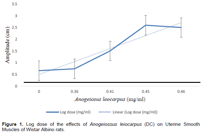
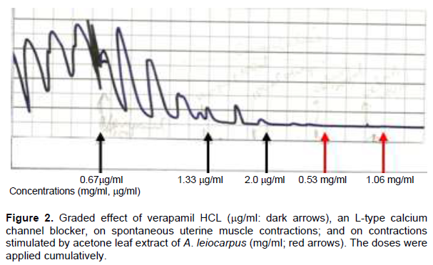
Acetone leaf extract of A. leiocarpus produced a concentration - dependent increase in uterine smooth muscle contraction, with 0.53 mg/ml as the lowest active concentration. The contraction was phasic in nature and affected predominantly the amplitude with little effect on the frequency component. The calculated effective concentration (EC50) value was 0.09 mg/ml (Figure 1.). Application of Verapamil HCL, an L-type calcium channel blocker to the perfusate caused inhibition of spontaneous uterine muscle contraction that was concentration-dependent (Figure 2), with complete abolition of contractions by 2.00 µg/ml. This concentration, that is, 2.00 mg/ml had previously been shown to abolish contractions of isolated uterine muscle tissue (Meisheri et al., 1981; Uchendu, 1999). The tissue did not respond to various concentrations of the extract (0.53 to 1.06 mg/ml) in the presence of this calcium channel blocker.
Effects of propranolol on contractions stimulated by the extract
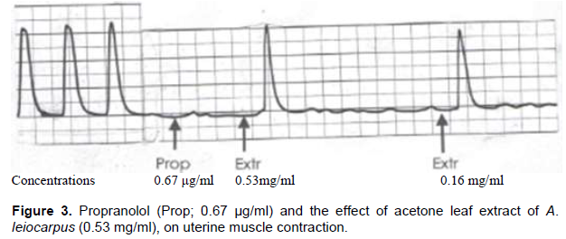
The addition of propranolol (0.67 µg/ml), a b-adrenergic receptor blocker to the perfusate, followed three min later by the extract (0.53 mg/ml) resulted in a single contractile force of maximum amplitude (30 mm in height), followed by a complete relaxation of the tissue that lasted for approximately 3.5 min. A second dose of the extract (0.16 mg/ml) added cumulatively (without washout) evoked a similar contractile response by the muscle preparation (Figure 3).
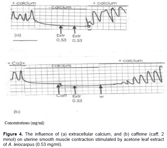
Effect of extracellular calcium [(Ca2+)] and caffeine (2 mmol) on contractions induced by acetone extract of A. leiocarpus
The effect of extracellular calcium on uterine contraction stimulated by acetone extract of A. leiocarpus is shown in Figure 4. The normal, spontaneous, uterine muscle contractions were abolished when CaCl2 was omitted in the physiological salt solution (Figure 4a). Application of the extract (0.53 mg/ml) in the Ca2+ free PSS did not elicit any contraction in the uterine muscle. Furthermore, the addition of caffeine (2 mmol), which is known to release Ca2+ via the calcium induced, calcium release (CICR) channel further suppressed the tissue response to the extract (Figure 4b). The spontaneous uterine muscle contractions were however, restored after washout and replacement with calcium containing physiological salt solution.
DISCUSSION
The uterus is a myogenic organ that is capable of contraction on its own without any external innervation. It contains functional muscarinic receptors that are responsive to cholinergic agents such as acetylcholine, eliciting prominent myometrial contractions (Paul et al., 2015). The present study revealed A. leiocarpus contracted the uterine muscle preparation in a graded manner under normal physiological conditions. The contractions were phasic in nature, and were dependent solely on influx of extracellular Ca2+ into the cell cytosol since the contractions were abolished when the uterine tissues were incubated in Ca2+-free physiological salt solution. This contention is further supported by the abolition of the extract-mediated contractions by verapamil, a well-known L-type Ca2+ channel blocker (Lacinová, 2005), even in a physiological salt solution containing Ca2+. This agrees with the report of other investigators that the uterine myocytes have a preponderance of voltage-operated Ca2+ channels and it is by massive movement of Ca2+ through these channels into the cell cytosol that myometrial contraction occurs (Sperelakis et al., 1992a, b; Datté et al., 1996; Luckas et al., 1999; Wray et al., 2003). The inability of the uterine muscle tissue to respond to caffeine, a stimulator of the Ca2+-induced-Ca2+-release (CICR) mechanism, in the present study, is evidence that the extract of A. leiocarpus was unable to trigger Ca2+ release from the intracellular storage site to support the Ca2+ pool that is required to initiate the contractile process. However, there are contractile agonists that are able to access Ca2+ from the sarcoplasmic reticulum of myocytes through inositol trisphosphate (IP3)-induced Ca2+ release (IICR) or through Ca2+-induced Ca2+release (CICR) mechanisms (Marc et al., 1986; Santo-Domingo and Demaurex, 2010), but the overall contribution from this site to the cytosolic Ca2+ pool have been reported to be minimal (Kupittayanant et al., 2002; Pehlivanoglu et al., 2013).
In the present study, the uterine muscle response to the extract after propranolol, even though single, was of sufficient amplitude (force) and could have risen due to inhibition or blunting of the b-adrenergic receptors by this non-selective antagonist, resulting in the accentuation of the force (amplitude) but not the frequency of the extract-mediated contraction. Previous studies have shown that propranolol, when administered alone, does not elicit uterine muscle contraction but will significantly reduce the inhibitory effect of isoprenaline, a non-specific b-adrenoceptor agonist, on myometrial contractility (Johansson and Andersson, 1980), a condition that is known to potentiate the effects of contractile agonists (Wang et al., 2010).
CONCLUSION
This study has shown that crude acetone leaf extract of A. leiocarpus contracted isolated uterine muscle strips of the rat in a concentration-related manner. The contractions had a requirement for Ca2+which was mobilized solely through influx from the extracellular compartment into the cell cytosol. Therefore, the claim by local farmers that fresh leaves of A. leiocarpus induce abortion in small ruminants (sheep and goats) has some scientific merits. Further study can focus on the in vivo effects of this medicinal plant on female reproduction.
CONFLICT OF INTERESTS
The authors have not declared any conflict of interests.
REFERENCES
|
Bradley KN, Flynn ERM, Muir TC, McCarron JG (2002). Ca2+ regulation in guinea-pig colonic smooth muscle: The role of the Na+-Ca2+ exchanger and the sarcoplasmic reticulum. Journal of Physiology |
|
|
Cao R, Rossetti G, Bauer A, CarIoni P (2015). Binding of the antagonist caffeine to the human adenosine receptor hA2AR in nearly physiological conditions. PLoS One. |
|
|
Chiroma M, Arastus W, Peter AM, Ijuptil C, Umar KS, Chukwuka U (2019). In-vitro Effect of Indomethacin on Uterine Muscle Contraction Stimulated by Acetone Leaf Extract of AnogeissusleiocarpusInternational. Journal of Livestock Research 9(7):24. |
|
|
CIOMS (2016). International Ethical Guidelines for Health-related Research Involving Humans Fourth Edition. Geneva, Biomedical Research. |
|
|
Datté J, Offoumou AM, Manda OM (1996). Uterotonic effects of hydromethanolic extract of Parquetina nigrescens (Periplocaceae) on spontaneous contractile activity in the isolated myometrium of pregnant rats. Journal of Ethnopharmacology. |
|
|
Ferré S (2008). An update on the mechanisms of the psychostimulant effects of caffeine. Journal of Neurochemistry. |
|
|
Ferré S (2016). Mechanisms of the psychostimulant effects of caffeine: implications for substance use disorders. Psychopharmacology (Berl). |
|
|
Glade MJ (2010). Caffeine-Not just a stimulant. Nutrition |
|
|
Hondeghem LM, Katzung BG (1984). Antiarrhythmic agents: The modulated receptor mechanism of action of sodium and calcium channel-blocking drugs. Annual Review of Pharmacology and Toxicology. |
|
|
Jensen WB (2007). The Original of the Soxhlet Extractor. Journal of Chemical Education 84(12)19-13. |
|
|
Johansson SRM, Andersson RGG (1980). Effects of b-adrenergic agonists on rat uterine motility and cAMP level in vivo. Acta Pharmacology and Toxicology 42:347-353. |
|
|
Kuo IY, Ehrlich BE (2015). Signaling in muscle contraction. Cold Spring Harb. Perspect. Biol. |
|
|
Kupittayanant S, Luckas MJ, Wray S (2002). Effect of inhibiting the sarcoplasmic reticulum on spontaneous and oxytocin-induced contractions of human myometrium. British Journal of Obstetrics and Gynaecology 109:289-296. |
|
|
Lacinová L (2005). Voltage-dependent calcium channels. General Physiology and Biophysics |
|
|
Li J, Jiang H, Wong WH (2010). Modeling non-uniformity in short-read rates in RNA-Seq data. Genome Biology. |
|
|
Luckas MJ, Taggart MJ, Wray S (1999). Intracellular calcium stores and agonist-induced contractions in isolated human myometrium. American Journal of Obstetrics and Gynecology 181:468-476 |
|
|
Marc S, Leiber D, Harbon S (1986). Carbachol and oxytocin stimulate the generation of inositol phosphates in the guinea pig myometrium. FEBS letters 201: 9-11. |
|
|
Martín P, Moncada M, Enrique N, Asuaje A, Valdez Capuccino JM, Gonzalez C, Milesi V (2014). Arachidonic acid activation of BKCa (Slo1) channels associated to the β1-subunit in human vascular smooth muscle cells. Pflügers Archiv: European Journal of Physiology |
|
|
Meisheri KD, Hwang O, van Breemen C (1981). Evidence for two separate Ca2+ pathways in smooth muscle plasmalemmae. The Journal of Membrane Biology 59:19 |
|
|
Motiejunaite J, Amar L, Vidal-Petiot E (2020). Adrenergic receptors and cardiovascular effects of catecholamines. Annales d'Endocrinologie (Paris). |
|
|
Paul J, Hua S, Roger S (2015). A Targeted Drug Delivery System for the Uterus. Reproductive Sciences. |
|
|
Rasola A, Bernardi P (2011). Mitochondrial permeability transition in Ca2+-dependent apoptosis and necrosis. Cell Calcium |
|
|
Santo-Domingo J, Demaurex N (2010). Calcium uptake mechanisms of mitochondria. Biochimica et Biophysica Acta (BBA)-Bioenergetics |
|
|
Sperelakis N, Inoue Y, Ohya Y (1992a). Fast Na+ channels and slow Ca2+ current in smooth muscle from pregnant rat uterus. Molecular and Cellular Biochemistry 114:79-89. |
|
|
Sperelakis N, Tohse N, Ohya Y (1992b). Regulation of calcium slow channels in cardiac muscle and vascular smooth muscle cells. Advances in Experimental Medicine and Biology 311:163-167. |
|
|
Steenen SA, van Wijk AJ, van Westrhenen R, de Lange J, de Jongh A (2015). Effects of propranolol on fear of dental extraction: Study protocol for a randomized controlled trial.Trials. |
|
|
Stull JT, Kamm KE, Vandenboom R (2011). Myosin light chain kinase and the role of myosin light chain phosphorylation in skeletal muscle. Archives of Biochemistry and Biophysics |
|
|
Uchendu CN (1999). Role of Ca2+ on uterine force stimulated by a glycoside from the root of Dalbergiasaxatilis. Indian Journal of Physiology and Pharmacology 43(2):171-178. |
|
|
Uchendu CN (2000). Biological activity of root extracts from Dalbergia saxatilis. Journal of Herbs, Spices and Medicinal Plants. |
|
|
Wang DW, Mistry AM, Kahlig KM, Kearney JA, Xiang J, George ALJ (2010). Propranolol blocks cardiac and neuronal voltage-gated sodium channels. Journal of Pharmacology and Pharmacotherapeutics 1:144. |
|
|
Wray S, Jones K, Kupittayanant S, Li Y, Matthew A, Monir-Bishty E (2003). Calcium signalling and uterine contractility. Journal of the Society for Gynecologic Investigation 10:252-264. |
|
|
Wray S, Kupittayanant S, Shmygol A, Smith RD, Burdyga T (2001). The physiological basis of uterine contractility: A short review. Experimental Physiology |
|
|
Zhang M, Abrams C, Wang L, Gizzi A, He L, Lin R, Chen Y, Loll PJ, Pascal JM, Zhang JF (2012). Structural basis for calmodulin as a dynamic calcium sensor. Structure |
|
Copyright © 2024 Author(s) retain the copyright of this article.
This article is published under the terms of the Creative Commons Attribution License 4.0


