Full Length Research Paper
ABSTRACT
Phytochemical screening of the methanolic fruit extract of Solanum macrocarpon was performed using standard method. Eighteen male albino mice, assigned into six groups (n=3) were used to determine the acute toxicity (LD50) of the extract. Haematological effect of the extract was determined using forty eight adult male rats assigned into four groups (A-D; n=12). The treatment groups received daily oral administration of the extract at doses of 400, 800 and 1600 mg/kg of body weight (bw) respectively for 21 days. The phytochemical screening of the extract revealed the presence of flavonoids, saponins, alkaloids, phenols, phytates, tannins, cyanides and terpenoids. The extract showed no mortality even at the dose of 5000 mg/kg bw. The highest treatment dose (1600 mg/kg) showed significant reduction in the white blood cell (WBC) count compared to rest of the treatment groups. There were no significant difference (p<0.05) in red blood cell (RBC), packed cell volume (PCV) and haemoglobin (Hb) levels of the treatment groups compared to control. Similarly, the mean cell volume (MCV), mean cell haemoglobin (MCH) and mean cell haemoglobin concentration (MCHC) results showed no significant difference from days 0 to 21 in all the treatment groups. Findings from this study suggest that except for the fact that the high dose of the extract antagonizes immunity; it has no serious adverse effect on the various haematological parameters, especially as it improves haemoglobin levels on prolonged administration.
Key words: Acute toxicity, blood profile, phytochemistry, Solanum macrocarpon.
INTRODUCTION
The usage of medicinal plants in West Africa is probably as old as the duration of human settlement in the region (Abdulrahman et al., 2010). As estimated by the World Health Organization, 80% of the global population in developing countries depends on traditional medicines mainly of plant origin with the steadily growing global market for traditional therapies standing at US$ 60 billion a year(World Health Organization, 2002).
According to Kariuki and Njoroge (2011), approximately 25% of all pharmaceutical products worldwide originated from traditional medicinal knowledge. In Nigeria, several thousands of plant species have been claimed to possess medicinal properties and have been employed in the treatment of many ailments (Okigbo and Mmeka, 2006).
Solanum macrocarpon, a tropical perennial plant of African ancestry belonging to the family Solanaceae. The genus Solanum is well known in traditional medicine (Burkhill, 2000; Grubben and Denton, 2004). The cultivated form of S. macrocarpon, known as ‘gboma’ in West Africa constitutes an important fruit and leaf vegetable. Local cultivars grown for the leaves are common throughout west and central Africa while the fruit types are mainly restricted to the humid coastal areas of West Africa (Bukenya-Ziraba and Bonsu, 2004). The leaves and fruits are cooked and consumed as a vegetable in soups and sauces and the taste is more or less bitter (Gbile and Adesina, 1988). The plant also serves as foliage for feeding livestock and is occasionally grown as an ornamental (Bukenya-Ziraba and Bonsu, 2004; Schippers 2001). Hematological parameters are important indices of physiological and pathological status for both animals and humans, thus playing a major role in disease investigation and diagnosis (Malomo, 2000; Adeneye et al., 2006). It has been documented that ingestion of some plant materials (either in the raw form or their extracts) having useful medicinal properties may cause anemia resulting from the sequestration of red blood cell (RBC) in spleen, impaired RBC production or primary bone marrow dysfunction (Cheeke, 1998; Mishra and Tandon, 2012). Therefore, this study was designed to investigate phytochemical constituents, acute toxicity and possible hematological effect of the methanolic S. macrocarpon fruit extract in order to ascertain its safety as a medicinal agent.
MATERIALS AND METHODS
Procurement of plant sample
The plant was cultivated at the agricultural farm of the University of Nigeria, Nsukka, and at maturity, unripe fruits were harvested and extracted in methanol for the study. The plant was identified at the herbarium of the Department of Plant Science and Biotechnology, University of Nigeria, Nsukka.
Preparation of S. macrocarpon fruit extract
The method of extraction was adapted from Uhegbu and Ogbuehi (2004) and Anaga et al. (2010). A total of 250 g of the powdered plant material was soaked in 500 ml of 99% methanol, stirred thoroughly and left to stand for 48 h. Afterwards, the mixture was filtered and evaporated to dryness at room temperature giving a 7.8% yield.
Phytochemical screening of extract (Qualitative and quantitative)
The flavonoids and cyanides contents of the extract were determined using the method described by Onwuka (2005) while that of alkaloids followed the gravimetric method of Harbone (1973). Phenols and saponins were determined using the method described by Obadoni and Ochuko (2001). The phytates was determined using the method described by Oberleas (1973). The method used for tannins determination followed that described by Pearson (1976).
Determination of terpenoids was done as described by Ezeonu and Ejikeme (2016). Briefly, two grams of the sample was weighed into a conical flask and 40 ml of chloroform solution was added and allowed to stand for 4 h. Thereafter the solution was filtered into a weighed crucible and oven dried. Then the weight of crucible with the extract was taken. Then 40 ml of absolute methanol was added and allowed for 2 h and then filtered into a weighed crucible, oven dried and re-weighed. The weight of the terpenoids was quantified in percentage as follows:
Terpenoid (%) = × 100
Determination of steroids was done as described by Mujeeb et al. (2014). Briefly, two grams of the sample was put into a test tube, and 50 ml of ethyl acetate was added and placed in a boiling water bath for 5 min. After cooling, the solution was filtered and mixed with an equal volume of chloroform which formed two layers. Two millilitres of the chloroform layer was pipetted into a test tube and 5 ml of water was added and adjusted to a pH of 7.0 using 0.1N NH4OH. Then the solution was filtered and allowed to stand for 5 min after which the absorbance was measured at 240 nm. Where: 2550 = extinction coefficient for steroids, df = dilution factor.
Acute toxicity studies of the plant extract
This was carried out by the method of Lorke (1983). The mice were separated into six groups (A, B, C, D, E and F) of three mice each. The first three groups (A, B, C) were administered the extract in graded doses of 200, 400 and 800 mg/k per kg body weight respectively in the first phase and observed for 24 h for signs of toxicity. Thereafter, the groups D, E, F were administered the extract at doses of 1000, 3000 and 5000 mg per kg body weight respectively and observed for signs of toxicity within the next 24 h.
Procurement and management of experimental animals
A total of eighteen male albino mice weighing 20-30 g were procured for the acute toxicity studies. On the other hand, forty eight male albino rats weighing 130 - 220 g were used to determine the effect of the extract on haematology. All the animals were procured from the Zoological Garden of the Department of Zoology and Environmental Biology, University of Nigeria, Nsukka. The animals were such as have no history of drug consumption and they were housed in clean rat cages under hygienic conditions in the experimental animal house. The animals had unrestricted access to feed (commercial growers mash Vital Feeds Nigeria) and clean water throughout the experiment. The animals were left for 14 days to acclimatize before the start of each experiment.
Experimental design
The study adopted an experimental approach and was approved by Institutional Animal Care and Use Committee of the Faculty of Veterinary Medicine, University of Nigeria, Nsukka (FVM-UNN-IACUC-2019-042). The acute toxicity study of the extract was carried out by the method of Lorke (1983). The mice were separated into six groups (A, B, C, D, E and F) of three mice each. The first three groups (A, B, C) were administered the extract in graded doses of 200, 400 and 800 mg/k per kg body weight respectively in the first phase and observed for 24 h for signs of toxicity. Thereafter, the groups D, E, F were administered the extract at doses of 1000, 3000 and 5000 mg per kg body weight respectively and observed for signs of toxicity within the next 24 h.
To study the effect of the extract on the haematological profile, the procured albino rats were divided into a control group (A) and three treatment groups (B, C and D) of twelve rats each. The treatment groups (B, C and D) received daily oral administration of the methanolic S. macrocarpon fruit extract at doses of 400, 800 and 1600 mg/kg of body weight respectively, while the control group received 1 ml/kg body weight of distilled water for the 21 days of the experiment.
Collection of blood samples
Blood samples were collected from each group before the start of the experiment (day 0) and at weekly intervals during treatment for the biochemical analyses. The blood samples were collected through the retro orbital plexus as described by Hoff (2000) and allowed to clot for about 30 min and afterwards centrifuged at 2000 rpm for 10 min. The sera obtained were then used to determine the biochemical parameters.
Determination of white blood cell (WBC)
This was determined using the method described by Sood (2006). The blood was sucked slowly and carefully up to 0.5 mark in the red diluting pipette. Immediately, the pipette was plunged into the diluting fluid and sucked up to 11 marks. Then, the ends of the pipette were gripped between the finger and the thumb and shaken thoroughly for 3 min. The diluted blood sample was loaded on a Neubauer counting chamber and all the cells in the four corner squares of the Neubauer chamber was counted using a light microscope at a high magnification of x 100. The number of white cells counted for each sample was calculated using the formula:
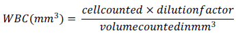
Where: dilution factor = 1: 20, and Number of large squares counted = 4
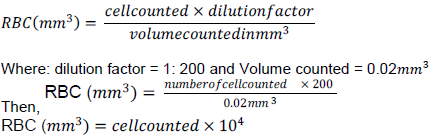
Determination of red blood cell count (RBC)
The method of RBC count was according to Sood (2006). The blood was sucked slowly and carefully up to 0.5 mark in the red cell diluting pipette. Immediately, the pipette was plunged into the diluting fluid and sucked up to 101 marks. Then, the ends of the pipette were gripped between the finger and the thumb and shake thoroughly for 3 min. The diluted blood sample was loaded on a Neubauer counting chamber and all RBC in the five groups of 16 small squares in the central area of the Neubauer chamber were counted using a light microscope at a high magnification of x 100. The number of red cells counted for each sample was calculated using the formula:
Blood haematocrit estimation (PCV)
This was determined by the microhaematocrit method as described by Coles (1986). A heparinized capillary tube was filled to approximately three fourth (3/4) of its length with well-mixed anticoagulated blood. The coloured end of the capillary tube was sealed with plasticin. The capillary tube was then placed into a microhaematocrit centrifuge set at 10,000 revolutions per minute (rpm) for 5 min. The height of the packed cell as well as the total height in millimeter was measured using the haematocrit reader. The packed cell volume was then calculated using the formula:

Haemoglobin estimation
Haemoglobin concentration was determined using the method of described by Sood (2006). The graduated tube was filled to the mark 2 on the red graduations with N/10 HCL using a dropper. Blood sample was sucked into the capillary pipette to 20 cm mark; the end of the pipette was wiped and blown into the acid in the mixing tube. The pipette was then rinsed by sucking up the acid and blowing it out back into the tube 3 - 4 times and then allowed to stand for 10 min for formation of acid haematin. Similarly, the acid haematin was diluted with distilled water in drop wise manner, mixed and the colour was compared with the standard by holding the comparator towards light. This was repeated until the colour of the diluted fluid matched with that of the standards in the comparator. From the level of the fluid in the mixing tube, the Hb was read out in gram percent.
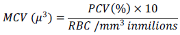
Estimation of mean cell volume (MCV)
The mean cell Volume (MCV) is the mean volume of the red cells. It was determined using the method as described by Baker et al. (2001). It was derived by calculation from packed cell volume (PCV) and RBC count as follows:
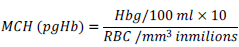
Estimation of mean cell haemoglobin (MCH)
This refers to the amount of haemoglobin present in the average erythrocyte. It was determined using the method as described by Baker et al. (2001). It was derived by calculation from the ratio of haemoglobin (Hb) and RBC count according to the following formula:
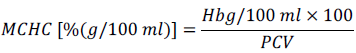
Estimation of mean cell haemoglobin concentration (MCHC)
The MCHC refers to the amount of haemoglobin in 100 ml of PCV, as opposed to the amount of haemoglobin in whole blood. It was determined using the method as described by Baker et al. (2001). It was derived by calculation from the ratio of hemoglobin (Hb) and PCV count according to the following formula:
Statistical analysis
The statistical analysis of data collected was done using the Statistical Package for Social Sciences (SPSS v.16). Analysis of Variance (ANOVA) was used to analyze the data while the Duncan Multiple Range Test (DMRT) was used to compare means. The results were presented as mean± S.EM with the level of significance set at p<0.05.
RESULTS
Result of phytochemical screening of the methanolic S. macrocarpon fruit extract
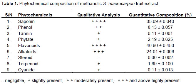
Table 1 shows the result of the qualitative and quantitative determination of phytochemicals present in the methanolic fruit extract of S. macrocarpon. It was observed that the quantity of flavonoids was highest in the plant extract followed by saponins and alkaloids. Phenols were moderately present, while phytate, tannin, cyanide and terpenoid were slightly present and steroids were negligible.
Acute toxicity studies of methanolic S. macrocarpon fruit extract
There was no mortality observed at the initial doses of 200, 400 and 800 mg/kg of the fruit extract after 24 h in phase one. Similarly, at higher dose levels of 1,000, 3,000 and 5,000 mg/kg (phase two), no death was recorded after 24 h, although some cage side behaviors such as restlessness and shivering were observed in the animals.
Effects of the methanolic fruit extract of S. macrocarpon on haematological profile
The result of the effects of the methanolic fruit extract of S. macrocarpon on blood picture of the albino rats is presented in Table 2. The result showed that there was no significant difference (p>0.05) in the WBC counts of the treated rats in all the weeks when compared with the control. However, in week 3, whereas the 400 and 800 mg/kg treatment doses showed significantly higher values of 1.78±0.10 and 1.80±0.06 respectively, the 1600 mg/kg treatment dose showed a significantly lower value of 0.90±0.10 when compared with the control. Considering the result across the weeks, whereas the rats administered 400 mg/kg of the extract showed no significant difference (p>0.05) in their WBC values from weeks 1 to 3 when compared with the value at week 0, those given 800 mg/kg showed a significant increase in week 3 (1.80±0.06) while those that received 1600 mg/kg had a significantly decrease value at week 2 (0.80±0.17).
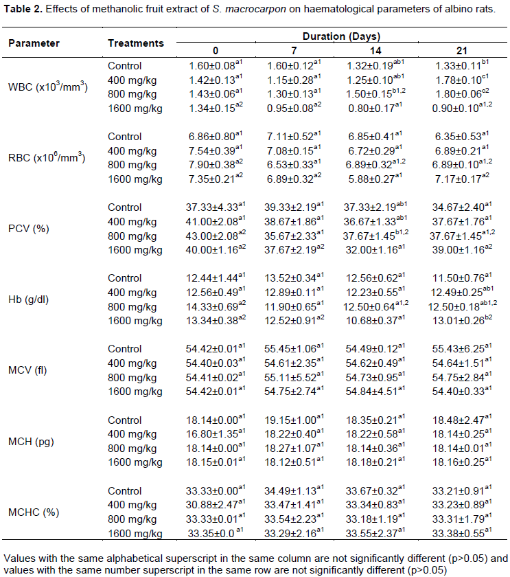
There was no significant difference (p>0.05) in the RBC of the treated rats when compared with the control in all the weeks. However, the treated rats were observed to obtain higher RBC levels than that of the control in week 3, with the highest treatment dose (1600 mg/kg) showing the highest RBC value (7.17±0.17). On the other hand, there was a general non-significant (p>0.05) decrease in the RBC of the treated rats across the treatment period. However, in week 1, the RBC of the rats administered 800 mg/kg of the extract decreased significantly (p<0.05) from its value in week 0, while that of the rats administered 1600 mg/kg increased significantly (p<0.05) in week 3 from its value in week 2.
There was no observed dose-dependent significant difference (p>0.05) in the PCV of the treated rats when compared with those of the control in all the weeks. However, it was noticed that the PCV of the rats administered 1600 mg/kg decreased minimally from that of the control in week 2. On the other hand, whereas there was an overall non-significant decrease (p>0.05) in the PCV of the treated rats across the treatment period, the 800 mg/kg dose level produced a significant decrease
(p<0.05) in the PCV of the rats in week 1, while the 1600 mg/kg dose level showed a significant increase (p<0.05) in week 3.
There was no observed dose-dependent, significant difference (p>0.05) in the haemoglobin of the treated rats when compared with control in all the weeks. However, the 1600 mg/kg treatment dose showed a significantly higher (p<0.05) haemoglobin value (13.01±0.26) than control in week 3. Whereas, the result showed an overall non-significant decrease (p>0.05) in the haemoglobin levels of the treated rats across the weeks, the 800 mg/kg treatment dose showed a significant decrease in haemoglobin levels in week 1, while the 1600 mg/kg dose showed a significant decrease in week 2 followed by a significant increase in week 3.
It was observed that the MCV of the treated rats was not significantly different (p>0.05) when compared with the control in all the weeks. Similarly, there was no significant difference (p>0.05) in MCV of the various treatment doses across the weeks. However, the observations show an increasing trend in the MCV of the treated rats across the duration of treatment with the 800 mg/kg treatment dose showing the highest increase from 54.41±0.02 in week 0 to 54.75±2.84 in week 3. The result showed that there was no significant difference (p>0.05) in the MCH of the treated rats when compared with the control in all the weeks. Similarly, there was no significant difference (p>0.05) in MCH of the various treatment doses across the weeks. However, whereas the 800 mg/kg treatment dose showed an almost constant value from 18.14±0.00 in week 0 to 18.14±0.01 in week 3, the 400 and 1600 mg/kg treatment doses showed a fluctuating non-significant (p>0.05) increases from 16.80±1.35 in week 0 to 18.14±0.25 in week 3 and from 18.15±0.01 in week 0 to 18.16±0.25 in week 3 respectively.
Furthermore, there was no significant difference (p>0.05) in the MCHC of the treated rats when compared with the control in all the weeks. Similarly, there was no significant difference (p>0.05) in MCHC of the various treatment doses across the weeks. However, whereas the 400 mg/kg treatment dose showed a progressive non-significant (p>0.05) decrease from 30.88±2.47 in week 0 to 33.23±0.89 in week 3, the 800 and 1600 mg/kg treatment doses showed a fluctuating non-significant (p>0.05) decrease from 33.33±0.01 in week 0 to 33.31±1.79 in week 3 and a non-significant (p>0.05) increase from 33.35±0.0 in week 0 to 33.38±0.55 in week 3 respectively.
DISCUSSION
Phytochemicals are substances found in edible fruits and vegetables that exhibit a potential for modulating human metabolism in a manner beneficial for the prevention of chronic and degenerative diseases (Tripoli et al., 2007). Thus the medicinal value of plants lies in the presence of these chemical substances that have a definite physiological action in the human body. The phytochemical analysis of the methanolic fruit extract of S. macrocarpon established the presence of such bioactive substances as flavonoids, saponins, alkaloids, phenols, phytates, tannins, cyanides, terpenoids and steroids in decreasing order of abundance. Sodipo et al. (2008) in their phytochemical analyses of S. macrocarpon L. aqueous fruit extract, reported copious presence of alkaloids and flavonoids, and slight presence of tannins, similar to the result of this present study. They, however, reported moderate presence of saponins in contrast to the result obtained in this study. Similarly, Dougnon et al. (2012), who carried out their study in Cotonou (Republic of Benin), reported a high presence of alkaloids, moderate presence of tannins, but absence of flavonoids and saponins in fruits of S. macrocarpon (Solanaceae) contrary to the results of this study.
The observed disparities in comparison with these past works could be explained on the basis of differences in the solvent of extraction (Parekh et al., 2005; Tatiya et al., 2011; Dent et al., 2013). It may also be as a result of differences in the geographical and environmental peculiarities. This is more so as past studies have implicated such factors in affecting the abundance and activities of the phytochemicals (Borokini and Ayodele, 2012; Ubani et al., 2012; Ullah et al., 2012). Flavonoids, saponins and alkaloids have been reported to exert a wide range of biological effects (Sandhar et al., 2011). According to Sudheesh et al. (1997), flavonoids extracted from Solanum melongena fruits showed significant hypolipidaemic action in normal and cholesterol-fed rats. Similarly, antioxidant, anti-inflammatory, hypocholesterolaemic and antimicrobial effects have been reported of flavonoids and saponins (Vinson et al., 1998; Francis et al., 2002; Cushnie and Lamb, 2005; Soetan et al., 2006; Chinedu et al., 2011; Mir et al., 2013). The bitter taste of S. macrocarpon and other eggplants is attributed to the presence of alkaloids, mainly glycoalkaloids (Chinedu et al., 2011). Alkaloids have several pharmacological activities including antihypertensive, antiarrhythmic, antimalarial, anticancer and antiseptic effects (Roberts and Wink, 1998; Soetan, 2008). Vohora et al. (1984) observed significant analgesic effect and some central nervous system depression with crude alkaloidal fraction isolated from S. melongena leaves.
Drugs and plant products are usually subjected to toxicity tests with experimental animals in order to predict safety or toxicity, and to provide guidelines for selecting safe doses in humans. Signs of toxicity include convulsions, tremors, protrusion of eye ball, and mortality (Rajurker et al., 2009). However, as no such signs were observed, the result of this present study, therefore, indicated no toxicity of the methanolic S. macrocarpon fruit extract. This confirms the safety of the plant for consumption as reported by Sodipo et al. (2011) who administered aqueous fruit extract of S. macrocarpon to hyperlipidemic rats. Nevertheless, cases of toxicity have been reported in tuberous Solanum spp. such as Solanum tuberosum (potato) due to the high presence of the glycoalkaloids, alpha-solanine and alpha-chaconine (Schippers, 2001; Langkilde et al., 2008).
Hematological parameters are important indices of physiological and pathological status for both animals and humans, thus playing a major role in disease investigation and diagnosis (Malomo, 2000; Adeneye et al., 2006). White blood cells (WBCs) are an important component of the host defence system, responsible for protection against bacteria, fungi, viruses, and other exogenous substances. The host defense system consists of an intricate cytokine network and progenitor cells, which maintain baseline myelopoiesis (formation of WBCs) and allow rapid adjustment in the rates of production of WBCs in response to acute and chronic stress (Ogawa, 1993; Stock and Hoffman, 2000). Increase in WBC is usually considered as a defensive mechanism by the immune system (Duru et al., 2013).Thus, the observed increases in WBC counts of the treated rats could be as a result of normal response of the animals’ defense system to foreign substances, in this case the plant extract. This is in line with the findings of Duru et al. (2013) who also observed an increase in WBC levels. However, it is possible that the extract had a suppressive effect on the WBCs at the highest dose. This is more so because while at dose levels of 400 and 800 mg/kg the WBC of the treated rats increased from 1.42±0.13 to 1.78±0.10 and 1.43±0.06 to 1.80±0.06 from weeks 0 to 3 respectively, at the 1600 mg/kg dose level, the WBC decreased progressively from 1.34±0.15 to 0.90±0.10.
The observations on the RBC count, PCV and haemoglobin (Hb) in this present study indicated no overall significant effect of the methanolic S. macrocarpon fruit extract on haematological parameters, although there are indications that there could be improvement with prolonged administration of the extract at high doses. This is because the highest dose (1600 mg/kg) administered, showed the highest RBC, PCV and Hb values in week 3.However, the results of this study portray the fact that the balance between the rate of production (erythropoiesis) and destruction of RBCs was not altered. Furthermore, as no adverse effect on serum bilirubin was observed, it is in agreement with the observation on RBC, and further confirms the absence of any haemolytic effect of the extract. In addition, the extract did not seem to stimulate erythropoietin release from the kidneys, which is the humoral regulator of RBC production (Oyedeji and Bolarinwa, 2012). RBCs and Hb are very important in the transfer of respiratory gases (Oyedeji et al., 2012). Hence the plant extract did not appear to affect the oxygen-carrying capacity of the blood. Low values of RBC and associated parameters (Hb and PCV) are indicative of anaemic conditions while very high values predict polycythemia. Thus, the present observation shows that the methanolic fruit extract of S. macrocarpon may not have the potential to induce anemia or polycythemia. Substances which significantly affect the values of RBC and associated parameters have effects on the bone marrow, kidney and haemoglobin metabolism (Young and Maciejewski, 1997; Oyedeji and Bolarinwa, 2012). Therefore, the treatment with the extract may not have had adverse effects on the bone marrow, kidney and haemoglobin metabolism, which is in line with the observations on the parameters studied. However, contrary to the findings of this study, Sodipo et al. (2012) reported a significant increase in haemoglobin (Hb), RBC count and PCV in triton-induced hyperlipidaemic rats treated with aqueous fruit extract of S. macrocarpon. Similarly, Duru et al. (2013) observed a significant increase in Hb and PCV when they incorporated powdered S. macrocarpon fruit into rat feed. Therefore, we can infer that the S. macrocarpon fruit in aqueous medium or in powdered form is more effective in improving haematological parameters than the methanolic extract.
The observed no significant effect of the methanolic S. macrocarpon fruit extract on the red cell indices (MCV, MCH and MCHC) were similar to those of Sodipo et al. (2012) and Duru et al. (2013), and implies that the incorporation of haemoglobin into RBC as well as the morphology and osmotic fragility of the RBC were not altered. Haemoglobin, RBC and PCV are associated with the total population of RBC while MCHC and MCH relate to individual RBC (Adebayo et al., 2010). Increased MCV and MCH values are indicative of macrocytic anaemia (Oyedeji et al., 2012). Thus, in addition, the methanolic S. macrocarpon fruit extract did not seem to have the potential to cause macrocytic anaemia in the treated animals.
In conclusion, the phytochemicals detected in this plant extract attest to the tremendous potentials of the plant. The acute toxicity study confirms the safety of its consumption. The results of this present study herein presented, revealed that the methanolic fruit extract of S. macrocarpon, at the administered doses and duration did not cause any serious adverse effect on haematological parameters. Thus, the extract did not impair the functional capabilities of the blood cells; hence we can assert that the fruits of the plant S. macrocarpon may not have any harmful effect on the consumers.
CONFLICT OF INTERESTS
The authors have not declared any conflict of interests.
REFERENCES
|
Abdulrahman FI, Akan JC, Sodipo OA, Onyeyili PA (2010). Effect of aqueous root-bark extract of Vitexdomina sweet on hematological parameters in rats. Journal of American Science 6:8-12. |
|
|
Adebayo AH, Abolaji AO, Opata TK, Adegbenro IK (2010). Effects of ethanolic leaf extract of Chrysophyl lumalbidum G. on biochemical and haematological parameters of albino wistar rats. African Journal of Biotechnology 9(14):2145-2150. |
|
|
Adeneye AA, Ajagbonna OP, Adeleke TI, Bello SO (2006). Preliminary toxicity and phytochemical studies of the stem bark aqueous extract of Musanga cecropioides in rats. Journal of Ethnopharmacology 105(3):374-379. |
|
|
Anaga AO, Ezeja MI, Amuzie CJ (2010). Investigation of the aqueous fruit extract of Lagenaria siceraria for pharmacological activities in vitro and in vivo. International Journal of Current Research 9:14-19 |
|
|
Baker FJ, Silverton RE, Pallister CJ (2001). Staining procedures. Introduction to Medical Laboratory Technology. |
|
|
Borokini TI, Ayodele AE (2012). Phytochemical screening of Tacca leontopetaloides (L.) Kuntze collected from four geographical locations in Nigeria. International Journal of Modern Botany 2(4):97-102. |
|
|
Bukenya-Ziraba R, Bonsu KO (2004). Solanum macrocarpon L. [Internet] Record from Protabase. Grubben, G. J. H. and Denton, O. A. (Eds.), PROTA (Plant Resources of Tropical Africa/Ressource Vegetales de l'AfriqueTropicale), Wageningen, Netherlands. |
|
|
Burkhill HM (2000). The Useful Plants of West Tropical Africa, 2nd Edition, Vol. 5, Royal Botanic Gardner, London, UK. |
|
|
Cheeke PR (1998). Natural toxicants in feeds, forages and poisonous plants.2 Edition. IL: Interstate Publishers, Danville |
|
|
Chinedu SN, Olasumbo AC, Eboji OK, Emiloju OC, Arinola OK, Dania DI (2011). Proximate and phytochemical analyses of Solanum aethiopicum L. and Solanum macrocarpon L. fruits.Research Journal of Chemical Sciences 1(3):63-71. |
|
|
Coles EH (1986). Veterinary Clinical Pathology, 4th Edition, W. B. Saunders Co., Philadelphia, USA. |
|
|
Cushnie TPT, Lamb AJ (2005). Antimicrobial activity of flavonoids. International Journal of Antimicrobial Agents 26(5):343-356. |
|
|
Dent M, Dragovic-Uzelać V, Penić M, BrnÄić M, Bosiljkov T, Levaj B (2013). The effect of extraction solvents, temperature and time on the composition and mass fraction of polyphenols in Dalmatian wild sage (Salvia officinalis L.) extracts. Food Technology and Biotechnology 51(1):84-91. |
|
|
Dougnon TV, Bankolé HS, Johnson RC, Klotoé JR, Dougnon G, Gbaguidi F, Assogba F, Gbénou J, Sahidou S, Atègbo JM, Rihn BH, Loko F, Boko M, Edorh AP (2012). Phytochemical screening, nutritional and toxicological analyses of leaves and fruits of Solanum macrocarpon Linn (Solanaceae) in Cotonou (Benin). Food and Nutrition Sciences 3:1595-1603. |
|
|
Duru M, Ugbogu A, Amadi B (2013). Effect of Solanummacrocarpon fruit on haematology, hepatic and renal function. Advances in Biochemistry 1(2):28-32. |
|
|
Ezeonu CS, Ejikeme CM (2016). Qualitative and Quantitative Determination of Phytochemical Contents of Indigenous Nigerian Softwoods. New Journal of Science, 2016: 9 pages. Article ID 5601327. |
|
|
Francis G, Kerem Z, Makkar HPS, Becker K (2002). The biological action of saponins in animal systems: a review. British Journal of Nutrition 88:587-605. |
|
|
Gbile ZO, Adesina SK (1988). Nigerian Solanum species of economic importance. Annals of the Missouri Botanical Garden 75:862-865. |
|
|
Grubben GJH, Denton OA (2004). Plant Resources of Tropical Africa II: Vegetables. Ponen and Looijenhv, Wageningen, Netherland. |
|
|
Harbone JB (1973). Phytochemical Methods: A guide to Modern Techniques of Plant Analysis. Chapman and Hall, New York. |
|
|
Hoff J (2000). Methods of blood collection in the mouse. Lab Animal 29(10):47-53. |
|
|
Kariuki AC, Njoroge GN (2011). Ethnobotanical and antimicrobial studies of some plants used in Kibwezi (Kenya) for management of lower respiratory tract infections. African Journal of Traditional, Complementary and Alternative Medicines 8(2):144-149. |
|
|
Langkilde S, Schrøder M, Stewart D, Meyer O, Conner S, Davies H, Poulsen M (2008). Acute toxicity of high doses of the glycoalkaloids, alpha-solanine and alpha-chaconine, in the Syrian golden hamster. Journal of Agricultural and Food Chemistry 56(18):8753-8760. |
|
|
Lorke D (1983). A new approach to practical acute toxicity testing. Archives of Toxicology 54:275-287. |
|
|
Malomo AO (2000). Toxicological implications of certriaxone administration in rats. Nigerian Journal of Biochemistry and Molecular Biology pp. 1533-1538. |
|
|
Mir MA, Sawhney SS, Jassal MMS (2013).Qualitative and quantitative analysis of phytochemicals of Taraxacum officinale. Wudpecker Journal of Pharmacy and Pharmacology 2(1):1-5. |
|
|
Mishra N, Tandon VL (2012). Haematological effects of aqueous extract of Ornamental plants in male Swiss albino mice. Veterinary World 5(1):19-23. |
|
|
Mujeeb F, Bajpai P, Pathak N (2014). Phytochemical evaluation, antimicrobial activity, and determination of bioactive components from leaves of Aegle marmelos. BioMed research international 2014. |
|
|
Obadoni BC, Ochuko PO (2001). Phytochemical studies and comparative efficacy of the crude extracts of some home static plants in Edo and Delta States of Nigeria. Global Journal of Pure and Applied Sciences 8:203-208. |
|
|
Oberleas D (1973). Phytates in Toxicants Occurring Naturally in Foods.National Academy of Sciences, Washington DC. |
|
|
Ogawa M (1993). Differentiation and proliferation of hematopoietic stem cells. Blood 81:2844-2853. |
|
|
Okigbo RN, Mmeka EC (2006). An appraisal of phytomedicine in Africa.King Mongkut's Institute of Technology Ladkrabang Science and Technology Journal 6:83-94. |
|
|
Onwuka GI (2005). Food Analysis and Instrumentation Theory and Practice.Naphthali Prints, Lagos, Nigeria. |
|
|
Oyedeji KO, Bolarinwa AF (2012). Effects of crude extracts of Portula caoleracea on haematological and biochemical parameters in albino rats.African Journal of Biomedical Research 15:41-47. |
|
|
Oyedeji KO, Bolarinwa AF, Owaduge DO (2012). Effects of some alcoholic beverages on haematological and plasma biochemical parameters in male albino rats. Research Journal of Medical Sciences 6(6):286-289. |
|
|
Parekh J, Jadeja D, Chanda S (2005). Efficacy of aqueous and methanol extracts of some medicinal plants for potential antibacterial activity. Turkish Journal of Biology 29:203-210. |
|
|
Pearson DA (1976). The Chemical Analysis of Foods, 7th Edition, Churchill and Livingstone, Edinburgh, London. |
|
|
Rajurker S, Rekhe DS, Maini S, Ravikanth K (2009). Acute toxicity studies of polyherbal formulation (Methiorep premix). Veterinary World 2(2):58-59. |
|
|
Roberts MF, Wink M (1998). Alkaloids: biochemistry, ecology and medicinal applications. Plenum Press, New York. |
|
|
Sandhar HK, Kumar B, Prasher S, Tiwari P, Salhan M, Sharma P (2011). A review of phytochemistry and pharmacology of flavonoids. InternationalePharmaceuticaSciencia 1(1):25-41. |
|
|
Schippers RR (2001). Domestication of indigenous vegetables for Sub-Saharan Africa. National Resource Institute, Chatham, U.K. Technical Report pp. 201-222. |
|
|
Sodipo OA, Abdulrahman FI, Akan JC, Akinniyi JA (2008). Phytochemical screening and elemental constituents of the fruit of Solanum macrocarpon Linn. Continental Journal of Applied Sciences 3:85-94. |
|
|
Sodipo OA, Abdulrahman FI, Sandabe UK, Akinniyi JA (2011). Effects of the aqueous fruit extract of Solanum macrocarpon Linn on the hematological parameters of triton-induced hyperlipidemic rats. African Journal of Pharmacy and Pharmacology 5(5):632-639. |
|
|
Sodipo OA, Abdulrahman FI, Sandabe UK (2012). Effects of the aqueous fruit extract of Solanum macrocarpon Linn. on hematological parameters of chronic triton-induced hyperlipidemic rats. Journal of Physiology and Pharmacology Advances 2(2):122-132. |
|
|
Soetan KO (2008). Pharmacological and other beneficial effects of antinutritional factors in plants-A review. African Journal of Biotechnology 7(25):4713-4721. |
|
|
Soetan KO, Oyekunle MA, Aiyelaagbe OO, Fafunso MA (2006). Evaluation of the antimicrobial activity of saponins extract of Sorghum bicolor L. Moench. African Journal of Biotechnology 5(23):2405-2407. |
|
|
Sood R (2006). Textbook of Medical Laboratory Technology.Jaypee Brothers Medical Publishers Ltd., New Delhi, India. |
|
|
Stock W, Hoffman R (2000). White blood cells 1: non-malignant disorders. Lancet 355:1351-1357. |
|
|
Sudheesh S, Presannakumar G, Vijayakumar S, Vijayalashmi NR (1997). Hypolipidemic effect of flavonoids from Solanum melongena, Plant foods for Human Nutrition 51:321-330. |
|
|
Tatiya AU, Tapadiya GG, Kotecha S, Surana SJ (2011). Effect of solvents on total phenolics, antioxidant and antimicrobial properties of Bridelia retusa stem bark. Indian Journal of Natural Products and Resources 2(4):442-447. |
|
|
Tripoli E, Guardia ML, Giammanco S, Majo DD, Giammanco M (2007). Citrus flavonoids: molecular structure, biological activity and nutritional properties - a review. Food Chemistry 104:466-479. |
|
|
Ubani CS, Oje OA, Ihekogwo FNP, Eze EA, Okafor CL (2012). Effect of varying soil minerals and phytochemical parameters on antibacterial susceptibility of Mitracarpus villosus ethanol extracts using samples from south east and south-southern regions of Nigeria. Global Advanced Research Journal of Microbiology 1(7):120-125. |
|
|
Uhegbu FO, Ogbuehi KJ (2004). Effect of aqueous extract (crude) of leaves of Vernonia amygdalina (Del.) on blood glucose, serum albumin and cholesterol levels in diabetic albino rats. Global Journal of Pure and Applied Sciences 10(1):189-194. |
|
|
Ullah N, Khurram M, Amin MU, Khan TA, Khayyam SU, Khan FA, Najeeb U, Ullah S (2012). Impact of geographical locations on Menthaspicata antibacterial activities. Journal of Medicinal Plants Research 6(7):1201-1206. |
|
|
Vinson JA, Hao Y, Su X, Zubik L (1998). Phenol antioxidant quantity and quality in foods: vegetable. Journal of Agricultural and Food Chemistry 48:3630-3634. |
|
|
Vohora SB, Kumar I, Khan MS (1984). Effect of alkaloids of Solanummelongena on the central nervous system.Journal of Ethnopharmacology 11(3):331-336. |
|
|
World Health Organization (WHO) (2002).World Health Organization fact sheet No. 271. World Health Organization, Geneva. |
|
|
Young NS, Maciejewski J (1997). The pathophysiology of acquired aplastic anemia. New England Journal of Medicine 336:1365-1372. |
|
Copyright © 2024 Author(s) retain the copyright of this article.
This article is published under the terms of the Creative Commons Attribution License 4.0


