Full Length Research Paper
ABSTRACT
This study determined plasma and liver tissue benzene, toluene, ethylene and xylene (BTEX), malondialdehyde (MDA), reduced glutathione (GSH), superoxide dismutase (SOD) and glutathione peroxidase (GPx) status in rats orally exposed to gasoline. Sixteen adult male Wistar rats (210.0 ± 20.0 g), distributed into control and test groups, of eight rats each were used. Rats in test group were administered 2.0 mLkg-1 body weight gasoline for sixty days, while control rats were given distilled water. The animals were sacrificed after exposure period, and relevant tissues collected and processed for analyses. BTEX concentrations in plasma and liver tissue homogenates were determined by gas chromatography with flame ionized detector (GC-FID), while SOD and GPx activities, as well as MDA and GSH levels were determined by standard spectrophotometric methods. The results showed a significantly (p<0.05) higher plasma and liver tissues BTEX concentrations in test rats, compared to the control. This shows that BTEX are among the gasoline hydrocarbons that are largely absorbed from the gastrointestinal tract (GIT), and distributed within the blood and liver tissues. Also, the levels of plasma and liver tissue MDA were significantly (p<0.05) higher, while SOD, GPx, and GSH status were significantly lower in test rats, compared respectively to control; an indication of gasoline-induced oxidative stress. These results suggest that the raised plasma and liver tissue MDA, and reduced SOD, GPx and GSH activities in test animals may be attributed to the raised tissue BTEX levels; concluding that BTEX, and/or their metabolites, are implicated in gasoline-induced oxidative stress in rats.
Key words: Gasoline, oxidative stress, benzene, toluene, ethylene and xylene (BTEX), rats.
INTRODUCTION
Gasoline is a highly flammable and volatile liquid that is commonly used widely as fuel and solvent for some domestic, industrial and transport activities in most societies. It evaporates rapidly to release vapours with high proportion of different hydrocarbon pollutants into the immediate environment. Hence, gasoline is one of the chemical substances that release different types of hydrocarbons into the environment (Verma and des Tombe, 2002; Caselli et al., 2010; Saxena and Ghosh, 2012). Particularly, benzene, toluene, and xylene (BTX) have been reported to be among the major gasoline’s chemical constituents of health concern (Periago and Prado, 2005). Exposure to gasoline is therefore likely to introduce different types of hydrocarbons into the body tissues (Egeghy et al., 2000; Rekhadevi et al., 2010; Tunsaringkarn et al., 2013). Typically, significant levels of toluene and xylene have been detected in the blood of both petrol station attendants and individuals who regularly refuel their vehicles (Tunsaringkarn et al., 2013, 2011). Assessment of the health risk associated with exposure to gasoline generated hydrocarbons may also be based on the extrapolations from data obtained from studies on laboratory animals.
It has been reported in the literature that exposure to several chemical agents, including gasoline constituents, produce various tissue toxicities in different species of animals, as well as human beings. Particularly, polycyclic aromatic hydrocarbons (PAHs) have been demonstrated to cause adverse effects on the immune system and neurodevelopmental processes at different stages of human development, as well as reduction in feotal growth rate (Perera et al., 2003; Tang et al., 2006). Also, evidence of a variety of developmental abnormalities, including reduced birth weight, impaired cognitive development, and increased susceptibility to procarcinogenic DNA damage, have reported to be associated with prenatal exposure to relatively low levels of PAHs (Perera et al., 2003; Tang et al., 2006; Dede and Kagbo, 2001; Gbadebo et al., 2009).
Toxicity effects of chemical agents on the body tissues are induced through different mechanisms, and oxidative stress tends to be one of the important underlying mechanisms implicated in chemical induced toxicities. For instance, exposures to gasoline and diesel have been reported to increase tissue lipid peroxidation product, malonyl dialdehyde (MDA) concentration, reduced catalase, superoxide dismutase and glutathione peroxidase activities, indication of oxidative stress induced toxicities in experimental animals (Li et al., 2003; Eriyamiemu et al., 2007). Among these diesel, gasoline and kerosene induced oxidative stress toxicity effects reported in our earlier studies include the hepatotoxicity, haematotoxicity, nephrotoxicity and reproductive toxicity 16-24. These toxicity effects are likely to be caused by the chemical substances (including hydrocarbons), or their metabolites, introduced by the solvents into the body.
In the course of industrial and domestic utilization, gasoline in liquid and gaseous forms commonly falls out and contaminates water and food substances in the environment. Individuals consuming such contaminated water and food substances are likely to be exposed to gasoline orally. This study was therefore designed to determine the hydrocarbons (BTEX) and oxidative stress markers status in the plasma and liver tissue of rats orally exposed to gasoline.
MATERIALS AND METHODS
Chemicals
Purge-and-trap-grade methanol used to rinse the glassware and prepare all standards was procured from Burdick and Jackson (Muskegon, MI). Analytical reagent kits for MDA and GSH assay, as well as reagents for potassium chloride phosphate buffer preparation, ethylenediamine tetra - acetic acid and sodium chloride were purchased from Sigma (St. Louis, MO, USA). Benzene, toluene, ethylene and xylene standards were purchased from Sigma-Aldrich Company with purity 99.9%. All other chemicals used in the microextratction and gas chromatographic analysis of samples were of standard analytical grade, and were obtained from Microliter Inc. (Suwanee, GA) and Agilent Technologies (Palo Alto, CA).
Animals handling and treatment
Sixteen male adult albino Wistar rats weighing 210±20 g were randomly distributed into two groups, of eight animals each. The animals were obtained from the Animal House of Biochemistry Department, University of Calabar, and were housed in plastic cages, with metallic top. The animals were kept under the prevailing normal tropical room temperature and humidity, and a 12-h light/dark cycle. Animals were allowed free access to standard laboratory animal feeds (growers’ pellets from Guinea Feeds, Benin, Nigeria) and water ad libitum. The rats were acclimatized for one week before the commencement of the experimental treatments. After the acclimatization period, rats in the test group were orally gavaged 2 mLkg-1 body weight of gasoline in sunflower oil as vehicle, daily for sixty (60) days; while rats in the control group were gavaged with 0.5 mL of distilled water since previous study showed no significant adverse effect of sunflower oil compared to distilled water (Uboh et al., 2013). The procedure adopted in this study for animal handling was in accordance with the guidelines on the care and well being of research animals of NIH (1985) (King and Wootton, 1956), and was approved by the Department of Biochemistry Ethics Committee.
Blood collection and preparation of liver homogenate
At the end of the 60 days of experimental treatment, the rats were sacrificed after 24 h fasting. Whole blood samples were collected by cardiac puncture into Ethylenediamine tetra-acetic acid (EDTA; pH 7.4) sample bottles using 5 ml syringe with needle, while the liver organ tissues were removed, rinsed in distilled water and refrigerated for 24 h. The blood samples were centrifuged using table top centrifuge (MSE, England), and plasma separated for hydrocarbons and oxidative stress markers analyses. The refrigerated liver organ tissues were excised, weighed and homogenized in Potassium Chloride (KCl, 10 mM) phosphate buffer (1.15%) with Ethylenediamine tetra - acetic acid (EDTA; pH 7.4) using laboratory. The homogenate obtained was centrifuged at 12,000 × g for 60 min to obtain the tissue supernatant which was also used to determine the hydrocarbons and oxidative stress markers status.
Determination of oxidative stress makers
The oxidative stress markers determined in this study included reduced glutathione (GSH), glutathione peroxidase (GPx), superoxide dismutase (SOD) and malondialdehyde (MDA) levels in plasma and liver tissue homogenate. The concentrations of GSH and MDA were determined spectrophotometrically following the methods described by King and Wootton (1956) and Wallin et al. (1993), respectively; while the activities of SOD and GPx were determined by the methods described by Paglia and Valentine (1967), respectively.
Determination of hydrocarbons concentrations
The concentrations of BTEX in the plasma and liver tissue homogenate supernatant were determined by gas chromatography with flame ionized detector (GC-FID) using modified Headspace-Solid Phase Microextraction technique described by Tunsaringkarn et al. (2004). In this technique, 0.2 g of sodium chloride was added to 0.5 mL of blood and liver tissue homogenate samples in the respective capped glass bottles. The contents of the respective sample bottles were vortexed for 15 s, controlled and absorbed by solid phase microextraction in water bath (50°C) with sonication for 20 min. The samples, so treated were then respectively injected into gas chromatograph (GC, Varian CP 3800) at temperature 220°C using flame ionization detector at 220°C (column CP-SIL5 CB, splitless). The samples were put in the oven for 10 min at the starting temperature of 50°C. The temperature was increased at 5°C min-1 until 90°C was reached. Then, again the temperature was increased at 30°C min-1 until 250°C was reached and stayed for 17 min. The quantity of blood benzene was analysed under relative intensity of chromatographic signal for 40 min. Following the validation method of Mahatein (http://www.reo06.net/home/images/upload/file/report/jurairat070509.pdf), the limit of quantitations (LOQs) of ten times of the standard deviation at minimum concentration was carried out. LOQs for benzene, toluene, ethylbenzene and xylene were 33.30, 16.70, 66.70 and 33.30 μgL-1 (ppb), respectively. Average coefficient of determination (r2) was 0.99 for the chemicals (m-and p-xylene appeared at the same peak).
Data analysis
Data were reported as the mean values and standard deviations (mean±SD). Statistical differences between the test and control groups were determined by Student’s t test, and a p-value less than 0.05 was considered statistically significant.
RESULTS
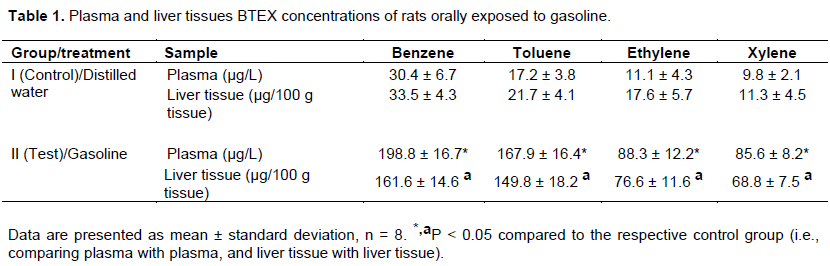
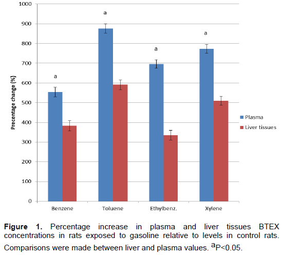
The results of this study are presented in Tables 1 and 2, and Figure 1. The plasma and liver tissue BTEX concentrations recorded for rats exposed to gasoline are presented in Table 1; while Table 2 shows the plasma and liver tissue SOD and GPx activities, as well as GSH and MDA concentrations of rats exposed to gasoline. Figure 1 presents the comparative percentage increase in plasma and liver tissue BTEX concentrations following oral exposure to gasoline. The results showed that the plasma and liver tissues concentrations of BTEX obtained for rats exposed to gasoline were significantly (p<0.05) higher, compared respectively to the control values. However, the percentage increase in plasma BTEX concentrations were significantly (p<0.05) higher, compared to the percentage increase in liver tissue BTEX concentrations in rats exposed to gasoline (Figure 1). This suggests that BTEX are among the gasoline hydrocarbons with higher GIT absorption rate; and that the blood partition coefficients of BTEX are likely to be higher than the liver tissues partition coefficients of BTEX in rat model, following oral exposure to gasoline.

It was also observed from the results of this study that the activities of SOD and GPx, as well as GSH concentration were significantly (p<0.05) lower, while MDA concentration was significantly (p<0.05) higher in rats exposed to gasoline, when compared respectively to the values recorded for rats in the control group (Table 2). This represents an indication of oxidative stress in rats orally exposed to gasoline.
DISCUSSION
This study was designed to determine the types and concentrations of hydrocarbons accumulated in the plasma and liver tissues, and oxidative stress status, in rats orally exposed to gasoline. In our previous studies, inhalation exposure to gasoline vapours/fumes was reported to produce oxidative stress related toxicities in several biological tissues19-24. The results of this study showed that oral exposure of rats to gasoline resulted in the accumulation of BTEX within the liver tissues and plasma fluid. However, the levels of BTEX accumulated in the plasma were observed to be higher than the levels accumulated in the liver tissues. The result of this present study, agrees with the report of Tunsaringkarn et al. (2013) on the correlation between haematological parameters and BTEX in gasoline station workers.
According to van Schooten et al. (1997), measurements of PAH in feces after oral administration of pure PAH indicated that only an insignificant proportion of the orally administered PAH is excreted in unchanged form. The results of the study therefore suggested rapid absorption and high bioavailability of PAH in blood after oral intake (Van de Wiel et al., 1993). The results of our present study suggested that BTEX contents of gasoline are readily absorbed from the GIT following oral dose ingestion. This observation indicates a higher bioaccumulation of BTEX in the blood than the liver tissues following oral ingestion of gasoline. BTEX have been reported to be among the major aromatic hydrocarbon constituents in the ambient air of gasoline and gas refill stations (Keprasertsup et al., 2003; Yimrungruang et al., 2008; Thaveevongs et al., 2010; Correa et al., 2012). These reports suggest that BTEX constitute a good proportion of the volatile hydrocarbon constituents of gasoline. However, National Institute for Occupational Safety and Health (NIOSH, 1990), Agency for Toxic Substances and Disease Registry (ATSDR, 2007), Weiselm (2010) and McHale et al. (2012) demonstrated that benzene is not directly toxic as a parent compound, but that it may be biotranformed into reactive metabolites by hepatic tissues metabolizing system.
Like other xenobiotics (Aseervatham et al., 2013; Sengupta and Banerjee, 2014), BTEX reported to be introduced into the body by gasoline in this study are likely to be metabolized into various metabolites in the liver. Hydrocarbons, including the aliphatic and aromatic hydrocarbons, which are known to be the major constituents of petroleum products, are basically metabolized in the liver tissue (Otitoloju and Olagoke, 2011; King et al., 2012). This may accounts for the comparative lower liver tissues BTEX levels reported in this study, against the plasma levels. It has been reported that benzene and toluene may readily be metabolized into such reactive metabolites that can pose several toxicity effects to the body tissues. Metabolism of these hydrocarbons are therefore likely to generate some reactive metabolites/free radicals into the body tissues (Otitoloju and Olagoke, 2011; King et al., 2012). Generation of these reactive metabolites may therefore be considered to be implicating factors in the hepatotoxicity, haematotoxicity, nephrotoxicity and reproductive toxicity earlier reported to be associated with exposure to gasoline (Uboh et al., 2005a, b, 2009, 2007a, b, c, 2008a, b, 2010, 2013; Otitoloju and Olagoke, 2011; King et al., 2012; Barath et al., 2010; Abubakar et al., 2013).
It is also recorded in this study that oral exposure to gasoline produced a significant increase in malondialdehyde (MDA) concentration, and decrease in GSH, GPx and SOD activities in the plasma and liver tissues. This increased MDA concentration recorded in this study supports the earlier literature report that MDA, a product of lipid peroxidation, is a useful biomarker for petroleum polycyclic aromatic hydrocarbons toxicity effects (Otitoloju and Olagoke, 2011; King et al., 2012). Also, literature reports document marked reduction in tissue glutathione (GSH) concentration, GPx and SOD activities, as well as increase in tissue MDA concentration as known indicators of oxidative stress (Eriyamremu et al., 2007). This study therefore indicated that oral exposure to gasoline is capable of inducing oxidative stress in rats. Also, it may be suggested that the reactive species that likely to be generated from BTEX metabolism may be responsible for the oxidative stress observed to be induced by exposure to gasoline in this study.
Toxicological effects of toxicants have been reported to be associated with interference between the toxicants and the cellular and sub-cellular processes, resulting in a disruption of the normal cellular metabolic activities (Rekhadevi et al., 2010; Perera et al., 2003; McHale et al., 2012). This study reports a strong correlation between liver tissue oxidative stress and accumulation of hydrocarbons in the plasma and liver tissues of rats orally exposed to gasoline. The result of this study also give strong indication that the hydrocarbons observed to be accumulated in the plasma and liver tissues of rats exposed to gasoline are likely to be among the causes of oxidative stress damage of the liver tissues. This observation supports the reports of previous studies on the oxidative stress effect of petroleum fractions (Eriyamremu et al., 2007; Uboh et al., 2007b; Aseervatham et al., 2013). This study disclosed how exposure to gasoline may results in the accumulation of BTEX in the plasma and tissues, suggesting BTEX to be responsible for the observed oxidative stress induced liver function impairment. It may then be postulated that oxidative stress is one of the mechanisms of action of petroleum (gasoline) hydrocarbon-induced liver dysfunction. Generally, benzene is a known carcinogen in humans, whereas, toluene, ethylbenzene and xylene are not known to be direct carcinogen in humans. High blood benzene levels have been reported in gasoline workers in Bangkok (Tunsaringkasrn et al., 2011). It may therefore be concluded that BTEX and their metabolites, are among the chemical agents responsible for gasoline-induced oxidative stress in rats.
CONFLICT OF INTERESTS
The authors have not declared any conflict of interests.
REFERENCES
|
Abubakar MB, Abdullah WZ, Sulaiman SA, Uboh FE, Ang BS (2013). Effect of honey supplementation on toxicity of gasoline vapor exposure in rats. International Journal of Applied Research in Natural Products 6(4):16-22. |
|
|
Aseervatham GSB, Sivasudha T, Jeyadevi R, Ananth DA (2013). Environmental factors and unhealthy lifestyle influence oxidative stress in humans-an overview. Environmental Science and Pollution Research 20(7):4356-4369. |
|
|
Agency for Toxic Substances and Disease Registry (ATSDR) (2007). Toxicological Profile For Benzene. U.S. Department Of Health And Human Services. Agency for Toxic Substances and Disease Registry, Atlanta, GA, 2007. |
|
|
Barath S, Mills NL, Lundbäck M, Törnqvist H, Lucking AJ, Langrish JP, Donaldson K (2010). Impaired vascular function after exposure to diesel exhaust generated at urban transient running conditions. Particle and Fibre Toxicology 7(1):19. |
|
|
Caselli M, de Gennaro G, Marzocca A, Trizio L, Tutino M (2010). Assessment of the impact of the vehicular traffic on BTEX concentration in ring roads in urban areas of Bari (Italy). Chemosphere 81(3):306-311. |
|
|
Correa SM, Arbilla G, Marques MR, Oliveira KM (2012). The impact of BTEX emissions from gas stations into the atmosphere. Atmospheric Pollution Research 3(2):163-169. |
|
|
Dede EB, Kagbo HD (2001).Investigation of acute toxicological effects of diesels fuel in rats (Rattus rattus) using Histopathological methods. Journal of Applied Sciences and Environmental Management 5(1): 83-84. |
|
|
Egeghy PP, Tornero-Velez R, Rappaport SM (2000). Environmentaland biological monitoring of benzene during self-service automobile refueling. Environmental Health Perspectives 108(12):1195-1202. |
|
|
Eriyamremu GE, Osagie VE, Omoregie SE, Omofoma CO (2008). Alterations in glutathione reductase, superoxide dismutase, and lipid peroxidation of tadpoles (Xenopus laevis) exposed to Bonny Light crude oil and its fractions. Ecotoxicology and Environmental Safety 71(1):284-290. |
|
|
Gbadebo AM, Taiwo AM, Ola OB (2009). Effects of crude oil and spent oil on Clarias garipinus: a typical marine fish. American Journal of Environmental Sciences 5(6):753-758. |
|
|
Keprasertsup C, Bashkin V, Wangwongwatana S, Pokethitiyook P, Adsavakulchai S, Towprayoon S (2003). Concentrations of MTBE, Benzene, Toluene, Ethylbenzene, and Xylene in Ambient Air at Gas Stations and Traffic Area in Bangkok. Proceedings of the 2nd Regional Conference on Energy Technology Towards a Clean Environment, Phuket, Thailand; 12-14: p 5032(O). |
|
|
King EJ, Wootton I (1956). Micro-analysis in Medical Biochemistry. J. & A. Churchill |
|
|
King MA, Sogbanmu TO, Osibona AO, Doherty VF, Otitoloju AA (2012). Toxicological evaluation and usefulness of lipid peroxidation as biomarker of exposure to crude oil and petroleum products tested against African catfish, Clarias gariepinus and hermit crab, Clibanarius africanus. Nature Environment and Polution Technology 11(1):1-6. |
|
|
Li N, Hao M, Phalen RF, Hinds WC, Nel AE (2003). Particulate air pollutants and asthma: a paradigm for the role of oxidative stress in PM-induced adverse health effects. Clinical Immunology 109(3):250-265. |
|
|
McHale CM, Zhang L, Smith MT (2012). Current understanding of the mechanism of benzene-induced leukemia in humans: implications for risk assessment. Carcinogenesis 33(2):240-252. |
|
|
National Institute of Health (NIH) (1985). Guide for the care and use of laboratory animals. DHEW Publication. Office of Science and Health Reports, Bethesda, U.S.A. |
|
|
National Institute for Occupational Safety and Health (NIOSH) (1990). National Occupational Exposure Survey (1981-83), Cincinnati, OH, 1990. |
|
|
Otitoloju A, Olagoke O (2011). Lipid peroxidation and antioxidant defense enzymes in Clarias gariepinus as useful biomarkers for monitoring exposure to polycyclic aromatic hydrocarbons. Environmental Monitoring and Assessment 182(1-4):205-213. |
|
|
Paglia DE, Valentine WN (1967). Studies on the quantitative and qualitative characterization of erythrocyte glutathione peroxidase. The Journal of laboratory and clinical medicine 70(1):158-169. |
|
|
Perera FP, Rauh V, Tsai WY, Kinney P, Camann D, Barr D, Dietrich J (2003). Effects of transplacental exposure to environmental pollutants on birth outcomes in a multiethnic population. Environmental Health Perspectives 111(2):201-205. |
|
|
Periago JF, Prado C (2005). Evolution of occupational exposure to environmental levels of aromatic hydrocarbons in service stations. Annals of Occupational Hygiene 49(3):233-240. |
|
|
Rekhadevi PV, Rahman MF, Mahboob M, Grover P (2010). Genotoxicity in filling station attendants exposed to petroleum hydrocarbons. Annals of Occupational Hygiene 54(8):944-954. |
|
|
Saxena P, Ghosh C (2012). A review of assessment of benzene, toluene, ethylbenzene and xylene (BTEX) concentration in urban atmosphere of Delhi. International Journal of the Physical Sciences 7(6):850-860. |
|
|
Sengupta P, Banerjee R (2014). Environmental toxins: Alarming impacts of pesticides on male fertility. Human and Experimental Toxicology 33(10):1017-1039. |
|
|
Tang D, Li TY, Liu JJ, Chen YH, Qu L, Perera F (2006). PAH-DNA adducts in cord blood and fetal and child development in a Chinese cohort. Environmental Health Perspectives 114(8):1297-1300. |
|
|
Thaveevongs P, Panyamateekul S, Prueksasit T (2010). Exposure risk assessment of volatile organic compounds (VOCs) of the workers at gas station in Bangkok. Engineering Journal 2(3):1-12. |
|
|
Tunsaringkarn T, Choochat N, Theppitaksak B (2004). Headspace -Solid Phase Microextraction for determination of benzene, toluene, ethylbenzene, xylene and MTBE in blood. Journal of Health Research 18(1):4959 |
|
|
Tunsaringkarn T, Soogarun S, Rungsiyothin A, Zapuang K, Chapman RS (2011). Health status of gasoline station workers in Pathumwan Area, Bangkok, Thailand, in 2004 and 2009. Journal of Health Research 25(1):15-19. |
|
|
Tunsaringkarn T, Zapuang K, Rungsiyothin A (2013). Correlation between blood cell parameters and BTEX exposure among gasoline station workers. Journal of Environmental and Occupational Science 2(1):15-20. |
|
|
Tunsaringkasrn T, Ketkaew P, Zapaung K, Rugsiyothin A, Taneepanichkul S (2011). Risk Ratio of Benzene, Toluene, Ethylbenzene, and Xylene (BTEX) Exposures and Their Relations to Biological Parameters of Gasoline Workers in Bangkok, Thailand. Applied Environmental Research 33(1):27-38. |
|
|
Uboh FE, Akpanabiatu MI, Alozie Y (2008). Comparative effect of gasoline vapours on renal functions in male and female albino wistar rats. Journal of Pharmacology and Toxicology 3(6):478-484. |
|
|
Uboh FE, Akpanabiatu MI, Atangwho IJ, Alozie Y (2008). Exposure to gasoline vapours: a potential risk factor for atherosclerosis in male and female rats. Journal of Pharmacology and Toxicology 3(8):600-609. |
|
|
Uboh FE, Akpanabiatu MI, Atangwho IJ, Ebong PE, Umoh IB (2007). Effect of gasoline vapours on serum lipid profile and oxidative Stress in hepatocytes of male and female rats. Acta Toxicologica 15(1):25-30 |
|
|
Uboh FE, Akpanabiatu MI, Ebong PE and Umoh IB (2007). Gender differences in the Haematotoxicity and weight changes associated with exposure to gasoline vapours in wistar albino rats. Acta Toxicologica 15(2):125-131 |
|
|
Uboh FE, Akpanabiatu MI, Ebong PE, Eyong EU and Ekaidem IS (2005b). Evaluation of to toxicological implications of inhalation exposure to kerosene and petrol fumes in rats. Acta Biol. Szegiend 49(3-4):19-22. |
|
|
Uboh FE, Akpanabiatu MI, Edet EE, Ebong PE (2010). Increase activity of serum total and prostatic acid phosphatase, alkaline phosphatase, gamma glutamyltransferase and testosterone level in rats exposed to gasoline vapours. Journal of Medicine and Medical Sciences 1(1):016-020. |
|
|
Uboh FE, Akpanabiatu MI, Ekaidem IS, Ebong PE, Umoh IB (2007a). Effect of Inhalation Exposure to Gasoline Vapours On Sex Hormones Profile In Wistar Albino Rats. Acta Endocrinologica (Buc) 3(1):23-30 |
|
|
Uboh FE, Akpanabiatu MI, Ndem JI, Alozie Y, Ebong PE (2009). Comparative nephrotoxic effect associated with exposure to diesel and gasoline vapours in rats. Journal of Toxicology and Environmental Health Sciences 1(4):68-74. |
|
|
Uboh FE, Ebong PE, Eka OU, Eyong EU, Akpanabiatu, MI (2005). Effect of inhalation exposure to kerosene and petrol-fumes on some anaemia-diagnostic indices in rats. Global Journal of Environmental Sciences 4(1):59-63. |
|
|
Uboh FE, Ufot SU, Eyong EU (2013). Comparative effect of withdrawal from exposure on gasoline and diesel induced nephrotoxicity in male albino wistar rats. Journal of Clinical Toxicology 3(170):2161-0495. |
|
|
Van de Wiel JAG, Fijneman PHS, Duijf CMP, Anzion RBM, Theuws JLG, Bos RP (1993). Excretion of benzo [a] pyrene and metabolites in urine and feces of rats: influence of route of administration, sex and long-term ethanol treatment. Toxicology 80(2-3):103-115. |
|
|
Van Schooten FJ, Moonen EJC, Van der Wal L, Levels P, Kleinjans JCS (1997). Determination of polycyclic aromatic hydrocarbons (PAH) and their metabolites in blood, feces, and urine of rats orally exposed to PAH contaminated soils. Archives of Environmental Contamination and Toxicology 33(3):317-322. |
|
|
Verma DK,Tombe KD (2002). Benzene in gasoline and crude oil: occupational and environmental implications. AIHA Journal 63(2):225-230. |
|
|
Wallin B, Rosengren B, Shertzer HG, Camejo G (1993). Lipoproteinoxidation and measurement of TBARS formation in a single microlitre peate; its use for evaluation of antioxidants. Analytical Biochemistry 208(1):10-15. |
|
|
Weisel CP (2010). Benzene exposure: an overview of monitoring methods and their findings. Chemico-Biological Interactions 184(1-2):58-66. |
|
|
Xin Z, Waterman DF, Hemken RW, Harmon RJ (1991). Effects of copper status on neutrophil function, superoxide dismutase, and copper distribution in steers. Journal of Dairy Science 74(9):3078-3085. |
|
|
Yimrungruang D, Cheevaporn V, Boonphakdee T, Watchalayann P, Helander HF (2008). Characterization and health risk assessment of volatile organic compounds in gas service station workers. Environment Asia 2:21-29. |
|
Copyright © 2024 Author(s) retain the copyright of this article.
This article is published under the terms of the Creative Commons Attribution License 4.0


