Full Length Research Paper
ABSTRACT
Trypanosomosis is a serious disease that causes a significant production loss in cattle. A cross sectional study was conducted in Jimma Horro District of Kellem Wollega Zone, Western Ethiopia to determine prevalence and associated risk factors of bovine trypanosomosis from October 2016 to October 2017. Blood samples from randomly selected 384 cattle of both sex and different age group were collected and examined with parasitological techniques. The overall prevalence of bovine trypanosomosis was 3.7% (14/384) in the study areas. The infection was highest due to Trypanosome congolense (50%) followed by Trypanosome vivax (28.6%) and Trypanosoma brucei (21.4%). Multivariable logistic regression analysis identified body condition as risk factors (P<0.05) for trypanosomosis in the district. However, there were no statistically significant difference observed among age groups, sex, skin color and different peasant associations (P> 0.05). The overall mean Packed Cell Volume (PCV) value was statistically significant difference between aparasitaemic and parasitaemic cattle (P< 0.05).The study showed that bovine trypanosomosis is one of the constraints to cattle production in Jimma Horro District. Hence, there is a need to create awareness about impact of disease on cattle production and appropriate control methods of trypanosomosis should be designed and implemented.
Key words: Bovine, Jimma Horro district, prevalence, risk factors, trypanosomosis.
INTRODUCTION
About 85% of the Ethiopian populations are engaged in the agricultural sector (Benti and Zewdie, 2014). The livestock subsector contributes about 16.5% of the national Gross Domestic Product (GDP) and 35.6% of the agricultural GDP. It also contributes 15% of export earnings and 30% of agricultural employment (Leta and Mesele, 2014). The country has the largest livestock population in Africa. In spite of the presence of huge ruminant population (59.5 million cattle, 30.7 million sheep and 30.2 million goats) (CSA, 2017), Ethiopia fails to optimally exploit resources due to a number of factors such as diseases, poor nutrition, poor husbandry practices and lack of government policies for disease prevention and control (Bekele et al., 2010). Among the animal diseases trypanosomosis is one of parasitic disease that hampering the livestock development in Ethiopia (Dumesa and Demessie, 2015).
Trypanosomosis is caused by unicellular, flagellated protozoan parasites which belong to the genus Trypanosoma which is found in the blood and other tissues of vertebrates including livestock, wild life and people (Gupta et al., 2003; Blood and Radostits, 2007; Gupta et al., 2009; Sharma et al., 2012; Singla et al., 2015). Bovine trypanosomosis covering 10 millions of square kilometers of potentially productive land, results in drastic reduction of animal production and productivity in Ethiopia (Kitila et al., 2016). The species of trypanosomes are known to exist in Ethiopia, which are pathogenic to cattle are Trypanosoma congolense, Trypanosoma vivax and Trypanosoma brucei. Those species are distributed mainly in tsetse belt region of the country. However, T. vivax is also found in areas outside of the tsetse belt, where it can possibly be transmitted by mechanical vectors of biting flies (Getechew, 2005). In Ethiopia, trypanosomosis is wide spread in domestic livestock in the Western, South and Southwestern lowland regions and the associated river systems (that is, Abay, Ghibe Omo and Baro/Akobo). About 220,000 Km2 of this region are infested with five species of tsetse flies namely Glossina pallidipes, Glossina morsitans, Glossina fuscipes, Glossina tachinoides and Glossina longipennis (NTTICC, 2004).
Besides Ethiopia trypanosomosis is a serious disease in domestic livestock that cause a significant negative impact in food production and economic growth in many parts of the world including Ethiopia (Kumar et al., 2012). African livestock producers are administering estimated 35 million US$ curative and prophylactic treatments annually (Holmes et al., 2004). The direct losses from trypanosomosis in livestock include mortality, morbidity, abortion, impaired fertility and the cost of implementing and maintaining trypanosomosis control operations (Juyal et al., 2005; Singh and Singla, 2013). Indirect losses stem from farmers responses to the perceived risk of the disease, including the reduction and in some cases, the exclusion of livestock from tsetse-infested grazing lands and reduced crop production due to insufficient animal draught power (Siyum et al., 2014). Tsetse transmitted animal trypanosomosis still remain as one of the largest cause of livestock production losses in Ethiopia (Kitila et al., 2017).
Trypanosomosis is one of the most important cattle problems in Jimma Horro District. This district is potential for cattle production but the district is infested with tsetse flies. As a result, the people suffer from low level of draught power and productivity that compromise the socio-economic and nutritional status of inhabitants.
Hence, knowing the current status of bovine trypanosomosis and its associated risk factors is important to reducing economic losses by parasite. To effectively control such losses and realize benefit from cattle resource, it is crucially important to study prevalence of bovine trypanosomosis and factors contributing to its occurrence. Furthermore, science-based interventions could be made available for policy makers and animal health extension personnel. There is no any study conducted previously in Jimma Horro District. Therefore, objective of study was to determining the prevalence and associated risk factors of bovine trypanosomosis in the Jimma Horro District.
MATERIALS AND METHODS
Study areas
The study was conducted from October 2016 to October 2017 in four selected peasant associations (Nedi Gudina, Hambash, Gombo and Burka Gudina) of Jimma Horro District, Kellem Wollega Zone in Western Ethiopia. This district is bounded by Begi district in North, Gawo Kebe district in East, Yamalogi Wolel district in South and Gidami district in West. The area is located at about 665 km west of Addis Ababa. The area is located at an elevation of 1400-1830m above sea level. The Topography of this district is characterized by Forest of Wolel Mountain and Dati Wolel Park. The main river in this district is Supe, Burar and Kumbabe. The climatic condition alternates with long summer rain fall (June to September), short rainy season (March to May) and winter dry season (December to February). The minimum and maximum annual rain fall and daily temperature range from 800 to 1200 mm and 15 to 25°C, respectively. Jimma Horro District is characterized by Dega (19.7%), Woyna dega (48.5%) and Kola (31.8%). Livestock population in area is estimated to be about 68,500 heads of cattle, 5,761 mules, 8,786 donkeys, 233 Horses 19,952 sheep, 13,575 goats and 69,975 species of poultry. The farmers in the area practice mixed farming system (JHDAO, 2O16).
Study population
Study population were zebu cattle kept under extensive traditional husbandry condition in selected peasant associations of Jimma Horro District of Kellem Wollega Zone in western Ethiopia. The animals were managed by grazing the communally owned pasture land throughout the year under the same agro-ecology without any additional supplementary feedings.
Study design
Cross-sectional study was conducted from October 2016 to October 2017 to determine the prevalence of bovine trypanosomosis and associated risk factors of the disease.
Sampling method and sample size determination
The study district was selected purposively based on history of parasite reports. Simple random sampling technique was used to select the peasant associations. Four peasant associations were sampled from Jimma Horro District based on number of cattle population. Sampling frame of cattle was taken from respective peasant associations. During sampling age, sex, skin color, body condition of cattle and peasant association were recorded. Since there was no previous study done in the area, the sample size was determined based on the expected prevalence of 50% and absolute desired precision of 5% at confidence level of 95%. As a result a total of 384 animals were needed to be sampled according to formula given by Thrusfield (2005).
Sample collection and parasitological examination
Buffy coat technique
A little sample of blood was collected from an ear vein using heparinized microheamatocrit capillary tube. One end of the heamatocrit tube containing whole blood sample was sealed with hematocrit clay. The heamatocrit tube was centrifuged at 12000 rpm for 5 min. The capillary tube was cut using a diamond tipped pen 1 mm below the buffy coat to include the upper most layers of the red blood cells and 3 mm above to include the plasma. The content of capillary tube was expressed on to slide, homogenized on to clean slide and covered with cover slip. The slide was examined under X40 objective and X10 eye piece for the movement of the parasites (Paris et al., 1982; Juyal and Singla, 2005).
Thin blood smear
Placed a drop of blood on clean slide and spread by using another clean slide at angle of 45°C air dried and fixed for 2 min in methyl alcohol, then immersed in Giemsa stain for 50 min (Cherenet et al., 2006). Drained and washed of excess stain using distilled water and allowed to dry by standing up right on the rack and was examined under microscope with oil emersion objective lens. In Giemsa stained smears the species were distinguished by their size, shape, position, location and size of the kinotoplast.
Measuring of packed cell volume
The capillary tubes containing blood samples were placed in microheamatocrit centrifuge with sealed end outer most. The samples were allowed to centrifuge at 12000 rpm for 5 min. Tubes were then placed in heamatocrit and readings were expressed as a percentage of packed cells to the total volume of whole blood. Animals with PCV <24% were considered to be anemic.
Data management and analysis
Data obtained from this study was recorded and stored in Microsoft® Excel for Windows 2010 and transferred to Statistical Package for the Social Sciences (SPSS) version 20.0. The prevalence of trypanosomosis in different variables (peasant association, body condition, skin color, sex and age) was analyzed by using logistic regression model. Student’s t-test was employed to compare the mean PCV of the parasitaemic and aparasitaemic animals. Associations between outcome (trypanosomosis) and explanatory variables (risk factors) for all units of analysis were investigated by using logistic regression model. The strength of the association between outcome and explanatory variables was assessed using the crude and adjusted odds ratios (OR). The explanatory variables (P≤0.25) were further checked for multicollinearity using the variance inflation factor (VIF) and tolerance factor (TF) before multivariable logistic regression analysis. Variance inflation factor values of greater than 3 or tolerance less than 0.1 were considered the cut-off points (Apeanti, 2016) for the collinearity diagnostics. Variables were also tested for interaction effects using cross-product terms. For all the analyses, confidence level (CL) is at 95% and P≤0.05 were set for significance.
RESULTS
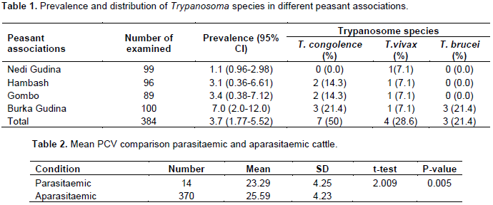
The overall prevalence of bovine trypanosomosis in the study areas was 3.7%. The prevalence in each peasant association was determined to be 1.1% in Nedi Gudina, 3.1% in Hambash, 3.4% in Gombo and 7.0% in Burka Gudina of Jimma Horro District. Trypanosome congolence was dominant species with proportion of 50%, followed by T. vivax (28.6%) and T. brucei (21.4%) in Table 1.
The mean PCV value for the parasitemic cattle was 23.29 +4.25 SD while the mean PCV value for the aparasitaemic cattle was 25.59+4.23 SD. There was statistically significant difference (P< 0.05) in mean PCV value between parasitaemic and aparasitaemic cattle (Table 2).
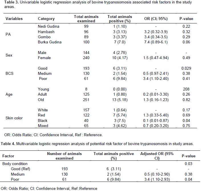
The highest (7.0%) and lowest (1.1%) prevalence of bovine trypanosomosis was recorded in Burka Gudina and Nedi Gudina peasant associations, respectively. However, there was no statistical significant difference (P>0.05) between prevalence of trypanosomosis and peasant associations. The prevalence of trypanosomosis was 5.18% in older age category than in adult age category (0.8%) cattle. The prevalence of trypanosomes infection between age group was not statistically significant difference (P>0.05). The prevalence of trypanosomosis was higher in female (4.17%) than male (2.78%) cattle, but there was no statistically significant difference (P>0.05). The highest prevalence of trypanosomosis was recorded in cattle with poor body condition cattle (9.84%). Moreover, variation in prevalence of trypanosomosis among the body condition was statistically significant (P<0.05). Poor body condition cattle being almost three times (OR=3.4) more likely to be infected with trypanosomes organisms compared to good body condition cattle. Highest prevalence of trypanosomosis was found in black skin color of cattle (7.5%), followed by red skin color (5.74%) and lowest in white skin color (0.64%) of cattle. However, there was no statistically significant difference (P>0.05) of skin color of cattle with prevalence of trypanosomosis (Table 3). Variables with a P-value less than 0.25 in the univariable analysis with no multicollinearity were entered into multivariable logistic regression model. No significant interactions between variables were detected. A Hosmer-Lemeshow goodness-of–fit value (P=0.98), indicated that the model fit the data. The final multivariable logistic regression model showed that body condition was independently associated with (P<0.05) bovine trypanosomosis (Table 4).
DISCUSSION
The present study showed that from a total of 384 randomly examined cattle, 14(3.7%) were positive for trypanosome parasite. Similar level of prevalence was reported by Teka et al. (2012), Dawit et al. (2015) and Fayisa et al. (2015), who reported that prevalence of 3.7% in Abaya district, 4.9% in Arbamich and 4.4% in Didesa District in Ethiopia, respectively. On the other hand, the prevalence of trypanosomosis reported in the current study is lower than the values reported by Olani and Bekele (2016) 7.8% in Lalo-Kile district; Fedesa et al. (2015) 7.1% in Darima District and Miruk et al. (2008) 20.4% in Wolyta and Dawero Zone of Southern Ethiopia; Siyum et al. (2014) 16.9% in Sayo District in Western Ethiopia; Yalew and Fantahun (2017) 21.5% in Bambasi woreda, Western Ethiopia and Kitila et al. (2017) 7.4% in Yayo District Iluababora zone of Western Ethiopia. This variation might be due to differences in environmental factors, breed and managemental system in study areas.
The present result shows that out of 14 positive cattle for trypanosomosis, T. congolonse (50%) was predominant species of trypanosome, followed by T. vivax (28.6%) and T. brucei (21.4%) in study area. This may be due to major cyclical vectors or Glossina species are more efficient to transmitters of T. congolonse than T. vivax and high number of serodems of T. congolonse as compared to T. vivax (Olani and Bekele, 2016). Moreover, T. vivax is highly susceptible to treatment while the problems of drug resistance are higher in T. congolonse, since T. congolonse is mainly confirmed in blood, while T. vivax and T. brucei invade tissue (Biyazen et al., 2014). This finding is consistent with some previous studies in different parts of Ethiopia (Begna et al., 2011; Biyazen et al., 2014; Kassaye, 2015; Tola et al., 2016; Kassaye and Tsegaye, 2016; Kitila et al., 2017). Similarly, T. congolonse was dominant species with a proportion of (69.7%) and followed by T. vivax (19.2%) and T. brucei (9.1) in Western Ethiopia also reported by Siyum et al. (2014) and Dawit et al. (2015).
The mean PCV value of trypanosome positive cattle was significantly lower (23.29 ± 4.25) than that of negative cattle (25.59 ± 4.23). The occurrence of positive animals with PCV of greater than 24% might be thought of as recent infection of the animals (Vanden and Rowlands, 2001). Low PCV value may not solely be due to trypanosomosis. However, these factors are likely risk for both parasitemic and aparasitemic cattle. Thus, the difference in mean PCV value between the parasitemic and aparasitemic cattle indicates that trypanosomosis is involved in reducing the PCV value in the infected cattle. This result was in line with Rowlands et al. (2001), who reported that the treatment resulted into an increase in PCV value of positive animals when PCV was less than 26%. Hence, the mean PCV was a good indicator for the health status of the herd in an endemic area. This result was also in agreement with previous report as anemia is the classical sign of the disease pathogenicity; the low PCV in parasitaemic animals could have contributed in reducing the mean PCV for cattle (Getachew et al., 2014; Efrem et al., 2013). Likewise, this result is in line with Mezene et al. (2014), who stated that parasitaemic animals had generally lower mean PCV value than aparasitaemic animals.
In the present study, body condition indicated that animals with poor body condition are three times more likely to be affected by trypanosomosis (OR= 3.4) than good body condition. This may be due to trypanosomosis results in progressive emaciation of the infected animals; never less, non-infected cattle under good condition have well developed immune status that can respond to any foreign protein better than those of non-infected cattle with poor body condition (Taylor et al., 2007). This finding is consistent with some previous studies in Ethiopia (Dawud and Molalegne, 2011; Girma et al., 2014; Getachew et al., 2014; Gona et al., 2016; Yalew and Fantahun, 2017) stated that prevalence of trypanosomosis was statistically significantly associated with body condition in cattle. This study finding is also in line with that of Bitew et al. (2011), Teka et al. (2012) and Fayisa et al. (2015), who reported that statistically significant association between prevalence of trypanosomosis and body condition in cattle. However, in contrary to this Abebayehu et al. (2011), Bekele and Nasir (2011), Tafese et al. (2012), Dawit et al. (2015) and Kitila et al. (2017) reported that body condition of cattle was not significantly associated with the prevalence of trypanosomosis in cattle.
In the present study, no statistically significant variation was observed in prevalence of bovine trypanosomosis among skin color of cattle. Comparison conducted between the different skin colors of cattle indicated that higher prevalence was observed in cattle’s having black skin color (7.5%) followed by 5.7% red and 4.62% mixed skin color. Tsetse flies by nature are attracted toward a black color, so in animals having black skin color there is high prevalence of trypanosomosis recorded (Teka et al., 2012; Gona et al., 2016). The prevalence of bovine trypanosomosis was no statistical significant difference (P>0.05) among sex, age groups of cattle and peasant association. This might be because of an equal chance of exposure cattle to the parasite and even distribution of the disease in the district. This result is in line with the previous study (Abebayehu et al., 2011; Bekele and Nasir, 2011; Tafese et al., 2012).
CONCLUSION AND RECOMMENDATIONS
Trypanosomosis is most important constraint for cattle production in Jimma Horro District. The present result showed that existence of T. congolonse, T. vivax and T. brucei were responsible for bovine trypanosomosis in study area. Body condition was statistically significance difference with prevalence of trypanosomosis in the district. However, age groups, sex, skin color and different peasant associations were not showed statistically significance difference. The mean PCV value of trypanosome cattle was significantly lower than negative cattle indicating the effect of trypanosomosis in lowering the PCV value. Thus awareness creation and appropriate control methods of trypanosomosis on its vectors and against the parasite should be designed and implemented.
CONFLICT OF INTERESTS
The authors have not declared any conflict of interests.
REFERENCES
|
Abebayehu T, Eset H, Berhanu M, Rahmeto A, Solomon M (2011). Mechanically transmitted bovine trypanosomosis in Tselamity wereda,Western Tigray, Northern Ethiopia, Agricultural Journal 6(1):10-13. |
|
|
Apeanti WO (2016). Contributing factors to pre-service mathematics teachers' e-readiness for ICT integration. International Journal of Research in Education and Science 2(1):223-238. |
|
|
Begna F, Abebe S, Bekele M (2011). Bovine Trypanosomosis in selected villages of Humbo District, Southern Ethiopia. Global Veterinaria 7:192-198. |
|
|
Bekele J, Asmare K, Abebe G, Ayelet G, Esayas G (2010). Evaluation of deltamethrinapplications in the control of tsetse and trypanosomosis in the southern rift valley areas of Ethiopia. Veterinary Parasitology 168:177-184. |
|
|
Bekele M, Nasir M (2011). Prevalence and host related risk factors of bovine trypanosomosis in Hawagelan district, West Wellega zone,Western Ethiopia, African Journal of Agricultural Research 6(22):5055–5060. |
|
|
Benti AD, Zewdie W (2014). Major reproductive health problems of indigenous Borena cows in Ethiopia. Journal of Advanced Veterinary and Animal Research 1(4):182-188. |
|
|
Bitew M, Amedie Y, Abebe A (2011). Prevalence of bovine trypanosomosis in selected areas of Jabi Tehenan district, West Gojam of Amhara regional state, North western Ethiopia. African Journal of Agricultural Research 6(1):141-144. |
|
|
Biyazen H, Duguma R, Asaye M (2014). Trypanosomosis: Its risk Factors, and anaemia in cattle population of Dale Wabera District of Kellem Wollega Zone, Western Ethiopia. Journal of Veterinary Medicine. Hindawi Publishing Corporation. |
|
|
Blood DC, Radostits OM (2007). Veterinary Medicine: A Text Book of Diseases of Cattle, Sheep, Pigs, Goats and Horses, Bailliere Tindall, 10th edition. |
|
|
Cherenet T, Sani RA, Speybroeck N, Panandam JM, Nadzr S, Van den Bossche P (2006). A comparative longitudinal study of bovine trypanosomiasis in tsetse-free and tsetse-infested zones of the Amhara Region, northwest Ethiopia. Veterinary Parasitology 140:251-258. |
|
|
Central Statistical Agency (CSA) (2017). Livestock and Livestock Characteristics, Agricultural sample Survey. Addis Ababa, Ethiopia. Statistical Bulletin 2(583):9-13. |
|
|
Dawit A, Alemayew T, Bekele K, Zenebe T, Kebede G, Kabeta T (2015). Prevalence of bovine trypanosomosis, and it's Associated risk factors in Abaya District, Borena Zone, Ethiopia. Natural Sciences 13(10):64-70. |
|
|
Dawud A, Molalegne B (2011). Epidemiological study of bovine trypanosomosis in Mao-komo Special District, Benishangul Gumuz Regional State, Western Ethiopia. Global Veterinary 6:402-408. |
|
|
Dumesa T, Demessie Y (2015). Review on tsetse transmitted bovine trypanosomosis in Ethiopia. European Journal of Applied Sciences 7(6):255-267. |
|
|
Efrem D, Bashatu F, Bacha B, Addisalem H, Misgana D (2013). Prevalence of bovine trypanosomosis in Lalo Kile District, Kelem Wollega Zone,Western Ethiopia. Acta Parasitologica Globalis 4:38. |
|
|
Fayisa G, Mandefro A, Hailu B, Chala G, Alemayehu G (2015). Epidemiological status and vector identification of bovine trypanosomiosis in Didesa District of Oromia Regional State, Ethiopia. International Journal of Nutrition and Food Sciences 4(3):373-380. |
|
|
Getechew A (2005). Review Article trypanasomasis in Ethiopia. Ethiopian Journal of Biological Sciences 27(1):1-8. |
|
|
Fedesa H, Assefa K, Tekalegn D (2015). Study on spatial distribution of tsetse fly and prevalence of bovine trypanosomosis and other risk factors: case study in Darimu District, Ilu Aba Bora Zone, Western Ethiopia. Journal of Pharmacy and Alternative Medicine P 7. |
|
|
Getachew S, Kabeta T, Abera Z, Deressa B (2014). Epidemiological survey of bovine trypanosomosis in Sayo District of Kellem Wollega Zone, Western Ethiopia. American-Eurasian Journal of Scientific Research 9(3):67-75. |
|
|
Girma K, Meseret T, Tilahun Z, Haimanot D, Firew L, Tadele K, Zelalem A (2014). Prevalence of bovine trypanosomosis, its vector density and distribution in and around Arbaminch, Gamogofa Zone, Ethiopia. Acta Parasitologica Globalis 5:1. |
|
|
Gona Z, Teshale A, Tilahun A (2016). Study on prevalence of bovine trypanosomosis and density of its vectors in three selected districts of Wolaita Zone, Southern Ethiopia. Journal of Veterinary Medicine and Animal Health 8(9):128-135. |
|
|
Gupta MP, Singla LD, Singh KB, Mohan R, Bal MS (2003). Recrudescence of trypanosomosis following administration of dexamthasone in bovines. Indian Veterinary Journal 80:360-361. |
|
|
Gupta MP, Kumar H, Singla LD (2009). Trypanosomosis concurrent to tuberculosis in black bucks. Indian Veterinary Journal 86:727-728. |
|
|
Holmes PH, Eisler MC, Geerts S (2004). Current chemotherapy of animal trypanosomiasis. In: Maudlin I, Holmes P.H. and Miles M.A. (eds). The Trypanosomiases. CABI, UK, pp.431-444. |
|
|
Jimma Horro District Agricultural Office (JHDAO) (2O16). Jimma Horro District Agricultural Office, annual report, pp. 55-65. |
|
|
Juyal PD, Singla LD (2005). Towards newer approaches for diagnosis and control measures of suura (due to Trypanosoma evansi) in livestock. In: Compendium of Winter School on Novel Approaches for Diagnosis and Control of Parasitic Diseases of Domestic and Wild Animals held from 07-27 October, 2005 at PAU, Ludhiana, pp. 294-305. |
|
|
Juyal PD, Singla LD, Kaur P (2005). Management of surra due to Trypanosoma evansi in India: an overview. In: Infectious Diseases of Domestic Animals and Zoonosis in India, Tandon V and Dhawan BN (Eds), Proceedings of the National Academy of Sciences India Section B: Biological Science 75(Special issue):109-120. |
|
|
Kassaye BK (2015). Prevalence of bovine trypanosomosis and apparent density of tsetse flies in Sayonole District Western Oromia, Ethiopia. Journal of Veterinary Science and Technology 6:254. |
|
|
Kassaye BK, Tsegaye D (2016). Prevalence of bovine trypanosomosis, tsetse density and farmers perceptions on the impact of control program in Kellem Wollega Zone, Western Oromia, Ethiopia. Journal of Veterinary Science and Technology 7:295. |
|
|
Kitila G, Kebede B, Guta D, Bekele F, Wagari M, Tilahun B, Jaleta D (2016). Epidemiological investigation of bovine trypanosomosis and its vector apparent densities in Yayo District Illuababora Zone, Western Oromia, Ethiopia. Research and Reviews: Journal of Veterinary Sciences 3:1-6. |
|
|
Kitila G, Kebede B, Guta D, Bekele F, Wagari M, Tilahun B (2017). Epidemiological investigation of bovine trypanosomosis and its vector apparent densities in Yayo District Illuababora Zone, Western Oromia, Ethiopia. Austin Journal of Veterinary Science and Animal Husbandry 4(1):1031. |
|
|
Kumar H, Gupta MP, Sidhu PK, Mahajan V, Bal MS, Kaur K, Ashuma VS, Singla LD (2012). An outbreak of acute Trypanosoma evansi infection in crossbred cattle in Punjab, Journal of Applied Animal Research 40(03):256-259. |
|
|
Leta S, Mesele F (2014). Spatial analysis of cattle and shoat population in Ethiopia: growth trend, distribution and market access. Springer Plus 3:310. |
|
|
Mezene W, Ahimedine B, Moti Y, Efrem D, Kumela L (2014). Bovine trypanasomosis and tsetse fly survey in Bure District, Western Ethiopia. Acta Parasitologica Globalis 5:95. |
|
|
Miruk A, Hagos A, Yacob HT, Asnake F, Basu AK (2008). Prevalence of bovine trypanosomosis and trypanocidal drug sensitivity studies on Trypanosoma congolense in Wolyta and Dawero zones of southern Ethiopia. Veterinary Parasitology 152:141–147. |
|
|
National Tsetse and Trypanosomiasis Investigation and Control Centre (NTTICC) (2004). Annual Report on Tsetse and Trypanosomosis Survey. Bedelle Ethiopia. |
|
|
Olani A, Bekele D (2016). Epidemiological status and vector identification of bovine trypanosomosis in Lalo-Kile District of Kellem Wollega Zone, Western Ethiopia. Journal of Veterinary Medicine and Research 3(2):1045. |
|
|
Paris J, Murray M, Mcodimba F (1982). A comparative evaluation of the parasitological technique currently available for the diagnosis of African Trypanosomosis in cattle, Acta Tropica 39:307-316. |
|
|
Rowlands GJ, Leak SGA, Peregrine AS, Nagda SM, Mulatu W, D'ieteren GDM (2001). The incidence of new and the prevalence and persistence of recurrent trypanosome infections in cattle in south-west Ethiopia exposed to a high challenge with drug-resistant parasites. Acta Tropica 79:149-163. |
|
|
Sharma P, Juyal PD, Singla LD, Chachra D, Pawar H (2012). Comparative evaluation of real time PCR assay with conventionalparasitological techniques for diagnosis of Trypanosoma evansi in cattle and buffaloes. Veterinary Parasitology 190:375-382. |
|
|
Singh V, Singla LD (2013). Trypanosomosis (Surra) in livestock. In: Veterinary Parasitology in Indian Perspective, Katoch R, Godara R and Yadav A (Eds), Satish Serial Publishing House, Delhi pp. 277-302. |
|
|
Singla LD, Singla N, Parshad VR (2015). Development of concurrent infection of notoedric mange in rabbits infected with Trypanosoma evansi. Scandinavian Journal of Laboratory Animal Science 41(2):1-6. |
|
|
Siyum G, Tadele K, Zelalem A, Benti D (2014). Epidemiological survey of bovine trypanosomosis in Sayo District of Kellem Wollega Zone, Western Ethiopia. American-Eurasian Journal of Scientific Research 9:67-75. |
|
|
Tafese W, Melaku A, Fentahun T (2012). Prevalence of bovine trypanosomosis and its vectors in two districts of East Wollega zone, Ethiopia, The Onderstepoort Journal of Veterinary Research 79:123-128. |
|
|
Taylor AM, Coop LR, Wall LR (2007). Veterinary Parasitology, 3rd ed. UK. Blackwell publishing pp. 44-102. |
|
|
Teka W, Terefe D, Wondimu A (2012). Prevalence study of bovine trypanosomosis and tsetse density in selected villages of Arbaminch, Journal of Veterinary Medicine and Animal Health 4(3):36-41 |
|
|
Thrusfield M (2005).Veterinary Epidemiology, 3rd Edn., Blackwell Publishing, England pp. 345-543. |
|
|
Tola M, Kebede B, Kitila G, Gezehegn E (2016). Prevalence of bovine trypanosomosis and its vector apparent density in Chora District of Illuababora Western Oromia, Ethiopia. Journal of Veterinary Medicine and Animal Health 8:64-71. |
|
|
Vanden BP, Rowlands GJ (2001).The relationship between the parasitological prevalence of trypanosomal infections in cattle and herd average packed cell volume, Acta Tropica 78(2):163-170. |
|
|
Yalew ST, Fantahun B (2017). Prevalence of Bovine Trypanosomosis and its Associated Risk Factors in Bambasi woreda, Western Ethiopia. Journal of Dairy, Veterinary and Animal Research 5(1):00132. |
|
Copyright © 2024 Author(s) retain the copyright of this article.
This article is published under the terms of the Creative Commons Attribution License 4.0


