ABSTRACT
A cross sectional study was carried out in Holeta Agricultural Research Center Dairy Farm, Oromia Regional State, Ethiopia to determine the status of gastrointestinal (GI) parasites on dairy cattle, reveal the level of severity of GI parasites on the basis of mean egg per gram of faces (epg) count through McMaster technique and identify the different gastrointestinal parasites dominated by flotation and sedimentation techniques. A total of 206 faecal samples were collected from purposively selected cows, bulls and heifers. The overall prevalence of GI parasites was found to be 87.9%. The average epg was found to be 179.8, indicating light level of parasite infection. The sex-wise prevalence revealed 85.0 and 15.0% in female and male animals, respectively. The qualitative faecal examination techniques, showed a prevalence of coccidia (56.3%), Fasciola spp. (26.2%), Paramphistomum spp. (10.2%), Bunostomum spp. (8.7%), Oesophagostomum spp. (8.3%), and Tricuris spp. (1.5%). With statistically significant difference (p<0.05), the prevalence was higher in milking cow (28.72%) than the rest of the animal categories and the lowest prevalence was observed on dry cow (12.15%). There was concurrent infection with two and more than two different GIT parasites with respective prevalence of 38.3 and 25.2%. The finding of the present study clearly suggests that GI parasites were higher in the farm with low severity, which contributes reduction in productivity. Hence, further and strengthened parasite control intervention is highly recommended taking into account the seasonality of parasite burden.
Key words: Animal category, breeds, epg, faeces, GI parasites, prevalence, Holeta
Ethiopia, located in Eastern Africa, is predominantly an agricultural nation. Animal production is practiced in all ecological zones of the country (Tegegne and Crawford, 2000). The total animal population for the country is estimated to be about 53.99 million cattle out of which, about 98.95%% of the total cattle in the country are local breeds and while the remaining are hybrid and exotic breeds that accounted for about 0.94 percent and 0.11 percent%, respectively (Central Statistical Agency (CSA), 2008).
Ethiopia’s great livestock potential is not properly exploited Ethiopia’s great livestock potential is not properly exploited due to many prevailing socio economic values and attitudes, traditional management methods, limited genetic potential and rampant disease (Etana, 2002). Parasitic infections are the major constraints for poor performance that causes great economic loss to dairy industry through retarded growth, low productivity and increased susceptibility of animals to other infections (Yadav et al., 2004).
The most important helminthes parasites in cattle include nematodes, trematodes and cestodes in which their impact is greater in small and large-scale farms of sub-Saharan Africa in general and Ethiopia in particular, due to the availability of wide range of agro-ecological factors suitable for diversified hosts and parasite species (Hailemariam, 2006).
In Holeta Agricultural Research dairy farm where the exotic, cross and local dairy cows and calves are managed under semi-intensive rearing system. Tthe prevalence, species composition and epidemiology of gastrointestinal (GI) parasites affecting dairy animals have not been investigated in detail in recent time. Hence, the objectives of this study were to determine the status of dry season GI parasites on dairy cattle of Holeta Agricultural Research Center dairy farm. Besides, the level of infection based on the mean epg count and the relationship between measurable parameters and GI parasites were assessed.
Study area
The present study was conducted in Holeta Agricultural Research Center dairy farm; in west Shoa Zone Wolmera District Oromia Regional State, Ethiopia. Wolmera is a district dominated by highland temperature. Holetais is an administrative town of Wolmera district in central Ethiopia, located in West Shoa zone surrounding Finfinne of the Oromia Region. It has latitude and longitude of 9°3′N 38°30′E and an altitude of 2391 m above sea level. The host town of the study area is Holeta Agricultural Research Center dairy farm of the Ethiopian Institute of Agricultural Research.
Study animal
The study was conducted on 206 (31 male and 175 female) Holstein Friesian cows, bulls and heifers maintained under intensive and semi-intensive management production system. Different age and sex groups were represented as much as possible. Selection was conducted irrespective of age and sex based on body condition. Their body conditions which score less than 1.5 were selected purposively.
Study design
A cross sectional study design was conducted in dry season from December to February 2015 to identify, assess and estimate the prevalence of GI parasites.
Sample size determination and sampling methodology
Sample size was calculated with an expected prevalence of 50%. The desired sample size for the study was calculated using the formula given by Thrusfield (2005) with 95% confidence interval and 5% absolute precision.
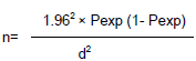
Where;
Pexp = expected prevalence,
d = absolute precision;
n =sample size.
206 animals were selected purposively from respective barns of the farm.
Sample collection and examination
Faecal sample collection
Faecal samples were collected per-rectum using plastic rectal gloves. Each sample was labeled with the animal number, date of collection, age, and sex in edible pen. The collected samples were kept in icebox with the lid tightly placed, and were transported to laboratory. Except on occasions where immediate processing was not possible, samples will be kept in four refrigerators and processed within 12 h.
Faecal sample examination
Faecal examination was done by different qualitative and quantitative examination technique for the presence of parasitic eggs and their level (Gupta and Singla, 2012). Qualitative faecal examination was carried out by sedimentation and floatation technique. The floatation fluid used in this study was saturated solution of sodium chloride (Nacl) prepared in the laboratory. The procedure given by Urquhart et al. (1996) was monitored using the above parasitological methods. Quantitative faecal examination was also conducted using McMaster egg counting technique. Severity of infection as obtained from the number of epg was determined as follows: ≤5000 epg is regarded as mild infection; 5000 to 10000 epg as moderate infection; and >10000 epg as severe infection (Jorden, 1994).
Data analysis
The entire data were entered into Microsoft Excel spread sheet and coded. Statistical analyses were performed using SPSS version 20 software packages. Percentage was used to calculate prevalence while Chi-square was used to calculate the degree of association between risk factors and prevalence of gastrointestinal parasites. In the analysis, a difference was taken as significant at p-value <0.05 and confidence level at 95%.
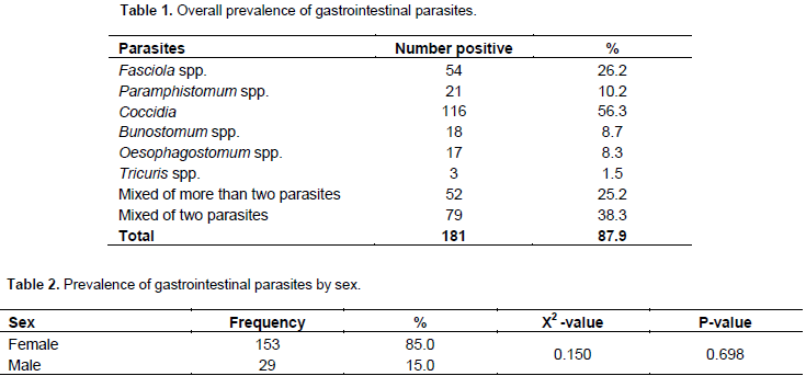
The result revealed an overall prevalence of 87.9%. The parasites encountered included Fasciola spp (26.2%), Paramphistomum spp (10.2%), coccidia (56.3%), Bunostomum spp (8.7%), Oesophagostomum spp (8.3%) and Tricuris spp (1.5%) (Table 1). Mixed infections with two parasites were also more common (38.3%) than infections with more than two parasites (25.2%) (Table 1). Out of examined female and male, 29 (15%) and 153 (85 %) were found to be positive, respectively. Accordingly, the prevalence of GI parasites in female was higher than male with no significant variation (p>0.05) (Table 2). Of all the positive animals, (14.9%) were bull, (12.15%) were dry cow, (18.23%) were heifer, (28.72%) were milking cow and (25.96%) were dry cow. Accordingly, the highest prevalence was recorded in pregnant category.

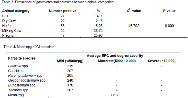
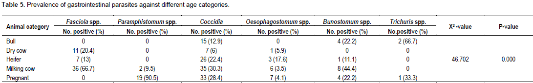
There were highly significant difference between animal categories on infection with GI parasites (p<0.05) (Table 3) The McMaster technique revealed an average epg value of 179.8. Similarly, the study showed that the mean epg of Oesophagostomum spp. is greater than the other GI parasites (Table 4). Animal category has a potential risk factors, which are associated with gastrointestinal parasites at the dairy farms. The highly prevalent parasite among different categories with an individual prevalence rate was Fasciola spp. 66.7%, coccidia 30.3%, and Bunostomum spp. 44.4% in milking cow, Paramphistomum spp. 90.5% in pregnant cow, Oesophagostomum spp. 17.6% in heifers, and Trichuris spp. 66.7 in bull (Table 5).
The overall prevalence of gastrointestinal parasites in the present study was 87.9%. This finding is higher than 41.2% prevalence by Epherem (2007) and 26.3% by Darsema (2009) in Western Amhara region, Ethiopia. In addition, Keyyu et al. (2006) reported an overall prevalence of 44.4 and 37.0% for large and small scale dairy cattle, respectively in Tanzania. This higher prevalence in the study area could be due to the fact that cattle from farmers surrounding farm have frequent exposure to the same communal grazing land that causes contamination of pasture, most favorable environmental condition for the development of larvae, variation in management and husbandry practices, climate, and management of pastures. In this study, lower prevalence of Fasciola spp. (26.2%) was obtained when compared with 34% prevalence reported by Tesfa (1994) in Gojjam with 61.97% prevalence. This difference may be due to variation in management system, absence of swampy area, effect of deworming and variation in the study period. The overall prevalence of coccidia in this study was 56.3%, which is higher than previous findings of 31.9% reported by Alemayehu et al., (2013) in Kombolcha. This variation is most likely attributed to the differences in agro-ecology and husbandry practices of the study animals and the resistance level of animal increase as age increase.
The prevalence of Paramphistomum spp found in the present study (10.2%) was lower than the finding by Fromsa et al (2011) in Jimma (45.2%), which reported 6.7% in dairy farm in Hawassa town. Many studies conducted in different parts of the world reported prevalence of 22% in Pakistan by Raza et al. (2009). This lower prevalence may be due to agro ecological factors and management system. In the current study, the prevalence of Oesophagostomum spp. was found to be 8.3% which is lower than (11%) as reported by Hiko and Wondimu (2011) in Haramaya University Dairy Farm. This difference could be due to, difference in deworming habit and higher susceptibility of exotic cattle in cross and local breeds. The lower prevalence of Bunostomum spp. (8.7%) and Trichuris (1.5%) in farm was due to the effect of deworming practice, which is given as three times per year and also, the study season in which most gastrointestinal parasites can’t resist dry environment. The study shows that gastrointestinal parasite in the study area is higher in female (85%) than male (15%). As reported by Ram (2009), the difference in parasite prevalence between sexes was due to the fact that,females are found to have higher infection rates due to their low immunity in gestation and lactation period. The co-infection pattern observed in this study showed that, dairy animals have a high chance of related exposure of different GI parasites.
In relation with animal category, the occurrence of GI parasites has a significant difference which is relatively lower in dry cow (12.15%) and higher in milking cow (28.75%), with a respective highly prevalent parasite species of Ascaris spp. (13.8%) and Fasciola spp. (66.7%). Such finding may be due to the fact that, in milking cow the immune response of the host to gastrointestinal nematodes is partially suppressed, leading to an increase in the population of the worms. According to the McMaster quantitative result, the mean epg of the overall gastrointestinal parasite encounter during the study period was 179.8 while the minimum and maximum epg scored was 176 for Bunostomum spp. and 240 for Oesophagostomum spp. Jorgen (1994) reported that, the epg level of the present study has a mild infection. This finding might be due to the effect of the seasonal deworming in the farm.
CONCLUSION AND RECOMMENDATIONS
The overall prevalence of gastrointestinal parasite in the study area indicated that, gastrointestinal helminthosis was found to be an important health problem due to its high prevalence and occurrence of polyparasitism. The majority of cattle were infected by more than one parasite type with some animals showing pure infection. Therefore, the farm is prone to health problems related to gastrointestinal helminthosis which might subsequently reduce the economic output from cattle production. Most of the animals examined during the present study relatively harbor low to moderate parasite eggs. In view of these conclusions, the following recommendations are forwarded;
1) Strategic parasitic control programs should be designed.
2) The role of veterinarians in giving professional advices regarding preventive and control measures against gastrointestinal helminthes should be prominent to prevent any abuses.
3) To improve the management and feeding condition of those cattle residing in the farm.
Further study on epidemiology and determinant factors for, the occurrence of helminthes parasites and implementation of appropriate control and prevention methods in GI parasites, identified the cause economic losses and diseases of animals in this study.
The authors have not declared any conflict of interests.
REFERENCES
|
Alemayehu A, Nuru M, Belina T, Mekibib B, Desta T, Tesfaye D (2013). Prevalence of bovine coccidia in Kombolcha district of South Wollo, Ethiopia. J. Vet. Med. Anim. Health 5(2):41-45.
|
|
|
|
Central Statistical Agency (CSA) (2008). Federal Democratic Republic of Ethiopia, Central Statistical Agency, agricultural sample survey. Available at:
View
|
|
|
|
|
Darsema G (2009). Epidemiological study on major gastrointestinal helminth parasites of calves in three cattle farms in the western part of Amhara Region, Ethiopia. Ethiop. Vet. J. 2:9-18.
|
|
|
|
|
Etana D (2002). Epidemiology of gastro-intestinal helminthiasis of Rift Valley goats under traditional husbandry system in Adami Tulu district, Ethiopia. SINNET: Ethiop. J. Sci.25:35-44.
|
|
|
|
|
Epherem W (2007). Prevalence of Bovine GI helminths in selected Dairy farms of Addis Ababa. DVM Thesis, JUCAVM, Jimma, Ethiopia.
|
|
|
|
|
Fromsa A, Meharenet B, Mekibib B (2011). Major trematode infections of cattle slaughtered at Jimma Municipality Abattoir and the occurrence of the intermediate hosts in selected water bodies of the zone. J. Anim. Vet. Adv. 10(12):1592-1597.
Crossref
|
|
|
|
|
Gupta SK, Singla LD (2012). Diagnostic trends in parasitic diseases of animals. In: Veterinary Diagnostics: Current Trends. Gupta RP, Garg SR, Nehra V and Lather D (Eds), Satish Serial Publishing House, Delhi, pp. 81-112.
|
|
|
|
|
Hailemariam T (2006). Ovine and bovine helminthiasis in Kelela, South Wollo. In: Proceedings of EVA Conference, Addis Ababa, Ethiopia, pp. 30-34.
|
|
|
|
|
Hiko A, Wondimu A (2011). Occurrence of nematodiasis in Holstein Friesian dairy Breed. J. Vet. Med. Anim. Health 3(1):6-10.
|
|
|
|
|
Jorgen H (1994). The epidemiology, diagnosis, and control of helminth parasites of ruminants: A handbook. Available at:
View
|
|
|
|
|
Keyyu JD, Kassuku AA, Msalilwa LP, Monrad J, Kyvsgaard NC (2006). Cross-sectional prevalence of helminth infections in cattle on traditional, small-scale and large-scale dairy farms in Iringa district, Tanzania. Vet. Res. Commun. 30(1):45-55.
Crossref
|
|
|
|
|
Ram C (2009).Prevalence of helminthes parasites in mules of Brick kiln of Lalitpur District, Napal.
|
|
|
|
|
Raza MA, Murtaza S, Bachaya HA, Hussain A (2009). Prevalence of Paramphistomum cervi in ruminants slaughtered in district Muzaffar Garh. Vet. J. 28(1):34-36.
|
|
|
|
|
Tegegne A, Crawford W (2000). Draft animal power use in Ethiopia. Draft Animal News, P 33.
|
|
|
|
|
Tesfa Y (1994). Bovine fasciolosis: Prevalence and economic importance assessment trial on cattle slaughtered at Bahirdar Municipal Abattoir. DVM Thesis, FVM, AAU, Ethiopia, P 39.
|
|
|
|
|
Thrusfield M (2005). Veterinary Epidemiology 3rd Ed., Blackwell Science Ltd., Oxford, UK, pp. 233-261.
|
|
|
|
|
Urquhart G, Aremour J, Duncan A, Dunn F, Jennis F (1996). Veterinary parasitology 2nd edition. The University of Glasgw, Black Well Science, Scotland. pp. 3-137. Available at:
View
|
|