Full Length Research Paper
ABSTRACT
Despite its endemicity and economic significance in Ethiopia, there was no sero-surveillance report of Peste des Petits Ruminants (PPR) in Bale Zone. With this regard the study was conducted in Dello Mena and MaddaWalabu districts of Bale zone with the objectives of determining the seroprevalence of PPR and associated risk factors. A cross sectional study was conducted from March 2017 to May 2017 on weaned goat and camel which were randomly selected from 5 kebeles of each study districts. Accordingly, serums collected from 768 animals (that is, 384 from each district) were tested using competitive enzyme linked immunosorbent assay. An overall seroprevalence of 12.9% was recorded with relatively higher seroprevalence in Dello Mena (13.8%) compared to Madda Walabu district (12%). From 4 putative risk factors investigated by the study, species (goat 19%; camel 2.5%; ï£2=43.623; p=0.000) and age (adult 16.9%; young 7.2%; ï£2= 15.472; p=0.000) revealed statistically significant association with PPR seropositivity on Chi-square analysis. In addition, multivariable logistic regression analysis indicated that goats were found more likely infected by the disease (Adjusted Odds Ratio=9.522; p=0.000), while adult animals more likely survive infection and become seropositive compared to young animals (Adjusted Odds Ratio= 2.713; p=0.000). PPR found very important health problem in the study areas, especially in goats and younger animals. Due to high sero-prevalence in the study area due attention should be given on the eradication of the disease via organized active surveillance and vaccination of unvaccinated segment of the population, especially younger animals, on annual base.
Key words: Camel, Dello Mena, goat, Madda Walabu, Peste des Petits ruminants, risk factors, seropositivity.
INTRODUCTION
The camel (Camelus dromedarius) and goats are an important livestock species well adapted to arid and semiarid environments of the world (Schwartz and Dioli, 1992). They are extremely important for the livelihood of the pastoralist communities and their cultural life (Asefa, 2000; Mohammed, 2008; Tefera and Gebreab, 2001).
In Ethiopia, they are densely populated in the lowland areas of the country. According to Central Statistical Agency (CSA) sample survey report of 2013 from the rural sedentary areas of the country, Ethiopian goat and camel population estimate stands at 24.06 and 0.92 million, respectively. Even if the country has large production potential, but is not making use of this huge potential attributed to different constraints among which disease stands in the front line (Samson and Frehwot, 2010).
Major diseases contributing to the poor performance of goat and camel in the sub-sector include Peste des Petits Ruminants (PPR), sheep and goat pox, camel pox (Baga), brucellosis and contagious caprine pleuropneumonia (CCPP). Epidemics of PPR threaten national livestock industries by high levels of morbidity and mortality in goat and morbidity in camel. This disease is remained as central issues related to food security, link between animal health and meat quality. Occurrence of such disease impact both poor and richer livestock producers by marginalizing them from higher price livestock markets and restricting their capacity for value-added trade. Failure of pastoral community to sell their goats and camels therefore can bring severe hard ship to a pastoral family with no other income sources of support (Hailu et al., 2015).
Even if the clinical PPR was suspected in 1977 in Afar region, the virus presence was confirmed with cDNA probeby 1994 (Pegram and Tereke, 1981; Roeder et al., 1994). Nowadays, the disease is recognized as responsible for mortality and morbidity across many regions of the country. Waret-Szkuta et al. (2008) reported an overall seroprevalence of 1.7, 15.3, 4.6, 8.0, 21.3, 15.3 and 1.8% in Oromia, Afar, Amhara, Benishangul Gumuz, Somalia, Tigray and South Nation and Nationalities Peoples (SNNP) region of Ethiopia. Most recently, an overall seroprevalence record of 30.9% from sheep and goat in pastoral and agro-pastoral area of Afar and Gambella region of Ethiopia has been reported (Megersa et al., 2011). Beside this, Abraham et al. (2005) have recorded 3% prevalence of PPR in Ethiopian camels.
Despite the fact that these studies give a clue about the sero-epidemiogical distribution of the disease in Ethiopia, the actual temporal and spacial distribution of the disease is not known in Bale zone which is crucial for progressive control and eradication program undergoing. Hence, this study was conducted to determine the sero-epidemiology of PPR that potentially affect goat and camel production system in Dello Mena and Madda Walabu districts of Bale zone, and to determine the risk factors exposed PPR in these districts.
MATERIALS AND METHODS
Description of the study area
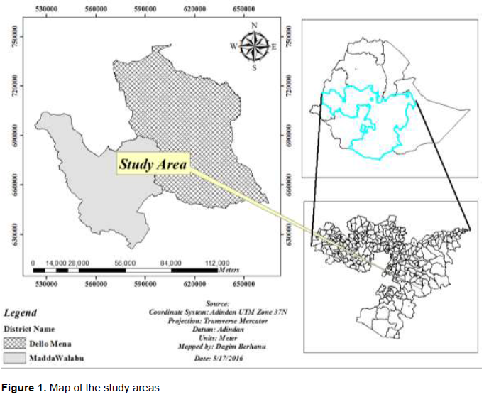
The study was conducted in Dello Mena and Madda Walabu districts of Bale Zone, Oromia Regional State, South Eastern Ethiopia. The study districts were selected purposely to represent the camel and goat rearing district of the zone based on their agricultural vocational activities and ecological conditions (Figure 1).
Dello Mena is located in the south western part of Bale zone at a distance of 555 km south east of Addis Ababa. It is bounded by Madda Walabu district in the south, Goba district in the north, Harena Buluk district in the west and south west, Berbere district in north east and Guradhamole in the east. It has a total area of 1,339 km2 which ranked the district 14th largest among the districts. The mean annual temperature of the district is 29.5°C, with the lowest and highest temperature being 21 and 38°C, respectively. The mean annual rainfall is 701.5 mm whereas the lowest and highest rainfall is 628 and 775 mm, respectively. The lowland area predominates with a narrow strip of high land area in the Northern part of Dello Mena district. Livestock rearing is the major way of life for the rural and lowland areas of district population. Accordingly, there are about 379,896 bovines, 15,743 sheep, 94,738 goats, 17,124 equines and 46,276 camels in the district (Dello Menna District Pastoral Development Office, 2016).
Madda Walabu district is located in the south western parts of the Bale zone. It is bounded by Guji zone in the west and south, Gura dhamole and Dello Mena districts in the east, and Harena Buluk district in the north. It has a total area of 8871 km2 which is ranked the district 1st largest among the zonal districts. It has distance of 196 km from district capital called Robe and 626 km from center of the country and the region, Addis Ababa. The mean annual temperature of the district is 30°C, with the lowest and highest temperature being 20 and 40°C, respectively. The mean annual rainfall is 600 mm whereas the lowest and highest rainfall is 400 and 800 mm, respectively. From early days, livestock rearing has played an important role in the life of district population. Similar to Dello Mena district, livestock rearing is the primary way of life for the rural and lowland areas of district population and there are about 243,250 bovines, 23,132 sheep, 72,487 goats, 12,674 equines, 50,392 camels, 10, 997 chickens in the district (Madda Walabu District Pastoral Development Office, 2016).
Study population
The target study population were goats and camels reared under extensive production system, pastoral and agro-pastoral system, in the selected districts of Bale zone. The status of PPR in Dello Mena and Madda Walabu districts was investigated in non-vaccinated camels and goats sampled from both districts.
Study design
A cross sectional type of study supported by questionnaire survey was conducted from March, 2017 to May, 2017 with the objective of determining the seroprevalence of PPR in goat and camel population, and associated exposing risk factors in the study areas.
Sampling method and determination of sample size
The sample size required for serological study was calculated according to the formula given by Thrusfield (2007) for systematic random sampling.

where n = required sample size, Pexp = expected prevalence, and d = desired absolute precision.
Since there is no reasonable research done in this area so far, the sample size was calculated at 95% CI, at 5% desired absolute precision and expected prevalence of 50%. Accordingly, the total numbers of sample required for this study were 384, however to increase precision the sample size were doubled to 768 and a total of 384 study animals were selected from each districts. Sample sizes for each species in each district were proportionally allocated. Accordingly, 257 (66.9%) goats and 127 (33.1%) camels from Dello Mena district and 227 (59%) goats and 157 (41%) camels from Madda Walabu district were sampled. During the actual sampling procedure, five kebeles were randomly selected out of the total 20 and 17 kebeles found in Madda Walabu and Dello Mena district, respectively. Then the study animals were selected systematically from each kebeles. Weaned and unvaccinated animals were included in the study.
The number of households (HHs) required for questionnaire survey was determined by the formula given by Arsham (2007) for survey studies.

where N= sample size and SE= standard error of the proportion.
Assuming the standard error of 4.1% at a precision level of 5%, and the confidence interval of 95%, 150 HHs owning goat and camel were selected by a simple random sampling technique for interview. While sampling the HHs the sampling frame was constructed in advance from the list obtained from both district pastoral development offices. Then the HHs were randomly selected from the sampling frame. In addition, the number of households selected per kebeles were fixed based on the proportion of HHs who owning goat and camel in each kebele.
Data collection
For the current study, both serological and questionnaire based data were collected to point out the disease spread pattern and factors governing its spread.
Questionnaire based data
Questionnaire survey was conducted in all selected kebeles of both districts to point out the general animal husbandry practice and animal health problems in the area. The questionnaires were prepared, pre-tested and adjusted by translating into local language (Afan Oromo) and administered by the interviewer.
Sera collection and testing
Animals were restrained by animal handlers and 10 ml of blood sample was collected from the jugular vein using plain vacutainer tubes with 18 to 20 gauge hypodermic needles. Vacutainer tubes with blood samples were then labeled with the species, sex, age, kebeles and districts of sampled animals. Then the collected blood id set tilted on a table overnight at room temperature to allow clotting and kept protected from direct sun light until the blood clotted and sera were separated. Then serum was filled into serum storage vials (cryovials) with appropriate identification and stored at -20°C until transported to National Veterinary Institute and the C-ELISA was performed.
A monoclonal antibody (MAb) based competitive enzyme linked immunosorbent assay (c-ELISA) (Diallo et al., 1989; OIE, 2013) was used for the detection of antibodies directed against the nucleoprotein of the PPR virus using approved competitive ELISA kit as described by Libeau et al. (1995). Briefly, the ELISA wells were coated with purified recombinant PPR nucleoprotein (NP); the samples to be tested and the controls were added to the microwells. Anti-NP antibodies, if present, form an antibody-antigen complex which masks the NP epitopes. An anti-NP-peroxidase (HRP) conjugate were added to the microwells and incubated. It fixes to the remaining free NP epitopes, forming an antigen-conjugate-HRP complex. After washing (to eliminate the excess conjugate), the substrate solution (TMB) was added and the resulting coloration depends on the quantity of specific antibodies present in the sample. Stop solution (sulfuric acid) was added to each well in order to stop the reaction. The optical density (OD) was read in an ELISA reader at 450 nm and the cut off points were calculated to validate the results. All sera with percentage inhibition (PI) > 50% was considered as positive. Sera with PI between 40 and 50% were considered doubtful and those sera with PI less than 40% were considered negative.
Data storage and analysis
Data generated from questionnaire survey and laboratory investigations were recorded and coded using Microsoft Excel spreadsheet (Microsoft Corporation) and analyzed using SPSS version 20 for Windows (Stata Corp. College Station, TX, USA). The seroprevalence of PPR was calculated as the number of seropositive samples divided by the total number of samples tested. To identify association of seropositivity with the potential risk factors was computed by Pearson’s Chi-square test and logistic regression analysis. A p value<0.05 was considered statistically significant.
RESULTS
Results from questionnaire survey
All respondents 150 (100%) in the study areas prioritize first enteritis and stomatitis syndrome as the cause of mortality in goats, while sudden death without specific sign as a cause of mortality in camels. One hundred and fifty (100%) respondents complain the occurrence of PPR in their goat flock and only 16.7% (25) of total respondents recognize and report the signs of PPR in camels. Regarding the temporal pattern of the disease in the area, the majority of the respondents (82.7%) revealed that the number of diseased animals, particularly goats, increase during the period between September and November. While the response of the rest respondents (17.3%) shows year round occurrence with pick prevalence between the February and April. Lack of drugs and vaccines (88.7%) and distance from modern services/clinics (11.3%) were the problem raised by the respondent. From the total respondents, 96.6% of them responded absence of annual vaccination for PPR in the areas. The results of other animal husbandry practice are shown in Table 1.

Seroprevalence of PPR
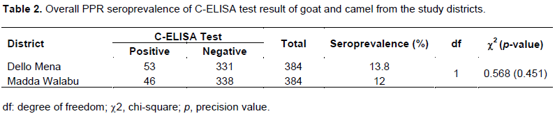
The overall seroprevalence of PPR estimated by C-ELISA test in the present study areas was 12.9%. In the current study, relatively higher seroprevalence of PPR was observed, both in goat and camel, in Dello Mena when compared with Madda Walabu district (Table 2).
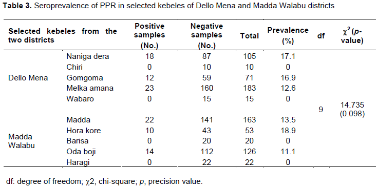
The present study also attempted to quantify within kebele seroprevalence of PPR in goats and camel in the study districts. Accordingly, from 5 kebeles of Dello Mena district included in the current investigation no PPR seropositivity was observed in two of them, Chiri and Wabaro. In other way, Gomgoma was the kebele with higher seroprevalence of the disease under investigation from Dello Mena district, while Hora kore (18.9%) was leading in PPR seropositivity among the 5 kebeles selected from Madda Walabu. However, no PPR seropositive goat and camel were found in 2 kebeles of Madda Walabu district. There was no significant association (p>0.05) between the selected kebeles of the study districts and PPR seropositivity (Table 3).
Chi-square analysis of association of the putative risk factors with PPR seropositivity
A Chi-square analysis revealed species and age were significantly associated (p<0.05) with PPR seropositivity among the putative risk factors considered during the study as indicated in Table 4.

Logistic regression analysis of putative risk factors associated with PPR seropositivity
Both univariable and multivariable logistic regression analyses were done on the proposed putative risk factors of PPR with the objective of measuring the strength of association between the putative risk factors and the disease. Consequently, two factors, species and age of animals, were found to be associated significantly with PPR both on univariable and multivariable logistic regression analysis (Table 5).

DISCUSSION
An over seroprevalence of 12.9% of PPR was estimated by C-ELISA testing Dello Mena and Madda Walabu in the current investigation. Species wise 19 and 2.5% seropositivity were observed in goat and camel sampled from the study areas, respectively. Wider ranges of PPR seroprevalence were reported by different authors from diverse part of the world including Ethiopia. Since almost all of those studies were conducted independently in each species, the compressions were made accordingly.
The seroprevalence estimated in goats by the present study is relatively higher than previously reported by Abraham et al. (2005) (9%) in different part of pastoral production system of Ethiopia. However, it is lower than the report made by Berihun et al. (2014) (47.5%), Mulindwa et al. (2011) (57.1%), Senyael et al. (2009) (46%) and Abdul-Dahiru et al. (2013) (55%) in southern part of Tigray, Uganda, Tanzania and Nigeria, respectively.
In line with the current finding of PPR seropositivity in camel, relatively higher result have been reported by Roger et al. (2001) (7.8%), El-Dakhly (2013) (23%) and Haroun et al. (2002) (14%) in Ethiopia, Libya and Sudan, respectively. In agreement with the current finding by Abraham et al. (2005) has reported PPR seroprevalence of 3% in camels. In addition, relatively close value of PPR seropositivity in camels have been reported by Timothy et al. (2015) (3.4%) and Ismail et al. (1992) (4.2%) in Nigeria and Egypt, respectively. Compared to the current study result, generally PPR seroprevalence result reported by other authors showed wide variability may be due to the different study design, Agro climatic conditions, cultural and social practice.
Concerning the putative risk factors, a range of explanatory variables were analyzed for their association with PPR seropositivity. Accordingly, species and age were found to be the important host factor significantly associated with PPR seropositivity. Higher seropositivity were recorded in the current study in goats (19%) when compared with 2.5% seroprevalence in camel, with the adjusted Odds Ratio=9.522 (p=0.000). This is due to the fact that goats are more prone to the disease than camel (OIE, 2016). In addition to species, age was found to be an important risk factor with the adjusted Odds Ratio=2.713 (p value of 0.000). Higher seropositivity was recorded in adult (16.9%) when compared with young animals (7.2%). Survival of the adults after infection and high mortality observed in the young stock may be the reason for this variation. In agreement with the current finding similar reports have been made by Berihun et al. (2014) and Abraham et al. (2005).
CONCLUSION
Relatively higher seroprevalence of PPR found in two pastoral districts of Bale Zone in south eastern part of Ethiopia was suggesting the disease could cause considerable economic losses through morbidity and mortality. The seasonal occurrence of the disease with pick during rainy season and lack of awareness on the exposing factors have been observed from questionnaire survey. The occurrence of the disease may cause restriction on the trade of animals and animal products internationally, affecting the export earnings of the country, thereby threatening the livelihood of the farmers and national agricultural economy. In conclusion, the prevailing PPR sero-positivity in the two districts indicates the importance of PPR in the pastoral production system of the study areas, especially in goats.
CONFLICT OF INTERESTS
The authors have not declared any conflict of interests.
ACKNOWLEDGMENTS
The author acknowledged Madda Walabu University, Research, Community Engagement and Technology Transfer Vice President office for parental support and encouragement in financial support for purchasing sample collection equipment and good will gesture extended to them during the request for all supports during the research. Their special thanks go to Dello Mena and Madda Walabu districts animal health technicians for their positive collaboration in mobilization of farmers during sample collection.
REFERENCES
|
Abdul-Dahiru E, Baba SS, Ambali AG, Egwu GO (2013). Seroprevalence of peste des petits ruminants among domestic small and large ruminants in semi-arid region of north eastern Nigeria.Veterinary World 6(10):807-811. |
|
|
Abraham G, Sintayehu A, Libeau G, Albina E, Roger F, Laekemariam Y, Abayneh D, Awoke KM (2005). Antibody seroprevalences against peste des petits ruminants (PPR) virus in camels, cattle, goats and sheep in Ethiopia. Preventive Veterinary Medicine70:51-57. |
|
|
Arsham H (2007). Questionnaire design and survey sampling. |
|
|
Asefa AA (2000).Review article: The camel, the prime source of food for human consumption in harsh arid and semiarid areas (Camelus dromedarius). Folia Veterinary 44:215-221. |
|
|
Berihun A, Daniel H, Kassaw A (2014). Seroprevalence of peste des petits ruminants in goats of southern Parts of Tigray Region. Global Veterinaria 12(4):512-516. |
|
|
Central Statistical Authority (CSA) (2012). Agricultural sample survey 2012/2013, Report on livestock and livestock characteristics. Central Statistical Agency of Ethiopia. Statistical Bulletin 570:12-16. |
|
|
Dello Menna District Pastoral Development Office (2016). Annual livestock population demographic pattern report. Unpublished. |
|
|
Diallo A, Barrett T, Barbron M, Subbarao SM, Taylor WP (1989). Differentiation of rinderpest and peste des petits ruminants viruses using specific cDNA clones. Journal of Virology Methods 23:127-136. |
|
|
El-Dakhly AT (2013). Serological Survey for Peste Des Petits Ruminants Virus (PPRv) in Camel from Different Regions in The West of Libya. International Journal of Science and Research 4(3):1455-1459. |
|
|
Hailu B, Alemayehu G, Sied N (2015).Participatory epidemiological studies of major trade constraint diseases of goats in selected districts of Afar Region. Journal of Biology and Agricultural Health Care 5(11). |
|
|
Haroun M, Hajer I, Mukhtar M, Ali BE (2002). Detection of antibodies against Peste des petits ruminants virus in sera of cattle, camels, sheep and goats in Sudan. Veterinary Research Communications 26(7):537-541. |
|
|
Ismail TM, Hassan HB, Nawal MA, Rakha GM, Abd El-Halim MM, Fatebia MM (1992). Studies on prevalence of rinderpest and peste des petits ruminants antibodies incamel sera in Egypt. Veterinary Medical Journal Giza 10:49-53. |
|
|
Libeau G, Prehaud C, Lancelot R, Colas F, Guerre L, Bishop DH, Diallo A. (1995). Development of a competitive ELISA for detecting antibodies to the Peste des petits ruminants virus using a recombinant nucleoprotein. Research on Veterinary Science 58:50-55. |
|
|
Madda Walabu District Pastoral Development Office (2016). Annual livestock population demographic pattern report. Unpublished. |
|
|
Megersa B, Biffa D, Belina T, Debela E, Regassa A, Abunna F, Rufael T, Stubsjøen SM, Skjerve E (2011). Serological investigation of Peste des Petits Ruminants (PPR) in small ruminants managed under pastoral and agro-pastoral systems in Ethiopia. Small Ruminats Research 97(1):134-138. |
|
|
Mohammed I (2008).Microscopic and post-mortem investigation of camel helminths in Somali pastoral areas, Jigjiga Zone, Ethiopia. DVM Thesis, Addis Ababa University, Faculty of Veterinary Medicine, Debrezeit, Ethiopia pp. 1-16. |
|
|
Mulindwa B, Ruhweza SP, Ayebazibwe C, Mwiine FN, Muhanguzi D, Olaho-Mukani W (2011). Peste des Petits Ruminants serological survey in Karamoja sub region of Uganda by competitive ELISA. Veterinary World 4(4):149-152. |
|
|
OIE (2013). Peste des petits ruminants. Chapter 2.7.11. In Manual of diagnostic tests and vaccines for terrestrial animal health. World Organization for Animal Health (OIE), Paris I and II). |
|
|
OIE (2016). Terrestrial Animal Health Code: |
|
|
Pegram RG, Tereke F (1981). Observation on the health of Afar livestock. Ethiopian Veterinary Journal 5:11-14. |
|
|
Roeder PL, Abraham K, Barrett T (1994). PPR in Ethiopian goats. Tropical Animal Health and Production 26(2):69-73. |
|
|
Roger F, Yesus MG, Libeau G, Diallo A, Yigezu LM, Yilma T (2001). Detection of antibodies of rinderpest and peste des petits ruminants viruses (Paramyxoviridae, Morbillivirus) during a new epizootic disease in Ethiopian camels (Camelus dromadarius). Revue de Médecine Vétérinaire 152:265-268. |
|
|
Samson L, Frehwot M (2010). Prevalence of small ruminant trypanosomosis and tsetse fly challenge in upper Didessa valley, Ethiopia. Global Veterinary 5(4):215-219. |
|
|
Schwartz HJ, Dioli, M (1992).The one humped camel in Eastern Africa. In: Schwartz, H.J., Dioli, M.(ed.):Pictorial Guide to Diseases, Health care and Management. Wakersheim, M. Germany pp. 1-29. |
|
|
Senyael SE, Kapaga A, Kivaria F, Tinuga D, Joshua G, Sanka P. (2009). Prevalence and distribution of Peste des petitis Virus in various districts in Tanzania. Veterinary Research Communication 10:927-936. |
|
|
Tefera M, Gebreab F (2001).A study on the productivity and diseases of camels in Eastern Ethiopia. Tropical Animal Health and Production 33:256-274. |
|
|
Thrusfield M (2007). Veterinary epidemiology 3rd Edition. Oxford, England: Blackwell Science Ltd. P 236. |
|
|
Timothy YW, Melvyn Q, Dalan B, Demo JUK, Hussaini GU, David S (2015). Seroprevalence of peste des petits ruminants virus antibodies in camels in Nigeria. empres-animal health. International Journal of Animal and Veterinary Advances 3(1):18-22. |
|
|
Waret-Szkuta A, Roger F, Chavernac D, Yigezu L, Libeau G, Pfeiffer DU, Guitián J (2008). Peste des Petits Ruminants (PPR) in Ethiopia: Analysis of a national serological survey. BMC Veterinary Research 4(1):34. |
|
Copyright © 2024 Author(s) retain the copyright of this article.
This article is published under the terms of the Creative Commons Attribution License 4.0


