Journal of
Veterinary Medicine and Animal Health

  • Abbreviation: J. Vet. Med. Anim. Health
  • Language: English
  • ISSN: 2141-2529
  • DOI: 10.5897/JVMAH
  • Start Year: 2009
  • Published Articles: 416

Review

Review of common causes of abortion in dairy cattle in Ethiopia

Dereje Tulu
  • Dereje Tulu
  • Ethiopian Institute of Agricultural Research, Tepi Agricultural Research Center, P. O. Box 34. Tepi, Ethiopia.
  • Google Scholar
Benti Deresa
  • Benti Deresa
  • School of Veterinary Medicine, College of Agriculture and Veterinary Medicine, Jimma University, P. O. Box 307, Jimma, Ethiopia.
  • Google Scholar
Feyisa Begna
  • Feyisa Begna
  • School of Veterinary Medicine, College of Agriculture and Veterinary Medicine, Jimma University, P. O. Box 307, Jimma, Ethiopia.
  • Google Scholar
Abiy Gojam
  • Abiy Gojam
  • School of Veterinary Medicine, College of Agriculture and Veterinary Medicine, Jimma University, P. O. Box 307, Jimma, Ethiopia.
  • Google Scholar


  •  Received: 18 August 2017
  •  Accepted: 08 November 2017
  •  Published: 31 January 2018

 ABSTRACT

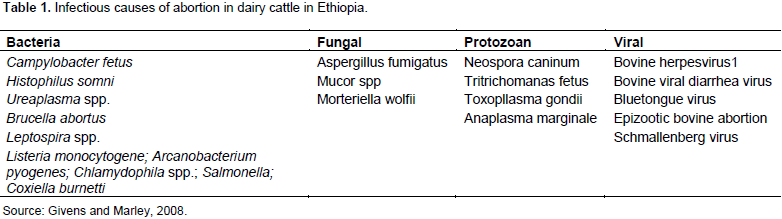
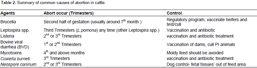
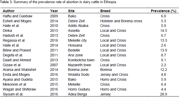
Abortion in dairy cattle may be caused by infectious and non-infectious agents. Infectious causes of abortion in dairy cattle include brucellosis, leptospirosis, listeriosis, Q fever, bovine viral diarrhea, mycotic abortion and neosporosis. Non-infectious causes of abortion in dairy cattle are genetic and non-genetic disorder. Risk factors associated with abortion in dairy cattle are genetic, environmental, management, geographical factors and infectious factors. Abortion in dairy cows brings about breeding and productive damages. Abortions cause significant economic loss to dairy farm. These losses can be attributed to loss of replacement calves, reduced milk production, costs of treatment, feeding of animals and premature culling of productive cows and heifers. Diagnosis of bovine abortion includes the collection of a complete history of the case and relevant epidemiological data and collected sample for analysis. However, determining the cause of bovine abortion is difficult as abortions are caused by numerous infectious and noninfectious factors. Status of abortion and breeds affected by abortion in Ethiopia were also reviewed.

Key words: Causes, abortion, dairy cattle, Ethiopia.


 INTRODUCTION

Ethiopia has the largest livestock population in Africa, with a total cattle population of 57.83 million. Out of this total cattle population, the female cattle constitute about 55.38% and the remaining 44.62% are male cattle. At present, about 99% of Ethiopia's national herd is of local breeds managed under extensive farming systems (CSA, 2016). The livestock contributes about 16.5% of the national Gross Domestic Product (GDP) and 35.6% of the agricultural GDP. It also contributes 15% of export earnings and 30% of agricultural employment (Leta and Mesele, 2014). However, the rate of urbanization is high, which places challenges on farmers and government to meet the demand for food (red meat and dairy products) for an increasing population. To increase livestock productivity and satisfy the increasing demand for livestock products, Ethiopia has given more attention to breed improvement, pasture development and animal health (Azage et al., 2001; Shapiro et al., 2015).
 
Ethiopia has paid considerable attention to livestock productivity (meat and milk) through breeding and health interventions to increase the contribution of livestock to economic growth as well as to meet the increasing local demands. The country has given priority on the development of dairying at farmer’s level to increase the supply of milk from smallholder dairy farms (Zegeye, 2003). However, reproductive health problems are becoming the major obstacles hindering this development plan (Adane et al., 2014; Ararsa and Wubshet, 2014). Among these, abortion is the main constraint in sector development plan to achieve its goal. Moreover abortion has direct impacts on reproductive performance of dairy cows (Lobago et al., 2006; Ernest, 2009). Abortion is defined as the termination of pregnancy between 42 and 260 days of gestation (Peter, 2000). The diagnosis of abortions is challenge to the farmers and veterinarian. There is sudden and dramatic increase of abortion in herds over a long period of time (Hossein-Zadeh, 2013).
 
Bovine abortion has infectious and none infectious causes (Hovingh, 2009). Infectious causes of abortion associated with abortion in cattle include viruses, bacteria, protozoa and fungus. The exact proportion of cases due to infectious agents is not known, but in 90% of cases in which an etiologic diagnosis is achieved, the cause is infectious (Parthiban et al., 2015). These pathogens can result in extensive economic losses, indicating the need for control measures to prevent infection or disease (Givens and Marley, 2008). Non-infectious factors such as genetic and non-genetic disorders have been reported in some investigations. The most important non genetic factors are heat stress, production stress, seasonal effect and seasonal changes (Hansen, 2002; Sani and Amanloo, 2007). The most important genetic disorders include chromosomal and single gene disorders and these disorders result in high abortion rate in cows and increased calf sterility.
 
Cow parity, sire effect, age at conception and abortion history could be some of the non-infectious maternal and paternal factors that cause abortion (Thurmond et al., 2005; Lee and Kim, 2007). Abortions have a highly negative impact on reproductive efficiency, resulting in significant economic losses for the cattle industry (De Vries, 2006). Spontaneous abortion of dairy cows is the most common problem that contributes substantially to low herd viability and decreasing production potential by reducing the number of potential female herd replacements and lifetime milk production, and by increasing costs associated with breeding and premature culling (Thurmond et al., 2005). The cost of abortion depends mainly on the time of gestation, milk production and time of insemination after parturition, the cost of nutrition, sperm costs, insemination time and labor costs (Rafati et al., 2010).
 
Late term abortions could also result in loss of potential replacement heifers, early culling of productive cows and loss in herd’s potential calf production (Carpenter et al., 2006). The prevalence rate of abortion varies in different production system and from place to place. Prevalence rate of abortion in Ethiopia range from 2.2 to 28.9% (Table 3) (Gizaw et al., 2007; Siyoum et al., 2016). This difference in prevalence rate may be due to variation in cattle breed and husbandry management system. Eshete and Moges (2014) indicated that incidence of abortion of more than 2 to 5% should be viewed seriously, efforts should be made to determine the causes and measures should be taken to control abortion. This paper reviews common cause of abortion, economic important and risk factor of abortion in dairy cattle.


 ABORTION IN DAIRY CATTLE

Abortion is the termination of pregnancy at a stage where the expelled fetus is of recognizable size ranging from 45 to 260 days of gestation and not viable (Peter, 2000). Sarder et al. (2010) also defined abortion as a condition in which fetus is delivered live or dead before reaching the stage of viability where the delivered fetus is visible by naked eyes. Some diseases that cause abortion in cattle, such as brucellosis, Leptospirosis are also zoonotic (Levett, 2005; De Vries, 2006). The important infectious agents that have been reported to cause abortion in cattle can be viral, bacterial, protozoa as well as several fungal species among others (Table 1) (Juyal et al., 2011). In addition, any disease causing high fever may also cause abortion (Radostits et al., 2007).


Common infectious causes of bovine abortion
 
Brucellosis
 
Brucellosis is an important disease of humans, and domestic and wild animals worldwide and is also a serious zoonosis (Mekonen et al., 2010). In female cattle, the disease is characterized by abortions storms in pregnant cattle, infertility, mastitis, retained placentae and arthritis (Radostits et al., 2007). Infected cow usually abort between the fifth and seventh month of pregnancy. Abortion due to brucellosis commonly occurs during the last trimester of pregnancy (Parthiban et al., 2015). All these manifestations lead to losses in the production system. Several species of the bacterium Brucella can cause brucellosis in cattle; however, Brucella abortus is the primary bovine pathogen (Godfroid et al., 2011). Brucellosis in cattle is spread by ingestion of contaminated pasture, feed and water, licking aborted foetuses or genital exudates from recently aborted cows or carrier cattle that have calved normally.
 
However, with infection through injured/intact skin, the mucosa at the respiratory system and conjunctiva frequently occurs (Acha and Szyfres, 2001; Degefa et al., 2011). While vaccination of cattle with strains S19 and RB51 has been the cornerstone of brucellosis control programmers in the developed world, adequate information on its occurrence in the developing world is lacking and the adoption of control programmers is still low (Godfroid et al., 2011). Several risk factors for bovine brucellosis have been reported. Among these are increased herd sizes, increased age, sex of the animal, husbandry practices such as animal confinement, contact with wildlife, geographical area and keeping different breeds in a herd (Muma et al., 2007; Tolosa et al., 2010; Matope et al., 2010; Mekonen et al., 2010).
 
Various techniques have been used to diagnose bovine brucellosis. These include the use of staining techniques, such as modified acid fast staining, culture and molecular techniques, such as polymerase chain reaction. However, in most epidemiological studies, serological tests, such as serum agglutination test (SAT), Rose-Bengal test (RBT), Buffered plate agglutination test (BPAT), fluorescence polarization assay (FPA) and ELISA, are often used. The limitations to the use of serological tests are false positives from vaccinated animals, cross-reactivity with other Gram-negative bacteria, and low sensitivity from tests such as SAT and RBT (OIE, 2009).
 
Leptospirosis
 
Leptospirosis is a contagious, bacterial disease of animals and humans. It is a globally important zoonotic disease caused by the pathogenic Gram negative bacteria of the genus, Leptospira (Bharti et al., 2003). The disease occurs worldwide, it is most common in temperate regions in the late summer and early fall and in tropical regions during rainy seasons (Tilahun et al., 2013). Although, the incidence of disease seems to have decreased in developed countries, it is apparently emerging rapidly as a significant public health problem in developing countries (Tangkanakul et al., 2000). All mammals appear to be susceptible to at least one species of Leptospira. The primary reservoir hosts for most Leptospira serovars are wild mammals, particularly rodents.
 
Reservoir hosts among domestic animals includes cattle, dogs, sheep and pigs and they may act as carriers for several months (temporary carrier) while rodents usually remain carrier throughout their life (permanent carrier) (Sophia, 2013). Rodents are therefore considered as the major reservoir of infection. The specific reservoir hosts vary with the serovar and the geographic region (OIE, 2005). In cattle, leptospirosis has been characterized by a wide variety of conditions including fever, icterus, hemoglobinuria, abortion and death. Cattle are the maintenance hosts for Leptospira serovar hardjo and Leptospira borgpeter-senii serovar hardjo, and incidental hosts for serovar pomona which is maintained in swine (Parthiban et al., 2015).
 
Leptospirosis can be transmitted either directly between hosts or indirectly in the environment. Leptospira species can be ingested in contaminated food or water, spread in aerosolized urine or water, or transmitted by direct contact with the skin. The organisms usually enter the body through mucous membranes or abraded skin. They may also be able to penetrate intact skin that has been immersed for a long time in water (Sophia et al., 2014). Leptospira species are excreted in the urine and can be found in aborted or stillborn fetuses, as well as in normal fetuses or vaginal discharges after calving (Levett, 2001). Leptospirosis of animals is investigated by direct and indirect laboratory methods. Direct methods are the isolation of the causative agent and the identification of Leptospira species antigens in tissue and body fluids using methods such as immunofluorescence staining, immunochemistry immune peroxidase staining, silver staining and various methods of polymerase chain reaction (PCR) (WHO, 2006).
 
Direct visualization of leptospirae in blood or urine by dark field microscopic examination has been used for diagnosis. But artefacts are commonly mistaken for leptospirae and the method has both low sensitivity and specificity (Vijayachari et al., 2001). For early diagnosis, serum is the optimal specimen. Urine from severely ill patients is often highly concentrated and contains significant inhibitory activity (Brown et al., 2003). The indirect methods of investigating leptospirosis are based on the detection of specific serum antibodies. These methods are either methods detecting serum antibodies without discriminating serovars, such as various ELISA tests, indirect immunofluorescence, the spot agglutination test or methods reliably identifying the infecting serovars, such as the microscopic agglutination test (MAT).
 
Microscopic agglutination test (MAT) is used as the ‘gold standard’ serological tests even though the test is very tedious and requires the maintenance of several leptospiral serovars in the laboratory. Also, the test requires the expertise personnel to read the results (Ooteman, 2006). Understanding the epidemiological features of leptospirosis is a critical step in designing interventions for reducing the risk of the disease transmission (Levett, 2001). Intervention strategies can target many points in the transmission cycle of leptospirosis. Although, little can be done in wild animals, leptospirosis in cattle is controlled through vaccination, prophylactic treatment of exposed cattle with antibiotics, quarantining of newly introduced cattle for at least 4 weeks, rodent control, regular serological testing, improved environmental hygiene, separating young animals from adults and safe artificial insemination (Dhanze et al., 2013).
 
Leptospirosis could result in a “storm of abortions” causing considerable economic losses from meat and milk reductions. Furthermore, these losses appear as more significant among cattle, because this animal species are considered less resistant than small ruminants (Tooloei et al., 2008). Abortion may occur several weeks after infection of the dam and is usually not associated with any obvious illness in the cow. Abortions due to serovar hardjo infection tend to occur sporadically. But abortion “storms” may occur as a result of infection with serovars pomona or grippotyphosa. Abortion storms are more common when the weather is wet and there is standing water that is contaminated with infective organisms (Bharti et al., 2011).
 
Abortion usually occurs during the last trimester of pregnancy but can occur from the 4th month onwards. Infertility and milk drop occurs only in pregnant or lactating cows because Leptospira organisms prefer pregnant uterus and lactating mammary gland to proliferate (Yadeta et al., 2016). Although, there is few documented information so far concerning the occurrence of leptospirosis in animals in Ethiopia, climatologic, socioeconomic and other factors are highly favorable for the occurrence and spread of the disease in the country. In Ethiopia, leptospirosis has been reported to occur in domestic animals working in Ethiopia, with incidences of 70.7% in cows (Yadeta et al., 2016). In the case of human leptospirosis, there is a pilot study in Wonji Hospital. According to Eshetu et al. (2004) from a total of 59 febrile patients attending the outpatient department of Wonji Hospital, 47.5% were positive for leptospirosis and the occurrence of the disease was more common in males than females.
 
Listeriosis
 
Listeriosis is an infectious disease of human and animals with a world-wide distribution. It manifests in three major clinical forms, meningoencephalitis, abortion and septicaemia (Hirsh et al., 2004). Listeriosis is caused by a member of the genus Listeria. Majority of the clinical cases are associated with Listeria monocytogenes infection. Only a few reported cases have been associated with L. ivanovii (Radostits et al., 2007). Listeria species are found widely throughout the environment. Listeria can be ingested with poorly preserved silage which is not fermented properly and is not acidic enough to kill the bacteria. It can be ingested via soil on the grass roots and also the placenta and discharges from the infected cow (Aderson, 2007). Listeria monocytogenes is a well-recognized cause of abortion, encephalitis and septicemia in cattle. Listeria ivanovii has also been implicated as a cause of abortion in cattle but occurs less frequently than L. monocytogenes. Abortion occurs after ingestion of L. monocytogenes contaminated feed and a resultant bacteremia.
 
Experimental studies have shown that after ingestion or parenteral injection of L. monocytogenes, the genital organs and foetus are invaded within 24 h of the onset of bacteremia. This results in abortion in 5 to 10 days (Radostits, 2007). Listeria infections and abortions usually develop in the late winter or early spring. Abortions are most commonly recognized in the last trimester of pregnancy and abortion storms can occur when all herd eat same batch of contaminated silage at same time (Yaeger et al., 2007). In abortion, the pathological picture depends on the stage of pregnancy. If it occurs in the early stages of the last trimester, the placenta is quickly invaded by the bacteria and the foetus dies as a result of septicaemia.
 
The dead foetus is expelled within 5 days and by this time autolytic changes cover the minor gross lesions produced by the organism. Metritis usually occurs and results in retention of the foetal membranes. If it occurs at a late stage, the offspring may be born in the normal way but is usually unable to survive. In the aborted foetus the lesions are less severe. Gross lesions are tiny pin-point yellow foci in the liver. Similar foci but visible only microscopically are seen in the lung, myocardium, kidney, spleen and brain. The bacteria can be demonstrated in the center of these focal areas (Thomson, 1988; Quinn et al., 2002). The organism is sensitive to a wide range of antibiotics. Culling infected animals should be advocated as they secrete the organisms in secretions and excretions, especially in the cases of mastitis.
 
Care in the use and preparation of silage is important as the pathogen grows luxuriantly at a pH greater than 5.5 (Walker, 2007). Farm management practices, such as improvement of nutritional status of animals and better housing conditions, can also be of some value in preventing disease (Dhama et al., 2017). In Ethiopia, few findings of L. monocytogenes have been reported, possibly due to lack of attention or resources (Molla et al., 2004). Study conducted by Seyoum et al. (2015) showed that prevalence of Listeria species was 28.4% and specifically that of L. monocytogenes was 5.6% from raw bovine milk and milk products from central highlands of Ethiopia. Other study conducted in Gondar on foods of animal origin indicated that 25% were positive for Listeria species and that of L. monocytogenes was 6.25% (Garedew et al., 2015)
 
Mycotic abortions
 
Mycotic abortion causes great economic losses to the individual farmer and cattle-breeding industry as a whole. Mycotic infection of the placenta is one of the most common causes of sporadic bovine abortion (Ali and Khan, 2006). Mycotic abortion is caused by different species of fungi and yeasts. About 35 different species of fungi have been known to cause abortion, Aspergillus fumigatus being the most commonly diagnosed casual organism which accounts for 60 to 80% of abortions. 20 to 35% of abortions have been attributed to fungal causes (Pal, 2015). Aspergillus fumigatus is the cause of over 70% mycotic abortions recorded in cattle, around the world (Ali and Khan, 2006) Abortion occurs when fungal spores enter a pregnant cow’s blood stream, settle at the junction of the maternal and foetal placentas, grow and attack the placental tissues (Walker, 2007). In general, fungal spores may be present in cattle feed. However, some feeds such as improperly preserved silage and hay that has been wet, contain many more spores than others. Storm of abortion occur in cattle when feeding with mouldy hay at same time.
 
The mycotic abortions were confirmed by isolation of Aspergillus fumigatus fungi from mouldy hay as well as from foetal abomasal contents (Chandranaik et al., 2014). Pregnancy in a cow with metabolic derangements from stress may predispose the pregnant cow to fungal infection. The incidence of the condition is high in late summer or early autumn, due to the presence of large number of fungal spores in pastures during this period (Ali and Khan, 2006). There is also evidence of a winter rise of disease incidence. The organism may cause abortion from 4 months to term. Other species of molds and yeasts have been associated with abortion (Parthiban et al., 2015). Any condition that reduces the cow’s resistance to infection increases the chances of mycotic abortion. Providing good health (via good management and nutrition) and not feeding moldy feeds can reduce the incidence. When possible, depending on the availability and demand decreases the period of confinement, decrease cow density and improve ventilation (Pal, 2015).
 
Query fever (Q fever)
 
Q fever is a zoonosis of worldwide distribution caused by Gram-negative intracellular bacteria Coxiella burnetii, which can infect arthropods, birds and animals (Cutler et al., 2007). Currently it is not possible to accurately estimate the true prevalence infection in domestic ruminants, due to lack of well-designed studies. However, there has been detection of C. burnetii in all five continents (except in New Zealand being the only country with a reported apparent prevalence of zero), with a wide range, in whatever kind. The apparent prevalence is slightly higher in cattle (20.0 and 37.7%) than in sheep and goats (around 15 to 25%) (Guatteo et al., 2011). Infections by C. burnetii in animal production are mostly asymptomatic, however, may be related to reproductive disorders such as abortion, stillbirths, repetition heat, low birth weight animals and metritis. Nevertheless, latter clinical manifestation appears to be unique in cattle, occurring during first three weeks after birth, with fetid vaginal discharge and/or increase in body temperature (Sheldon et al., 2006).
 
In most cases, abortion occurs in late pregnancy which range from 3 to 80% with unspecified characteristic clinical signs of infection with C. burnetii (Angelakis and Raoult, 2010). Aborted fetuses appear normal but infected placentas exhibit intercotyledonary fibrous thickening and discolored exudates, which are not specific to Q fever (Arricau-Bouvery and Rodolakis, 2005). Coxiella burnetii can also be recovered from milk for up to 32 months. Furthermore, there may be shedding bacteria in the urine, semen and vaginal discharge mucus. An important factor related to abortion rates in herds is the temperature, since fewer abortions take place between months of November and December. However, this occurrence increases gradually from January to February, decreasing again in March (Cantas et al., 2011). A relevant issue is infestation of cattle by ticks during months when temperature is higher.
 
Previous studies have shown that ticks seem to play an important role in the dissemination of bacteria in animals, especially wild, believing it to be an important factor in the transmission to domestic animals (Psaroulaki et al., 2006). On the other hand, a recent study developed in the Netherlands, after three years of an outbreak of Q fever, researchers investigated the role ticks play in the transmission C. burnetii, showing that actual risk of this infection by ticks is negligible. Moreover, for future risk assessments, it might be relevant to sample more ticks in the vicinity of previously C. burnetii infected goat farms and to assess whether C. burnetii can be transmitted transovarially and transstadially in Ixodus ricinus ticks (Sprong et al., 2011). Few studies conducted in Ethiopia indicated that 6.5% seroprevalence of C. burnetii was observed in Addis Ababa abattoir workers. Also, the existence of antibody against C. burnetii was reported in goats and sheep slaughtered at Addis Ababa abattoir, and its peri-urban zones. A seroprevalence of 31.6% of C. burnetii was recorded in cattle in South Eastern Ethiopian pastoral zones of the Somali and Oromia regional states (Gumi et al., 2013).
 
Bovine viral diarrhea (BVD)
 
Bovine viral diarrhea is a disease caused by bovine viral diarrhea virus (BVDV). Bovine viral diarrhea is one of the most important diseases of cattle worldwide (Almeida et al., 2010). It is an important cause of diarrhea, reproductive problems and reduced milk yield in affected herds (Lindberg and Houe, 2005). This is a Pestivirus in the family Flaviviridae that is closely related to border disease virus of sheep and classical swine fever virus of pigs (OIE, 2004). The disease occurs worldwide and infections may be subclinical in some animals (Lindberg and Houe, 2005). Bovine viral diarrhea virus can be persistent in infected animal and wild animal asymptomatic while shedding large amount of virus throughout their life time (Nelson et al., 2016).
 
Bovine viral diarrhea virus is transmitted by direct contact with saliva, faeces, semen, urine, tears and milk of infected cattle, or by in utero infection of fetuses (Radostits et al., 2007). Infection of naive pregnant cows and heifers may lead to abortion and other reproductive disorders, such as early embryonic death (the death of a conceptus within the first 2 months after conception) in the first 45 days, fetal death and mummification (Kabongo and Van Vuuren, 2004). Infection during the first trimester of pregnancy will cause storm of abortions approximately one month prior to parturition. Infection during the second trimester will often lead to a higher risk of birth defects and less abortion, and this is more common in beef cattle than dairy breeds. But in final trimester of pregnancy, there are no more effects (Van Campen, 2010).
 
The other effects of BVDV are birth of calves with congenital defects, calves with poor growth rates, and increased average age at first calving in affected herds (Heuer et al., 2007). The virus has also been shown to depress ovarian function in infected heifers by disrupting gonadal steroidogenesis, and impairing the quality of oocytes produced (Fray et al., 2000; Altamarand et al., 2013). Infection from day 9 to 45 of gestation results in reduced conception rates and infertility, early embryonic death and infertility. From days 45 to 75 of gestation, infection with BVDV will result in abortions, intrauterine growth retardation, and calves with congenital defects especially of the nervous system. Infection in late gestation (125 to 285) results in birth of normal calves with neutralizing antibodies (Grooms, 2004). This virus has a high affinity for leucocytes and reduces their numbers in infected animals. This immunosuppression potentiates the effects of other pathogens, including abortifacient ones, such as Neospora caninum (Bjorkman et al., 2000; Konnai et al., 2008).
 
Among the risk factors for BVDV infection in cattle are increased age and the origin of the animal (Mainar-Jaime et al., 2001); pasturing and increased herd sizes; and dam factors, such as high BVDV titres at calving and increased parity (Munoz-Zanzi et al., 2003). In addition, the use of artificial insemination breeding technique without the institution of biosecurity measures on the farm has been shown to increase the risk of BVDV spread by 2.8 times, most likely due to contamination of the herd through contaminated insemination equipment and personnel (Almeida et al., 2013). Several methods have been developed to detect BVDV infection in cattle (OIE, 2008). These include virus isolation in bovine tissue culture (kidney, lung, testis and turbinate cells), immunohistochemistry to detect virus antigen in tissue, nucleic acid detection by polymerase chain reaction, and serological tests, such as virus neutralization and enzyme-linked immune sorbent assay (ELISA).
 
Samples collected for analysis include: bulk milk to determine the herd status, individual milk, serum and plasma samples to determine individual animal sero-status, as well as tissue samples for immunohistochemistry. Serological tests, such as ELISA, are commonly employed in explorative studies since they can be used to determine the sero-status of large numbers of animals sampled in a population (OIE, 2008). In Ethiopia, few studies conducted on the disease indicated that 9.6, 16.6 and 6.11% seroprevalence of BVDV was reported in dairy cattle herds in Jimma, south western Shoa, and West Shoa, respectively (Nigussie et al., 2010). Seroprevalence of 11.7% of BVDV was also reported in breeding and dairy farms of southern and central Ethiopia (Asmare et al., 2012). There is no study conducted to determine the rate of persistent of infection caused by BVDV in Ethiopia.
 
Neosporosis
 
Neosporosis is a disease caused by Neospora caninum. This is a protozoan coccidian parasite that structurally resembles and is genetically related to Toxoplasma gondii (Silva et al., 2007). There are two species of Neospora currently recognized: N. caninum which causes clinical disease in dogs, cattle, sheep, equines and many wild animal species, and Neospora hughesi, which has been associated with reproductive losses and myoencephalitis in horses. Dogs are the definitive hosts of N. caninum and cattle are among the intermediate hosts (Hall et al., 2006; Fernandez et al., 2006). Cattle become infected by ingestion of feed and water contaminated by oocysts shed in dog faeces, or by congenital infection (Jenkins et al., 2002; Pan et al., 2004). This parasite has been reported to be the most important cause of abortion and neonatal mortality in beef and dairy cattle populations worldwide including Ethiopia (Murray, 2006; Silva et al., 2007; Asmare et al., 2012).
 
Abortions in cattle due to N. caninum occur from 3 months of gestation but are most common from 5 to 6 months of pregnancy. Neospora can be associated with sporadic abortions, endemic or abortion storms in cows have been reported. Other signs presented by infected cattle are foetal resorption, mummification, autolysis and stillbirth, and some calves are born alive with neuromuscular defects, while other calves are apparently healthy but persistently infected (Dubey and Schares, 2006). The incidence of abortion is often repeated in subsequent pregnancies, and congenital/vertical transmission from seropositive dams to their offspring is important in the epidemiology of neosporosis (Dubey et al., 2007). Reported risk factors for bovine abortions due to N. caninum include geographical location, breed, exposure to dogs or wild carnivores, and pregnant heifers (Dubey and Schares, 2006; Asmare et al., 2013). Various methods have been used to diagnose neosporosis in animals.
 
These include histopathology of tissues from aborted foetuses and still-births, parasite isolation from sacrificed animals, inoculation in mice, molecular techniques such as polymerase chain reaction, and oocyst recovery from dog faeces. However, serology (ELISA and immuno-fluorescent antibody test [IFAT]) is the most common technique used to diagnose neosporosis since it can be done ante-mortem and postmortem. Serology is useful in epidemiological studies since it can be used to reliably test exposure and infection in large animal populations (Dubey and Schares, 2006; Silva et al., 2007). In Africa, reports on neosporosis are limited; however, the available information is in line with global understanding of the protozoan that underscores the relevance of the N. caninum to the dairy sector (Ghalmi et al., 2012). The general seroprevalence of this disease globally ranges from 1.9 to 39.7% (Njiro et al., 2011; Ayinmode and Akanbi, 2013).
 
 
Recent studies confirmed that neosporosis is prevailing in dairy cattle of Ethiopia (Asmare et al., 2012; Asmare et al., 2013). However, the available published information comparing different pathogens exposure vis-à-vis reproductive disorders is limited to a single article based on the data from central and southern part of the country (Asmare et al., 2013). Few studies conducted in Ethiopia indicated that seroprevalence of 17.2% of N. caninum was reported in breeding and dairy farms of southern and central Ethiopia and 13.3% seroprevalence was also recorded in intensive or semi-intensively managed dairy and breeding cattle of Ethiopia (Asmare et al., 2012, 2013). Neosporosis appears to be a highly prevalent and widely distributed infectious cause of bovine reproductive disorders in urban and peri-urban smallholder farms, commercial dairy farms and breeding cattle in Ethiopia (Asmare et al., 2013). N. caninum is common in dairy cattle and is probably a more important cause of abortion in dairy cattle in Ethiopia than other infection cause of abortion (Asmare et al., 2012). The general control and prevention of causes of abortion in dairy cattle summary in Table 2.


Risk factors of abortion
 
Several causative factors, including external, maternal and genetic factors, have been reported for abortion in dairy cattle. These include heat stress, season, milk production, cow parity, serum progesterone level after conception, the inseminating bull, twin pregnancy and the herd (Lee and Kim, 2007). However, other investigations have reported that milk production and cow parity were not associated with abortion (Moore et al., 2005). Parity status and breed were significant factors affecting the incidence of abortion (Yakubu et al., 2015). However, Haileselassie et al. (2011) reported that parity status had no significant effect on the incidence of abortion. Factors that have been reported to increase the risk of abortion in dairy cattle herds include: being a heifer; being a cow of more than 10 years old; feeding on communal pastures; lack of vaccination against abortifacient diseases, hygiene, animal management and reproductive problems such as retained placentae, dystocia, uterine prolapse and stillbirth in the previous pregnancies (Waldner and Garcia, 2013; Waldner, 2014).
 
Risk factors such as environmental (nutrition, temperature extremes and toxins, among others), management (crowding and use of natural mating), geographical factors and infectious factors, with infections contributing up to 90% of the abortions also reported (Konnai et al., 2008; Mekonen et al., 2010). Environmental high temperature may affect inside-pens temperature and performance of dairy cattle. Omori et al. (2014) reported that hyperthermia during pregnancy causes abortion in dairy cattle. Environmental temperature also affect the level of aflatoxin in the feed given to animals where above tolerable level could be a predisposing stress factor; aflatoxin is more often found in fodder grown in warm and humid climates which support growth of moulds. It has been suggested that aflatoxin lowers resistance to diseases and interferes with vaccine-induced immunity (Diekman and Green, 1992). In another study, third-trimester abortion was reported after cattle consumed mouldy peanuts (Ray et al., 1986).
 
 
Normal annual abortion rate were cited to be 3 to 5% once cows are above 42 days of pregnancy (Hovingh, 2009), or similarly, an observable 2 to 5% in most dairies (Kirk, 2003). While some suggest the annual abortion rate should be less than 3% in dairy, others believe this is not typical. This difference may arise from the fact that many abortions may be due to early embryonic death where cows are identified as pregnant and then found to be open without visible signs of an abortion. As a consequence, many early abortions may go undetected or even dismissed as an unsuccessful insemination rather than a failed pregnancy (Carpenter et al., 2006). A low rate of abortions from 2 to 5% per 100 pregnancies per year is usually considered within the expected rate as sporadic abortions occur in any herd. However, occurrence of several abortions in a short period or high rate of abortions warrants investigation to detect the cause and take control measures (Esheti and Moges, 2014; Al Humam, 2014).


 ECONOMIC IMPORTANT OF ABORTION IN CATTLE

Abortion is one of the most important major reproductive health disorders of dairy cows in the world including Ethiopia in terms of economic impact (James and Rushton, 2002; Regassa and Ashebir, 2016). Abortions cause significant economic loss, especially those occurring during late gestation. These losses can be attributed to loss of replacement of calves, reduced milk production, costs of treatment, feeding of animals and premature culling of productive cows and heifers (Carpenter et al., 2006; Abdelhadi et al., 2015). The cost of abortion varies according to effective factors such as the time of gestation, milk production, days in milk, the time of insemination after parturition, the cost of nutrition, sperm costs, insemination time and labor costs, which differ from region to region. Abortions during early pregnancy result in increased days open (De Vries, 2006; Hovingh, 2009).
 
Different values were reported for the cost of abortion ranging from $90 to $2333 based on different studies. These differences are caused by the stage of gestation in which the abortion occurs and by the differences in factors such as predicted cow performance, breeding and replacement decisions, feed and milk price and the stage of lactation (De Vries, 2006; Lee and Kim, 2007; Hovingh, 2009). Estimates of the cost of an abortion to a producer range from $90 to $1,900 (Peter, 2000; Kirk, 2003), depending on when pregnancy and occurred and differences in predicted cow performance, prices, and breeding and replacement decisions. Hanson et al. (2003) stated that losses were $200 million per year in California herds.
 
Each case of abortion in dairy cattle has been estimated to lead to losses of about US $500 to $900. Per case, the cost of abortion has been estimated at $640 (Thurmond and Picanso, 1990) and from $600 to $800 (Eicker and Fetrow, 2003). Pfeiffer et al. (1997) estimated the cost of an abortion caused by N. caninum infections at $624 in New Zealand. Peter (2000) documented a cost of $600 to $1,000 per midterm abortion. Weersink et al. (2002) estimated the cost of an abortion, including reproductive loss and reduced milk yield at $1,286 in Canada. In addition, some of the causes of abortion, such as Brucella abortus, Toxoplasma and Leptospira, are zoonotic, thus posing a risk to human health (Carpenter et al., 2006; Murray, 2006). However, no reports are available on the estimation of the economic impact of bovine abortion in Ethiopia.


 DIAGNOSIS OF ABORTION

General principles in the diagnosis of abortion in dairy animals include the collection of a complete history of the case and relevant epidemiological data, such as recent introductions into the farm, determination of the number of animals affected, examination of the breeding, health and feeding records, careful examination of the affected dam(s), and collection of the expelled fetus and placenta for pathological and microbial examination. Furthermore, samples such as paired serum samples, urine, milk and vaginal swabs can also be collected for analysis. The results are then collated and analyzed to reach a diagnosis (Radostits et al., 2007). However, the diagnostic rate in bovine abortions is very low due to the diverse range of pathogens involved, as well as the fact that factors affecting the dam, fetus and placenta may be involved (Murray, 2006; Ernest, 2009).
 
Abortion also often follows an initial infection which may have occurred for several weeks or months; the etiology often is not detectable by the time the abortion occurs. The high cost of laboratory work to aid in the diagnosis of bovine abortion also compounds the problem (Carpenter et al., 2006; Murray, 2006). Diagnosis of the cause of bovine abortion is difficult as abortions are caused by many infectious and noninfectious factors (Miller, 1987; Jamaluddin et al., 1996). It has been demonstrated in numerous surveys that many abortions occur due to endemic infectious which are normally present in cattle populations world-wide (Kim et al., 2002).
 
The diagnosis of abortions often presents a challenge to the farm owner and the veterinarian in charge. A sudden and dramatic increase in the abortion rate in a herd is more commonly seen, although a gradual increase may be noted over a long period of time. For this reason, prompt and thorough action is required if abortions occur at any rate. Well-arranged records of a herd is often of benefit during the investigation of abortion problems (Al Humam, 2014). However, it is important to note that the causes of abortion in cattle are numerous and thus, their diagnosis is often challenging (Murray, 2006; Ernest, 2009). Epidemiological tools could help in narrowing down the field of investigation for a better interpretation of laboratory results (Markusfeld, 1997).
 
Status of abortion in dairy cows in Ethiopia
 
Ethiopia has various agro ecological zones, which have contributed to the evolution of different agricultural production systems (Beruktayet and Mersha, 2016). Husbandry systems, variation in cattle breed and environmental factors greatly influence the spread of the cause of abortion (Mekonen et al., 2010). Thus, the prevalence of abortion varies in different production system, cattle breed and agro ecological zones (Esheti and Moges, 2014). Studies on major reproductive problems of cows in different parts of Ethiopia have shown the occurrence of abortion in cattle. Study conducted by Haftu and Gashaw (2009) on major clinical reproductive health problems of dairy cows in and around Bako of West Ethiopia showed that 6.0% (n=217) of dairy cows are affected by abortion.
 
A study of the major reproductive health disorders of dairy cows in ILCA and Almaz dairy farms in Ada’a district, Debre Zeit town in East Shoa showed that 5.3%(n=245) of cows had abortion problem (Esheti and Moges, 2014). Other study conducted in Addis Ababa Milk showed major reproductive disorders in cross breed dairy cows under small holding indicating an overall prevalence of 5.9% (n=384) of abortion problems (Haile et al., 2010). A study conducted by Dinka (2012) showed that 14.6% (n=300) of dairy cattle was affected by abortion based on questionnaire interviews in and around Assella in Central Ethiopia. A retrospective study by Hadush et al. (2013) revealed that 6.7% (n=711) of the cows had abortion problem from dairy cows in three selected farms in Debre Zeit town. Another study conducted using questionnaire and observational survey in urban and peri urban area of Hossana indicated 2.6% (n=390) prevalence of abortion in dairy cattle (Haile et al., 2014).
 
A study in and around Bedelle showed a prevalence of 13.9% (n=302) of abortion in South west Ethiopia (Bitew and Prased, 2011) and 8.7% (n=370) prevalence in selected sites of Arsi zone (Degefa et al., 2011). A prevalence of 9.1% (n=231) abortion was reported at Kombolcha town in north east Ethiopia by Dawit and Ahmed (2013). A study conducted by Gizaw et al. (2007) and Ararsa and Wubishet (2014) also reported 2.2 (n=403) and 12.2% (n=409) in Nazareth town of central Ethiopia and Borena zone in southern Ethiopia, respectively. A prevalence of 6.4% (n=1013) abortion was also recorded in dairy cattle in and around Mekelle, Tigray (Mekonnin et al., 2015) and 5.9% (n=372) of prevalence of abortion was reported in Bako Livestock Research Farm (Ayana and Gudeta, 2015).
 
A study conducted by Regassa et al. (2016) on major factors influencing the reproductive performance of dairy farms in Mekelle city, Tigray reported 13.3% (n=798) prevalence of abortion. Recent reports from Adea Berga (Siyoum et al., 2016), Horro Gudru (Wagari and Shiferaw, 2016) and Wolaita Sodo town (Enda and Moges, 2016) indicated that the prevalence of 28.9 (n=97), 4.4 (n=402) and 4.8% (n=104) of abortion was recorded in cattle, respectively. The incidence of abortion of more than 2 to 5% should be viewed seriously, and efforts should be made to determine the causes so that proper methods of control can be instituted (Mainar-Jaime et al., 2005; Esheti and Moges, 2014). Abortion problem is the most common in dairy cows (Gizaw et al., 2007). In order to reduce these problems and their risk factors, formulation of strategic control measures including health education on the cause of abortion transmission, treatment and control has to be introduced (Dinka, 2012).


 CONCLUSION

Abortion is one of the most important reproductive health problems of dairy cows in Ethiopia in terms of economic impact. Both infectious and non-infectious agents may cause abortion in cattle. Non-infectious factors are genetic and non-genetic disorders. The non-genetic causes of abortion are heat stress, production stress, seasonal effect and seasonal changes. The common infectious causes of abortion in cattle include brucellosis, leptospirosis, listeriosis, Q fever, bovine viral diarrhea, mycotic abortion and neosporosis. These causes can result in extensive economic losses, showing the need for control measures to prevent abortion. Whereas, the infectious causes of abortion have been a primary focus of attention, and non-infectious cause of abortion is actually more common in endemic situations.
 
Several risk factors associated with abortion are genetic, environmental, management, geographical and infectious factors. Incidence of abortion in Ethiopia ranges from 2.2 to 28.9%, efforts should be made to determine the causes and measures should be taken to control abortion. Prevention should be focused on accurate records keeping and collection of samples for laboratory analysis and using good biosecurity practices that inhibit the introduction and spread of infectious causes of abortion and using vaccination programs could limit abortion occurrence. There should be maintenance of the general health and immune function of animals by providing a balanced feed, clean water and a clean and dry environment. It was suggested that detail epidemiological study on cause of abortion in cattle should be undertaken.


 CONFLICT OF INTERESTS

The authors have not declared any conflict of interests.


 REFERENCES

Abdelhadi F, Abdelhadi S, Niar A, Benallou B, Meliani S, Smail N, Mahmoud D (2015). Abortions in cattle on the level of Tiaret Area Algeria. Glob. Vet. 14:638-645.


Acha N, Szyfres B (2001). Brucellosis in zoonosis and communicable diseases common to humans and animals. 3rd Ed., Pan. America Health Organization Washington, D.C., USA. pp. 40-62.


Adane H, Yisehak T, Niguse T (2014). Assessment of major reproductive disorders of dairy cattle in urban and per urban area of Hosanna, Southern Ethiopia. Anim. Vet. Sci. 2:135-141.


Anderson ML (2007). Infectious causes of bovine abortion during mid- to late-gestation. Theriogenology 68:474-486.2007.


Al Humam N (2014). An epidemic of abortion in a commercial dairy farm in the eastern region, kingdom of Saudi Arabia. Glob. J. Dairy Farming Milk Prod. 2(3):074-080.


Ali R, Khan IH (2006). Mycotic abortion in cattle. Pak. Vet. J. 26(1):44-46.


Almeida L, Kaiser GG, Mucci NC, Verna AE, Campero CM, Odeon AC (2013). Effect of bovine viral diarrhoea virus on the ovarian functionality and in-vitro reproductive performance of persistently infected heifers. Vet. Microbiol. 165:326-332.
Crossref


Almeida L, Miranda IC, Hein HE, Neto WS, Costa EF, Marks FS, Rodenbusch CR, Canal CW, Corbellini LG (2013). Herd-level risk factors for bovine viral diarrhoea virus infection in dairy herds from Southern Brazil. Res.Vet. Sci. 95:901-907.
Crossref


Altamarand EA, Kaiser GG, Mucci NC, Verna AE, Campero CM, Odeon AC (2013). Effect of bovine viral diarrhoea virus on the ovarian functionality and in-vitro reproductive performance of persistently infected heifers. Vet. Microbiol. 165:326-332.
Crossref


Angelakis E, Raoult D (2010). Q fever. Vet. Microbiol. 140(4):297-309.
Crossref


Ararsa DB, Wubishet Z (2014). Major reproductive health problems of indigenous Borena cows in Ethiopia. J. Adv. Vet. Anim. Res. 1(4):182-188.
Crossref


Arricau-Bouvery N, Rodolakis A (2005). Is Q fever an emerging or reemerging zoonosis? Vet. Res. 36(3):327-349.
Crossref


Asmare K, Regassa F, Robertson LJ, Martin AD, Skjerve E (2012). Reproductive disorders in relation to Neospora caninum, Brucella spp. and bovine viral diarrhoea virus sero status in breeding and dairy farms of central and southern Ethiopia. Epidemiol. Infect. 141(8):1772-1780.
Crossref


Asmare K, Regassa F, Robertson LJ Skjerve E (2013). Seroprevalence of Neospora caninum and associated risk factors in intensive or semi-intensively managed dairy and breeding cattle of Ethiopia. Vet. Parasit. 193:85-94.
Crossref


Ayana T, Gudeta T (2015). Incidence of Major Clinical Reproductive Health Problems of Dairy Cows at Bako Livestock Research Farm over a Two-Year Period (September 2008-December 2010), Ethiopia. Anim. Vet. Sci. 3(6):158-165.
Crossref


Ayinmode AB, Akanbi IM (2013). First report of antibodies to Neospora caninum in Nigerian cattle. J. Infect. Dev. Countries 7:564-565.
Crossref


Azage T, Tsehay R, Alemu G, Hizkias K (2001). Milk recording and herd registration in Ethiopia. In Proceedings of the 8 Annual Conference of the Ethiopian Society of Animal Production (ESAP), 24- 26 August 2000, Addis Ababa, Ethiopia. pp. 90-104.


Beruktayet W, Mersha C (2016). Review of Cattle Brucellosis in Ethiopia. Acad. J. Anim. Dis. 5(2):28-39.


Bharti AR, Nally JE, Ricaldi JN, Matthias MA, Diaz MM, Lovett MA (2003). Leptospirosis, a zoonotic disease of global importance. Lancet Infect. Dis. 3:757-771.
Crossref


Bitew M, Prased S (2011). Study on major reproductive health problems in indigenous and cross breed cow in and around Bedelle, South west Ethiopia. J. Anim. and Vet. Adv. 10:723-727.
Crossref


Bjorkman C, Alenius S, Manuelson U, Uggla A (2000). Neospora caninum and bovine viral diarrhoea virus infections in Swedish dairy cows in relation to abortion. Vet. J. 159:201-206.
Crossref


Brown PD, Carrington DG, Gravekamp C (2003). Direct detection of leptospiral material in human postmortem samples. Res. Microbiol.154:581-586.
Crossref


Cantas H, Muwonge A, Sareyyupoglu B, Yardimci H, Skjerve E (2011). Q fever abortions in ruminants and associated on-farm risk factors in northern Cyprus. Vet. Res. 17(1):7-13.
Crossref


Carpenter TE, Chrie` M, Andersen M, Wulfson L, Jensen A, Houe H, Greiner M (2006). An epidemiologic study of late-term abortions in dairy cattle in Denmark. Prev. Vet. Med. 77:215-229.
Crossref


Chandranaik BM, Rathnamma D, Earanna N, Shivashankar BP, Kalge RS, Srinvasababu T, Kanaka S, Gangadharaiah HK, Muniyellappa HK, Raveendrahegde P, Venkatesha MD (2014). Abortion Storm in Cattle Due to Aspergillus Fumigatus Contaminated Feed in Drought Hit Southern Karnataka. Indian Vet. J. 91(11):82-84.


Central Statistical Agency (CSA) (2016). Livestock and Livestock Characteristics, Agricultural sample Survey. Addis Ababa, Ethiopia. Statistical Bull. 2(583):9-13.


Cutler SJ, Bouzid M, Cutler RR (2007). Q fever. J. Infect. 54(4):313-318.
Crossref


Dawit T, Ahmed S (2013). Reproductive health problems of cows under different management systems in Kombolcha, North east Ethiopia, Hawassa University, School of Veterinary Medicine, Hawassa, Ethiopia. Available at: 

View


De Vries A (2006). Economic value of pregnancy in dairy cattle. J. Dairy Sci. 89: 3876-85.
Crossref


Degefa T, Duressa A, Duguma R (2011). Brucellosis and some reproductive problems of indigenous Arsi cattle in selected Arsi zones of Oromia Regional State, Ethiopia. Glob. Vet. 7:45-53.


Diekman DA, Green ML (1992). Mycotoxins and reproduction in domestic livestock. J. Anim. Sci. 70:1615-1627.
Crossref


Dinka H (2012). Reproductive performance of crossbred dairy cows under smallholder condition in Ethiopia. Int. J. Livest. Prod. 3(3):25-28.


Dhama K, Karthik K, Tiwari R, Shabbir MZ, Barbuddhe S, Malik SV, Singh RK (2015). Listeriosis in animals, its public health significance (food-borne zoonosis) and advances in diagnosis and control: a comprehensive review. Vet. Quarterly 35(4):211-235.
Crossref


Dhanze H, Kumar M, Mane BG (2013). Epidemiology of leptospirosis, An Indian perspective. J. Food Borne Zoonotic Dis. 1(1):6-13.


Dubey JP, Schares G (2006). Diagnosis of bovine neosporosis. Vet. Parasitology. 140:1-34.
Crossref


Dubey JP, Schares G, Ortega M (2007). Epidemiology and control of neosporosis and Neospora caninum. Clin. Microbiol. Review. 20:323-367.
Crossref


Eicker S, Fetrow J (2003). Eicker, S., & Fetrow, J. (2003). New tools for deciding when to replace used dairy cows. In Proc. Kentucky Dairy Conf., Cave City, KY. Univ. Kentucky, Lexington. pp. 33-46.


Enda W, Moges N (2016). Major Reproductive Health Problems in Dairy Cows in Wolaita Sodo Town in Selected Farms in Ethiopia. Eur. J. Biol. Sci. 8(3):85-90.


Ernest H (2009). Common Causes of Abortions. Virginia cooperative extension publication. pp. 404-288.


Eshete G, Moges N (2014). Major reproductive health disorders in cross breed dairy cows in Ada'a District, East Shoa, Ethiopia. Glob. Vet. 13(4):444-449.


Eshetu Y, Simone K, Tsehaynesh M, Dawit W, Bethelehem N, Neway G, Belachew D. Eduard J (2004). Human leptospirosis, in Ethiopia: A pilot study in Wonji Hospital, Ethiopia. J. Health Dev. 18:48-51.


Fernandez E, Arna’iz-Seco I, Burgos M, Rodriguez-Bertos A, Aduriz G, Ferna’ndez- Garcı’a A, Ortega-Mora L (2006). Comparison of Neospora caninum distribution, parasite loads and lesions between epidemic and endemic bovine abortion cases. Vet. Parasitol. 142:187-191.
Crossref


Fray MD, Mann GE, Clarke MC, Charleston B (2000). Bovine viral diarrhea virus: Its effect on ovarian function in the cow. Vet. Microbiol. 77:185-194.
Crossref


Garedew L, Taddese A, Biru T, Nigatu S, Kebede E, Ejo M, Fikru A Birhanu T(2015). Prevalence and antimicrobial susceptibility profile of listeria species from ready-to-eat foods of animal origin in Gondar Town, Ethiopia. BMC Microbiol. 15:100
Crossref


Givens MD, Marley MS (2008). Infectious causes of embryonic and fetal mortality. Theriogenology pp. 1-16.


Gizaw M, Bekana M, Abayneh T (2007). Major reproductive health problems in smallholder dairy production in and around Nazareth town, Central Ethiopia. J. Vet. Med. Anim. Health 5(4):112-115.


Godfroid J, Scholz HC, Barbier T, Nicolas C, Wattiau P, Fretin D, Whatmore AM, Cloeckaert A, Balsco JM, Moryon I, Saegerman C, Muma JB, Al Dahouk S, Neubauer H, Letesson JJ (2011). Brucellosis at the animal/ecosystem/human interface at the beginning of the 21st century. Prev. Vet. Med. 102:108-113.
Crossref


Grooms DL (2004). Reproductive consequences of infection with bovine viral diarrhoea virus. Vet. Clin. North Am. Food Anim. Pract. 20:5-19.
Crossref


Guatteo R, Seegers H, Taurel AF, Joly A, Beaudeau F (2011). Prevalence of Coxiella burnetii in domestic ruminants: A critical review. Vet. Microbiol. 149 (2):1-16.
Crossref


Gumi B, Firdessa R, Yamuah L, Sori, T, Tolosa T, Aseffa A, Zinsstag J, Schelling E (2013). Seroprevalence of Brucellosis and Q-Fever in Southeast Ethiopian Pastoral Livestock. J. Vet. Sci. Med. Diagn. 2:1.
Crossref


Hadush A, Abdella A, Ragassa F (2013). Major Prepartu and postpartum Reproductive problems of dairy cattle in central Ethiopia. J. Vet. Med. Anim. Health 5: 118-123.


Haftu B, Gashaw A (2009). Major Reproductive Health Problems of Dairy Cows in and around Bako, West Ethiopia. Ethiopian J. Anim. Prod. 9 (1):89-98.


Haile A, Kassa T, Mihret M, Asfaw Y (2010). Major Reproductive Disorders in Crossbred Dairy Cows under Small holding in Addis Ababa Milk shed, Ethiopia. World J. Agric. sci. 6:412-418.


Haile A, Tsegaye Y, Tesfaye N (2014). Assessment of major reproductive isorders of dairy cattle in urban and per urban area of Hosanna, Southern Ethiopia. Anim. Vet. Sci. 2 (5):135-141.


Haileselassie M, Kalayou S, Kyule M Belihu K (2011). Effect of Brucella infection on reproduction conditions of female breeding cattle and its public health significance in Western Tigray, Northern Ethiopia. Vet. Med. Int. 2011: 354943.
Crossref


Hall CA, Reichel MP, Ellis JT (2006). Performance characteristics and optimisation of cut-off values of two enzyme-linked immunosorbent assays for the detection of antibodies to Neospora caninum in the serum of cattle. Vet. Parasitol. 140:61-68.
Crossref


Hansen PJ (2002). Embryonic mortality in cattle from the embryo's prospective. J. Anim. Sci. 80 (2):33-44.
Crossref


Heuer C, Healy A, Zerbini C (2007). Economic effects of exposure to bovine viral diarrhoea virus on dairy herds in New Zealand. J. Dairy Sci. 90:5428-5438.
Crossref


Hirsh DC, Machachlan NJ, Walker RL (2004).Veterinary Microbiology. 2nd ed. Malaysia: Blackwell publishing. pp. 185-189.


Hanson T, Bedrick EJ, Johnson WO, Thurmond MC (2003). A mixture model for bovine abortion and foetal survival. Stat. Med. 22:1725-1739.
Crossref


Hossein-Zadeh GN (2013). Effects of main reproductive and health problems on the performance of dairy cows: A review. Spanish J. Agric. Res. 11(3):718-735.
Crossref


Hovingh E (2009). Abortions in dairy cattle. Common causes of abortions. Virginia Coop. Virginia Polytechnic Institute and State University, Blacksburg.


Jamaluddin AA, Case JT, Hird DW, Blanchard PC, Peauroi JR, Anderson ML (1996). Dairy cattle abortion in California: Evaluation of diagnostic laboratory data. J. Vet. Diag. Investig. 8:210-218.
Crossref


James AD, Rushton J (2002). The economics of foot and Mouth Disease. Rev. Sci. Tech. off. Int. Epiz. 3:637-644.


Jenkins M, Baszler T, Bjo¨rkman C, Schares G, Williams D (2002). Diagnosis and seroepidemiology of Neospora caninum-associated bovine abortion. Int. J. Parasitol. 32:631-636.
Crossref


Juyal PD, Bal MS, Singla LD (2011). Economic impact, diagnostic investigations and management of protozoal abortions in farm animals. In: All India SMVS' Dairy Business Directory 11:39-46.


Kabongo N, Van Vuuren M (2004). Detection of bovine viral diarrhoea virus in specimens from cattle in South Africa and possible association with clinical disease. J. South Afr. Vet. Assoc. 75:90-93.
Crossref


Kim J, Lee J, Lee B, Park B, Yoo H, Hwang W, Shin N, Kang M, Jean Y, Yoon H, Kang S, Kim D (2002). Diagnostic survey of bovine abortion in Korea: with special emphasis on Neospora caninum. J. Vet. Med. Sci. 64:1123-1127.
Crossref


Kirk JH (2003). Infectious abortions in dairy cows. Available at: http://www.vetmed.ucdavis.edu/vetext/INF-DA/Abortion.html


Konnai S, Mingal CN, Sato M, Abes NS, Venturina A, Gutierrez CA, Sano T, Omata Y, Cruz CL, Onuma M, Ohashi K (2008). A survey of abortifacient infectious agents in livestock in Luzon, the Philippines, with emphasis on the situation in a cattle herd with abortion problems. Acta Trop. 105:269-273.
Crossref


Lee J, Kim HI (2007). Pregnancy loss in dairy cows: The contributing factors, effect on reproductive performance and the economic impact. J. Vet. Sci. 8(3):283-288.
Crossref


Leta S, Mesele F (2014). Spatial analysis of cattle and shoat population in Ethiopia: growth trend, distribution and market access. Springer Plus 3:310.
Crossref


Levett PN (2001). Leptospirosis, Clin. Microbiol. Rev. 14(2):296-326.
Crossref


Levett PN (2005). Leptospirosis. In: Principles and Practice of Infectious Diseases. Eds., G. Mandell, J. Bennett and R. Dolin. pp. 2789-2794.


Lindberg A, Houe H (2005). Characteristics in the epidemiology of bovine viral diarrhea virus (BVDV) of relevance to control. Prev. Vet. Med. 72:55-73.


Lobago F, Bekana M, Gustafsson H, Kindahl H (2006). Reproductive performances of dairy cows in small holder production system in Selalle, Central Ethiopia. Trop. Anim. Health Prod. 38:333-342.


Mainar-Jaime RC, Berzal-Hervanz B, Arias P, Rojo-Va'zquez FA (2001). Epidemiological pattern and risk factors associated with bovine viral diarrhea virus in a non-vaccinated dairy cattle population from the Astorias region of Spain. Prev. Vet. Med. 52:63-73.


Mainar-Jaime RC, Mu-oz PM, de Miguel MJ, Grilló MJ, Marin CM, Moriyón I, Blasco JM, (2005). Specificity dependence between serological tests for diagnosing bovine brucellosis in Brucella-free farms showing false positive serological reactions due to Yersinia enterocolitica O9. Canadian Vet. J. 46:193-196.


Markusfeld NO (1997). Epidemiology of bovine abortions in Israeli dairy herds. Prev. Vet. Med. 31:245-255.


Matope G, Bhebe E, Muma JB, Lund A, Skjerve E (2010). Herd level factors for Brucella seropositivity in cattle reared in smallholder dairy farms in Zimbabwe. Prev. Vet. Med. 94:213-221.


Mekonen H, Kalayou S, Kyule M (2010). Serological survey of bovine brucellosis in Barka and Orado breeds (Bos indicus) of western Tigray, Ethiopia. Prev. Vet. Med. 94:28-35.


Mekonnin AB, Harlow C, Gidey G, Tadesse D, Desta G, Gugssa T, Riley S (2015). Assessment of Reproductive Performance and Problems in Crossbred (Holstein Friesian X Zebu) Dairy Cattle in and Around Mekelle, Tigray, Ethiopia. Anim. Vet. Sci. 3(3):94-101.


Miller R (1987). Diagnosing the cause of bovine abortion. Bovine Practitioner 22:98-101.


Molla B, Yilma R, Alemayehu D (2004) Listeria monocytogenes and other Listeria species in retail meat and milk products in Addis Ababa, Ethiopia. Ethiop. J. Health Dev. 18:131-212.


Moore DA, Overton MW, Chebel RC, Truscott ML, BonDurant RH (2005). Evaluation of factors that affect embryonic loss in dairy cattle. J. Am. Vet. Med. Assoc. 226:1112-1118.


Muma JB, Samui KL, oloya J, Munyeme M, Skjerve E (2007). Risk factors of brucellosis in indigenous cattle reared in in livestock-wildlife interface area in Zambia. Prev. Vet. Med. 80:306-317.


Munoz-Zanzi CA, Hietala SK, Thurmond MC, Johnson WO (2003). Quantification, risk factors and health impact of natural congenital infection with bovine viral diarrhoea virus in dairy calves. Am. J. Vet. Res. 64:358-365.


Murray RD (2006). Practical approach to infectious bovine abortion diagnosis. In: Proceedings of the 24th World Buiatrics Conference, Nice, France.


Nelson DD, Duprau JL, Wolff PL, Evermann JF (2016).Persistent bovine viral diarrhea virus infection in domestic and wild small ruminants and camelids including the mountain goat (Oreamnos americanus). Frontiers Microbial. 6:1415.


Njiro SM, Kidanemariam AG, Tsotets AM, Katsande TC, Mnisi M, Lubisi BA, Patts AD, Baloyi F, Moyo G, Mpofu J, Kalake A, Williams R (2011). A study of some infectious causes of reproductive disorders in cattle owned by resource poor farmers in Gauteng Province, South Africa. J. South Afr. Vet. Assoc. 82:213-218.


Nigussie Z, Tariku M, Tefera S, Tadele T, Fekadu R (2010). Seroepidemiological study of bovine viral diarrhea (BVD) in three agroecological zones in Ethiopia. Trop. Anim. Health Prod. 42(3):319-321.


OIE (Office International des Epizooties), (2005). Institute for International Cooperation in Animal Biology. Iowa State University, College of Veterinary Medicine, Available at: 

View.


OIE (World Organization of Animal Health), (2008). Manual of diagnostic tests and vaccines for terrestrial animals. pp. 698-711.


OIE (World Organization of Animal Health), (2009). Manual of diagnostic tests and vaccines for terrestrial animals. 2-14:5-35


Omori H, Otsu M, Suzuki A, Nakayama T, Akama K, Watanabe M ,Inoue N (2014). Effects of heat shock on survival, proliferation and differentiation of mouse neural stem cells. Neurosci Res. 79:13-21.


Ooteman M, Vago A, Koury M (2006). Evaluation of MAT, IgM ELISA and PCR methods for the diagnosis of human leptospirosis. J. Microbiol. Method 65:247-257.


Pal M (2015). Growing Role of Fungi in Mycotic Abortion of Domestic Animal. J. Bacteriol. Mycol. 2(1):1009.


Pan Y, Jansen GB, Duffield TF, Hietala S, Kelton D, Lin CY, Peregrine AS (2004). Genetic susceptibility to Neospora caninum infection in Holstein cattle in Ontario. J. Dairy Sci. 87:3967-3975.


Parthiban S, Malmarugan S, Murugan M, Johnson S, Rajeswar J, Pothiappan P (2015). Review on Emerging and Reemerging Microbial Causes in Bovine Abortion. Int. J. Nutr. Food Sci. 4(4-1):1-6.


Peter AT (2000). Abortions in dairy cows: New insights and economic impact. Adv. Dairy Technol. 12:233


Pfeiffer DU (2002). Basic concepts of Veterinary epidemiology, evidence-based Veterinary Medicine. Veterinary epidemiology-an introduction. P 4.


Psaroulaki A, Hadjichristoudoulou C, Loukaides F, Soteriades E, Konstantinidis A, papastergiou P, ioannidou M C, Tselentis Y (2006). Epidemiological study of Q fever in humans, ruminant animals, and ticks in Cyprus using a geographical information system. Euro. J. Clin. microbial. Infect. Dis. 25(9):576-586.


Quinn PJ, Carter ME, Markey BK, Donnelly DJC, Leonard FC (2002). Veterinary Microbiology and Microbial Diseases USA: Blackwell science. pp. 72-75.


Radostits OM, Gay CC, Hinchcliff KW, Constable PD (2007). Veterinary Medicine. A Text book of Diseases of Cattle, Sheep, Pigs, Goats and Horses, 10th Ed. W.B., Saunders, London. pp. 963-985.


Rafati N, Mehrabani Y, Hansonb TE (2010). Risk factors for abortion in dairy cows from commercial Holstein dairy herds in the Tehran region. Prev. Vet. Med. 96:170-178.


Ray AC, Abbitt B, Cotter SR, Murphy MJ, Reagor JC, Robinson RM, West JE, Whitford HW (1986). Bovine abortion and death associated with consumption of aflatoxin-contaminated peanuts. J. Am. Vet. Med. Assoc. 188(10):1187-1188.


Regassa T, Ashebir G (2016). Major Factors Influencing the Reproductive Performance of Dairy Farms in Mekelle City, Tigray, Ethiopia. J. Dairy Vet. Anim. Res. 3(4):88.


Sani MB, Amanloo H (2007). Heat stress effect on open days in Holstein dairy cattle in Yazd province, Iran. 3rd Cong of Animal Science, Mashhad, Iran. P 85.


Sarder MJ, Moni MI, Aktar S (2010). Prevalence of reproductive disorders of cross breed cows in the Rajshahi district of Bangladesh. J. Agric. 8:65-75.


Seyoum T, Woldetsadik A, Mekonen K, Gezahegn A, Gebreyes A (2015). Prevalence of Listeria monocytogenes in raw bovine milk and milk products from central highlands of Ethiopia. J. Infect. Dev. Ctries 9(11):1204-1209.


Shapiro BI, Gebru G, Desta S, Negassa A, Nigussie K, Aboset G, Mechal H (2015). Ethiopia livestock master plan. ILRI Project Report. Nairobi, Kenya: International Livestock Research Institute (ILRI).


Sheldon IM, Lewis GS, LeBlanc S, Gilbert RO (2006). Defining postpartum uterine disease in cattle. Theriogenology 65(8):1516-1530.


Silva D, Lobato J, Mineo T, Mineo J (2007). Evaluation of serological tests for the diagnosis of Neospora caninum infection in dogs: Optimization of cut off titers and inhibition studies of cross-reactivity with Toxoplasma gondii. Vet. Parasitol. 143:234-244.


Siyoum T, Yohannes A, Shiferaw Y, Asefa Z, Eshete M (2016). Major reproductive disorders on Jersey breed dairy cattle at Adea Berga dairy farm, West Shewa Zone, Oromia Region, Ethiopia. Ethiop. Vet. J. 20(1):91-103.


Sophia H (2013). Leptospirosis in dogs in Lima, Peru. Description of changes in serology, hematology, blood chemistry and urinalysis before and after one month of treatment. Available at: http://epsilon.slu.se.


Sprong H, Tijsse-Klasen E, Langelaar M, De Bruin A, Fonville M, Gassner F, Takken W, Van Wieren S, Nijhof A, Jongejan F, Maassen B, Scholt J, Hovius W, Hemil Hovius K, Spiltalská E, van Duynhoven T (2011). Prevalence of Coxiella burnetii in ticks after a large outbreak of Q fever. Zoon. Public Health 58(4):1-7.


Tangkanakul W, Tharmaphornpil P, Plikaytis BD, Bragg S, Poonsuksombat D, Choomkasien P, (2000). Risk factors associated with leptospirosis in Northeastern Thailand, Am. J. Trop. Med. Hyg. 63:204-208.


Thurmond MC, Branscum AJ, Johnson WO, Bedrick EJ, Hanson TE (2005). Predicting the probability of abortion in dairy cows: a hierarchical Bayesian logistic-survival model using sequential pregnancy data. Prev. Vet. Med. 68:223-239.


Tolosa T, Bezabih D, Regassa F (2010). Study on seroprevalence of bovine brucellosis, and abortion and associated risk factor. Bull. Anim. Health Prod. Afr. 58:236-247.


Tooloei M, Abdollapour G, Karimi H, Hasanpor A (2008). Prevalence of serum antibodies against six leptospira serovars in sheep in Tabriz, North-western Iran. J. Anim. Vet. Adv. 7:450-455.


Van Campen H (2010). Epidemiology and control of BVD in the U.S. Vet. Microbiol. 142(1-2):94-98.


Vijayachari P, Sugunan AP, Umapathi T (2001). Evaluation of darkground microscopy as a rapid diagnostic procedure in leptospirosis. Indian J. Med. Res. 114:54-58.


Wagari A, Shiferaw J (2015). Major Reproductive Health Problems of Dairy Cows at Horro Guduru Animal Breeding and Research Center, Horro Guduru Wollega Zone, Ethiopia. Available at: 

View


Waldner CL (2014). Cow attributes, herd management, and reproductive history events associated with abortion in cow-calf herds from Western Canada. Theriogenology 81(6):840-848.


Waldner CL, García G (2013). Cow attributes, herd management, and reproductive history events associated with the risk of non-pregnancy in cow-calf herds in Western Canada. Theriogenology 79(7):1083-1094.


Walker RL (2007). Mycotic bovine abortion. Current therapy in large animal theriogenology.2nd ed., St. Louis: Elsevier. pp. 417-419.


World Health Organization (WHO) (2006). Guidelines for the prevention and control of leptospirosis.Zoonosis Division, National Institute of Communicable Diseases, 22-Sham Nath Marg, Dehli.


World Organization of Animal Health (OIE) (2009). Manual of diagnostic tests and vaccines for terrestrial animals. 2(14):5-35.


World Organization of Animal Health (OIE) (2004). Manual of Diagnostic Tests and Vaccines for Terrestrial Animals. www.oie.int.


World Organization of Animal Health, (OIE) (2008). Manual of diagnostic tests and vaccines for terrestrial animals. pp. 698-711.


Yadeta W, Bashahun GM, Abdela N (2016). Leptospirosis in Animal and its Public Health Implications: A Review. World Appl. Sci. J. 34(6):845-853.


Yaeger MJ, Holler LD (2007). Bacterial causes of bovine infertility and abortion. In: Youngquist RS, Threlfall WR, editors. Current therapy in large animal Theriogenology. 2nd ed., St. Louis: Elsevier. pp:389-399.


Yakubu A, Awuje AD, Omeje JN (2015). Omparison of multivariate logistic regression and classification tree to assess factors influencing prevalence of abortion in Nigerian cattle breeds. Plant Sci. 25(6):1520-1526.


Zegeye Y (2003).Challenges and opportunities of livestock marketing in Ethiopia. In: Proceedings of the 10th annual conference of Ethiopian Society of Animal Production (ESAP), 22- 24 August 2002 held in Addis Ababa, Ethiopia, 7:47-54.


          */?>