Full Length Research Paper
INTRODUCTION
Sheep and goats were the first livestock to be domesticated in central Asia, over 10,000 years ago, and are both currently widespread throughout the world and they are kept mainly for milk, meat, fiber, leather and showing or as pets (Bates, 2012). Ethiopia owns a considerable potential of small ruminants which are estimated to be 30.7 million sheep and 30.2 million goats (CSA, 2017). These animals play a significant role in food security and food self-sufficiency of rural households in the country. Apart from this, they are important sources of foreign currency to the country through export of meat and skin to the Middle East countries. Despite their numerical importance, the productivity of small ruminants in Ethiopia is still low due to poor management, diseases and feed scarcity.
Gastrointestinal nematode (GIN) infections are among the major diseases affecting the productive and reproductive performance of sheep and goats in Ethiopia (Asmare et al., 2016). Infections with GIN severely affect small ruminant health and compromise their productivity and reproductive performances (Baker, 2001; Suarez et al., 2009) and can be a major cause of economic losses in small ruminant production (Coop and Kyriazakis, 2001). GIN infections are a world-wide problem for both small- and large-scale farmers, but their impact is higher in sub-Saharan Africa in general and in Ethiopia in particular due to the availability of a wide range of agro-ecological factors suitable for diversified hosts and parasite species (Regassa et al., 2006).
The morbidity and mortality effects of GIN results from the parasites’ feeding activities or physical presence, migration and associated host immune response, abomasal hypertrophy, and blood and protein loss. Anemia, a decrease in the red blood cell (RBC) mass, results consequent to blood and protein loss. It is determined by measuring the packed cell volume (PCV that is hematocrit), the amount of hemoglobin in the blood, and the erythrocyte count (Thrall et al., 2012). The PCV value of normal goats ranges from 22 to 38% (Jackson and Cockcroft, 2002).
Over the years, several studies have been conducted in Ethiopia to assess the distribution of GIN infections in small ruminants. According to the available published reports, the prevalence of GIN infection is very high ranging from 24.7 (Aga et al., 2013) to 98.89% (Asha and Wossene, 2007). However, most of the previous studies were conducted in extensively managed sheep and goats and no information is available about the status of GIN in semi-intensively managed farms currently emerging in the country. Hence the objective of this study was to determine the prevalence and intensity of GIN infection, and to identify the major genera in semi-intensively managed sheep and goats.
MATERIALS AND METHODS
Study area
The present study was conducted in KALHARI private sheep and goats farm which is located between 6°45' N latitude and 38°20' E longitude in Dale district, Sidama zone, Southern Nations Nationalities and Peoples Regional State (SNNPR), Ethiopia. The annual mean maximum and minimum temperature of Dale district is 25.4 and 14.5°C, respectively.
Study population
The study population is composed of all adult female goats and black head Somali sheep purchased for breeding purpose from Negelle Borana pastoral area. The animals were raised under semi-intensive management in the farm. They were mainly provided with concentrates and silage and allowed to graze in the compound for some hours during day time.
Study design and sampling methods
A cross-sectional study design was employed to determine the prevalence of GIN nematodes in the farm. The sample size was determined according to the formula given by Thrusfield (2005) considering an expected prevalence of 97.4 and 94.4% for sheep and goats, respectively (Aragaw and Gebreegziabher, 2014). In this study since the population size is small (350 goats and 150 sheep) the required sample size was adjusted according to Thrusfield (2005). Accordingly, a total of 132 goats and 60 sheep were selected following a systematic random sampling technique.
Study methodology
Faecal sample collection and examination
About 5 to 10 g of faecal sample was collected once from each study animal directly from the rectum or during defecation in a screw-capped universal bottle (Hendrix, 1998). The samples were labelled with the required information and transported in cool box soon to Hawassa University, Faculty of Veterinary Medicine Parasitology laboratory for analysis. Those samples which were not examined on the same day were stored at 4°C and examined the next days. In the laboratory, the samples were processed by McMaster technique to detect the presence of GIN and determine faecal egg count (FEC) following the procedure described by Zajac and Conboy (2012) and Hansen and Perry (1994). The intensity of infection was categorised as light (50-800 FEC), medium (801-1,200 FEC) and heavy (>1,200 FEC) according to Hansen and Perry (1994) given for a mixed infection in small ruminants.
Faecal culture and larval identification
Faecal culture and identification of larvae were done according to Hansen and Perry (1994). Faecal samples from positive animals were cultured on Petri dish and then larvae (L3) were recovered by means of Baermann technique after 14 to 21 days of culture at room temperature (25°C). The recovered larvae were examined under 40x magnification and identification to the genus level was done on the basis of morphological characteristics (Zajac and Conboy, 2012).
PCV determination in goats
Due to limitation of resource and time constraint, blood collection for PCV determination was done only from goats. About 2 ml of blood sample was collected from jugular vein of goats into EDTA coated tubes. Then, the samples were transported with ice box to Parasitology and Pathology Laboratory, Faculty of Veterinary Medicine, Hawassa University for determination of PCV (Coffin, 1995). The PCV value obtained from examined goats was compared with the normal value (22-38%) set for the species (Jackson and Cockcroft, 2002).
Data management and analysis
All the data collected were entered into the Microsoft Excel, coded and analysed using STATA software for Windows version 11.0. Chi- square test was used to compare the difference in prevalence between sheep and goats. The difference in mean FEC of GIN between sheep and goats and mean PCV value between parasitaemic and aparasitaemic goats was evaluated by t-test. Statistical significance was set at p<0.05 with 95% confidence level.
RESULTS
Prevalence and faecal egg counts
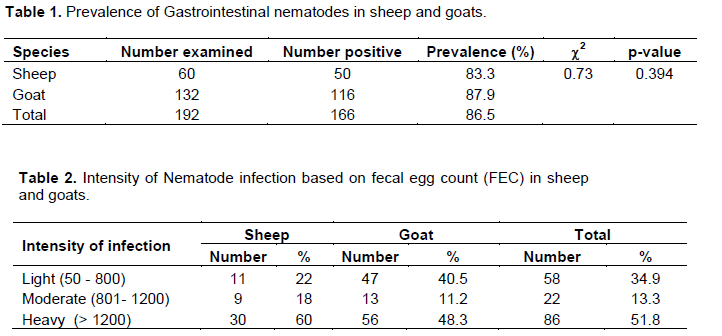
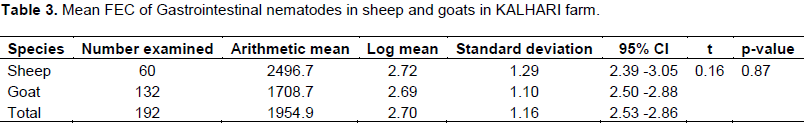
Out of the total 60 sheep and 132 goats examined, 83.3% (n=50) and 87.9% (n=116) sheep and goats, respectively were affected with one or more genera of GIN. No significant (p>0.05) variation was noted in the prevalence of GIN infection between sheep and goats (Table 1). The faecal egg counts (FEC) varied from 0 to 20,850 in sheep while 0 - 10,100 in goats. Categorization of the intensity of infection based on FEC revealed that 60% of sheep and 48.3% of goats were heavily infected (Table 2). The difference in mean FEC between sheep and goats was not significant (p>0.05) (Table 3).
Gastrointestinal nematodes identified
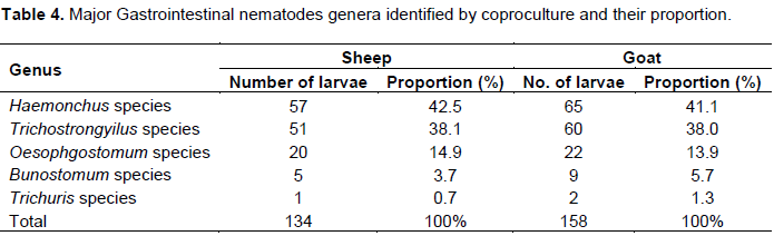
The result of coproculture in sheep and goats revealed a higher proportion of Haemonchus followed by Trichostrongylus, Oesophagostomum, Bunostomum and Trichuris species (Table 4).
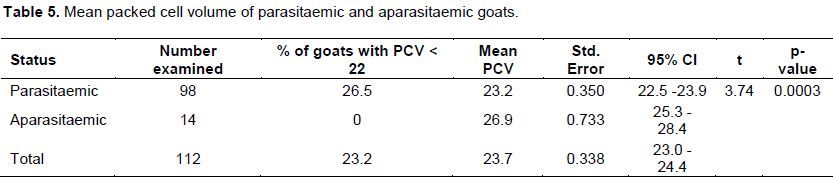
Assessment of PCV values in goats
The PCV value of 112 goats was measured to evaluate its correlation with GIN infection. Accordingly, it was found that the mean PCV of parasitaemic goats (23.2 ± 0.35) was significantly (p<0.05) lower than that of aparasitaemic animals (26.9 ± 0.73). Moreover, it was noted that 26.5% of parasitaemic goats have PCV value less than 22% while all aparasitaemic animals have PCV values 22% or above (Table 5).
DISCUSSION
In the present study, a high prevalence of GIN infection was recorded in both sheep (83.3%) and goats (87.9%) in KALHARI farm. This finding is higher than the prevalence reported by much of previous studies which ranges between 47.2 to 75.3% in sheep and 34.2 to 84.1% in goats in different parts of the country (Abebe et al., 2010; Admasu and Nurlign, 2014; Yimer et al., 2016; Yimer and Birhan, 2016; Ahmed et al., 2017; Derso and Shime, 2017; Getachew et al., 2017). In contrast, higher prevalence than the present had been reported by other studies (Tefera et al., 2009; Aragaw and Gebreegziabher, 2014; Wondimu and Gutu, 2017). This variation might be due to differences in agro-ecological conditions and management system.
The present study showed lack of significant variation (p>0.05) both in the prevalence and intensity of infection between sheep and goats. This is because both sheep and goats had equal chance of exposure to the infective larvae on pasture as all of the study animals were sampled from the same farm and kept under similar management conditions. In agreement to this finding, some of previous studies have also reported absence of significant prevalence differences between the two species (Tefera et al., 2009; Admasu and Nurlign, 2014; Aragaw and Gebreegziabher, 2014; Yimer and Birhan, 2016; Ahmed et al., 2017). In contrast to these, other researchers (Emiru et al., 2013; Belina et al., 2017; Derso and Shime, 2017; Getachew et al., 2017; Wondimu and Gutu, 2017) found significant difference in prevalence between sheep and goats. Similarly, unlike the present study, Abebe et al. (2010) observed significantly higher mean FEC of GIN in sheep than goats.
In contrast to previous studies (Regassa et al., 2006; Tefera et al., 2009; Admasu and Nurlign, 2014) which reported relatively lower proportion of massive infection in sheep and goats, this study demonstrated that a higher proportion of sheep (60%) and goats (48.3%) examined were heavily infected. Consistent to the current finding, a higher proportion of massive infection has also been documented in other studies in the country (Abebe et al., 2010; Ahmed et al., 2017; Getachew et al., 2017). Observation of heavy intensity of infection in the present study may be attributed to lack of regular deworming practice in the farm and consequently increased contamination of grazing pasture with eggs excreted by infected animals. The other possible explanation for the high faecal egg counts is the observation of Haemonchus species in more substantial proportion of affected animals. These parasites are very prolific, every single parasite capable of laying thousands of eggs daily and this continues for several successive months as long as environmental factors are favourable (Radostits et al., 2007).
As mentioned above, Haemonchus species was the most abundant parasite identified in both sheep and goats in the current study followed by Trichostrongylus species, Oesophagostomum species, Bunostomum species and Trichuris species. The preponderance of Haemonchus in the present study is entirely in agreement with the recently conducted systematic review of GINs of small ruminants in Ethiopia which revealed Haemonchus contortus as the most prevalent parasite in sheep and goats (Asmare et al., 2016). Indeed, it is one of the most pathogenic nematode parasites in ruminants implicated in widespread morbidity and mortality of sheep and goats (Taylor et al., 2007) and thus warrants special attention in gastrointestinal parasite control programs.
Due to limitations of resource, measurement of PCV was carried out. Analysis of the mean PCV revealed a significantly lower value in goats affected with GIN than in those not infected. This could be linked to the blood feeding habit of Haemonchus species that was recorded in a higher proportion in the infected animals.
CONCLUSION
This study revealed a high prevalence and intensity of GIN infection in sheep and goats in the study farm without significant difference between the two species. Perhaps this is attributed to lack of regular deworming practices in the farm. Indeed, the study provides substantial evidence that GIN could have serious impact on the productivity of the animals and profitability of the farm. Thus, strategic deworming of the animals using most effective anthelmintics and improvement of management practices are required so as to reduce losses associated with the parasites and ensure the profitability of the farm.
CONFLICT OF INTERESTS
The authors have not declared any conflict of interests.
REFERENCES
|
Abebe R, Gebreyohannes M, Mekuria S, Abunna F, Regassa A (2010). Gastrointestinal nematode infections in small ruminants under the traditional husbandry system during the dry season in southern Ethiopia. Tropical Animal Health and Production 42:1111-1117. |
|
|
Admasu P, Nurlign L (2014). Prevalence of Gastrointestinal Parasites of Small Ruminants in Kuarit District, North West Ethiopia. African Journal of Basic and Applied Sciences 6 (5):125-130. |
|
|
Aga TS, Tolossa YH, Terefe G (2013). Epidemiology of gastrointestinal nematodes of Horro sheep in Western Oromiya, Ethiopia. Journal of Veterinary Medicine and Animal Health 5(10):296-304. |
|
|
Ahmed J, Duguma A, Regassa D, Belina D, Jilo R (2017). Gastrointestinal Nematode Parasites of Small Ruminants and Anthelmintics Efficacy Test in Sheep of Haramaya District, Eastern Ethiopia. Animal and Veterinary Sciences 5(3):39-44. |
|
|
Aragaw K, Gebreegziabher G (2014). Small Intestinal Helminth Parasites in Slaughtered Sheep and Goats in Hawassa, Southern Ethiopia. African Journal of Basic and Applied Sciences 6(2):25-29. |
|
|
Asha A, Wossene A (2007). Gastrointestinal tract nematodosis of small ruminantsin three different agro-ecological zones in southern Ethiopia. Ethiopian Veterinary Journal 11(1):83-94. |
|
|
Asmare K, Sheferaw D, Aragaw K, Abera M, Sibhat B, Haile A, Kiarad H, Szonyie B, Skjervef E, Wielande B (2016). Gastrointestinal nematode infection in small ruminants in Ethiopia: A systematic review and meta-analysis, Acta Tropica 160:68-77. |
|
|
Bates P (2012). External Parasites of Small Ruminants: A Practical Guide to their Prevention and Control. CAB International, UK pp1-2 |
|
|
Belina D, Giri A, Mengistu S, Eshetu A (2017). Gastrointestinal Nematodes in Ruminants: The Parasite Burden, Associated Risk Factors and Anthelmintic Utilization Practices in Selected Districts of East and Western Hararghe, Ethiopia. Journal of Veterinary Science and Technology 8:433. |
|
|
Coffin DL (1995). Manual of Veterinary and Clinical Pathology. 3rd ed., Comst. Pub. Ass. Inc. Ithaca. New York, USA pp. 115-157. |
|
|
Coop RL, Kyriazakis I, (2001). Influence of host nutrition on the development and consequences of nematode parasitism in ruminants. Trends in Parasitology 17(7):325-330. |
|
|
Central Statistical Agency (CSA) (2017). Report on Livestock and Livestock Characteristics (Private Peasant Holdings). Agricultural Sample Survey 2016/17 (2009 E.C.), Federal Democratic Republic of Ethiopia, Central Statistical Agency (CSA), Addis Ababa, Ethiopia. |
|
|
Derso S, Shime A (2017). Small ruminant GIT Parasites in Enemay district, Ethiopia: Prevalence and risk factors. Online Journal of Animal and Feed Research 7(3):65-71. |
|
|
Emiru B, Amede Y, Tigre W, Feyera T, Deressa B (2013). Epidemiology of Gastrointestinal Parasites of Small Ruminants in Gechi District, Southwest Ethiopia. Advances in Biological Research 7(5):169-174. |
|
|
Getachew M, Tesfaye R, Sisay E (2017). Prevalence and Risk Factors of Gastrointestinal Nematodes Infections in Small Ruminants in Tullo District, Western Harerghe, Ethiopia. Journal of Veterinary Science and Technology 8:428. |
|
|
Hansen J, Perry B (1994). The epidemiology, diagnosis and control of helminth parasites of ruminants. ILRAD. Nairobi, Kenya P 171. |
|
|
Hendrix CM (1998). Diagnostic Veterinary Parasitology, 2nd ed. pp. 239-264. |
|
|
Jackson PGG, Cockcroft P D (2002). Clinical Examination of Farm Animals. Blackwell Science Ltd. P 302. |
|
|
Radostits OM, Gay CC, Hinchcliff KW, Constable PD (2007). Veterinary Medicine: A text book of the disease of cattle, horses, sheep, pigs and goats. 10thed. Elsevier, London pp. 1541-1563. |
|
|
Regassa F, Sori T, Dhuguma R, Kiros Y (2006). Epidemiology of Gastrointestinal Parasites of Ruminants in Western Oromia, Ethiopia. International Journal of Applied Research in Veterinary Medicine 4(1):51-57. |
|
|
Suarez VH, Cristel SL, Busetti MR (2009). Epidemiology and effects of gastrointestinal nematode infection on milk productions of dairy ewes. Parasite 16:141-147. |
|
|
Taylor MA, Coop R L, Wall, RL (2007). Veterinary Parasitology, 3rd ed. Blackwell Publishing Ltd, Oxford, UK P 2080. |
|
|
Tefera M, Batu G, Bitew M (2009). Prevalence of Gastrointestinal Parasites of Sheep and Goats in and around Bedelle, South-Western Ethiopia. The Internet Journal of Veterinary Medicine 8(2). |
|
|
Thrall MA, Weiser G, Allison, RW and Campbell TW (2012). Veterinary Hematology and Clinical Chemistry, 2nd ed. John Wiley & Sons, Inc. pp. 75-80. |
|
|
Thrusfield M (2005). Veterinary epidemiology. 3rd ed. Singapore, Black well Science pp. 228-246. |
|
|
Urquhart GM, Armour J, Dunlan JL, Dunn AM, Jennings W (1996). Veterinary Parasitology, 2nd ed. Blackwell science Ltd, Oxford, London, UK P 307. |
|
|
Wondimu A, Gutu S (2017). Gastrointestinal nematodes of small ruminants in Guto Gida District, East Wolloega, Ethiopia. Journal of Veterinary Medicine and Animal Health 9(5):83-87. |
|
|
Yimer A, Sissay D, Nazir S (2016). Prevalence and associated risk factors of gastrointestinal nematodiasis in small ruminants in North East Ethiopia. Journal of Animal Research 6(2):165-170. |
|
|
Yimer A, Birhan E (2016). Prevalence and Identification of Gastrointestinal Nematodes of Small Ruminants in Northern Ethiopia. Middle-East Journal of Scientific Research 24(8):2602-2608. |
|
|
Zajac A M, Conboy G A (2012). Veterinary Clinical Parasitology 8th edition, American Association of veterinary parasitologists, Black well publishing. |
|
Copyright © 2024 Author(s) retain the copyright of this article.
This article is published under the terms of the Creative Commons Attribution License 4.0


