Full Length Research Paper

ABSTRACT
The objective of the work was to evaluate two ELISA’s for the detection of anti-Map antibodies. 460 serum and fecal samples of bovines (n=267), ovines (n=140), and goats (n=53) were evaluated. Serums were used for detecting anti-Map antibodies using two different ELISAs, one in-house ELISA made with low-molecular weight proteins (<100 kD) from the antigen ultra-filtrate of Map strain 3065, and a commercial ELISA for paratuberculosis diagnostics. DNA was extracted from faeces to carry out PCR-N IS900. Sensitivity (Se), specificity (E) and Kappa (K) were calculated to estimate concordance between tests. The in-house ELISA made from low-weight map proteins showed a Se of 87%, E of 96% and K of 0.85, while the commercial ELISA showed Se=55%, E=97% and K=0.576, when compared to the PCR-N IS900 test. When comparing both ELISAs, Se was 50%, E was 97%, and K= 0.393. The in-house ELISA made from <100 kD proteins had a better Se and E than the commercial ELISA; concordance between them was low. Low-weight proteins (<100 kD) obtained from the antigen ultra-filtrate of Map strain 3065 are a good candidate for using as antigen in ELISA for the diagnosis of Johne’s disease
Key words: Paratuberculosis, ELISA, diagnostic.
INTRODUCTION
Johne’s disease is a chronic disease that affects domestic ruminants causing granulomatous enteritis. The etiological agent is Mycobacterium avium subsp. paratuberculosis (Map). Infection with Map is predominantly subclinical; livestock producers realize the presence of the disease when diarrhea and emaciation become apparent (Bates et al., 2019, Fry et al., 2008; Eda et al., 2006). Johne´s disease has an important impact on animal production by generating a reduction in milk and meat production, early discarding of animals and low fertility with predisposition for mastitis (Eda et al., 2006). With this disease, carcasses have a lower commercial value and there is an increase in control program costs (Soto et al., 2002). Mycobacterium avium subsp. paratuberculosis is excreted intermittently, and animals are infected by consuming feed and water contaminated with paratuberculous microorganisms shed by infected animals. Mycobacterium avium subsp. paratuberculosis concentration in faeces may surpass 108 colony forming units (CFU)/g (Valentin; 2002).Young animals less than six months of age are the most susceptible to infection, calves that are in subclinical stages of the disease shed the bacilli in faeces without showing the characteristic signs of the disease (Park et al., 2006).
The disease distribution is worldwide, especially in domestic ruminants confined rearing conditions; prevalence of the disease varies between 5 and 55% (Mathevon et al., 2017; Martínez et al., 2012; Pinedo et al., 2008; Park et al., 2006).
Transversal epidemiological studies have been carried out in states of the Mexican Republic (Chihuahua, Aguascalientes, Coahuila, Durango, Guanajuato, Hidalgo, Querétaro, Sinaloa, Jalisco, Chiapas, Veracruz, San Luis Potosí, Puebla) in units livestock production of cattle, sheep and goats, to determine the health status in relation to Johne´s disease of herds and flocks, the results obtained show that the disease is distributed throughout the country and the individual prevalence ranges from 1 to 32.37%, livestock production unit it is from 1 to 88.87% (Gallaga et al., 2017, Guzmán et al., 2016; Milián-Suazo et al., 2015, Morón-Cedillo et al., 2015).
Diagnosis of Johne’s disease in domestic ruminants is done through methods based on the detection of the bacillus in faecal samples or the detection of antibodies in serum or milk samples (Chaubey et al., 2019).
Cell-mediated immunity decreases with the progression of the disease and when this happens, humoral immunity becomes measurable and can be detected by serological tests (Mon et al., 2012, Shin et al., 2008). Among the available tests for determining antibodies against Map the most used are based on Enzyme-Linked Immunoassays (ELISA). There are several commercial ELISA tests available in the market and many studies have determined their sensitivity and specificity, which range between 45 to 80% and 80 to 90%, respectively (Chaubey et al., 2019 Mathevon et al.,2017, Speer et al., 2006, Klausen et al., 2003). ELISAs are considered a good option for diagnosing Johne’s disease because it is easy to implement in the laboratory, results are obtained rather quickly, and their interpretation is simple. Nevertheless, there are several limiting factors for their implementation or use in animal health diagnostics laboratory such as high cost and importation times (Martinez et al., 2012). Johne’s disease serological diagnosis by ELISA uses PPA-3 protoplasmic antigen obtained from M. avium strain 18. Currently, a mixture of proteins is used which includes the sonicated extract of purified Map antigen and protoplasmic antigens obtained from Map strains. Through sequencing and whole-genome analysis of Map, several specific proteins have been detected, which are considered immunoreactive and viable candidates for their use as antigens in serological tests to detect anti-Map antibodies (Mon et al., 2012). The use of antigenic protein cocktails or low molecular weight polyproteins obtained from Map, that have specific epitopes can be an interesting alternative to increase the possibility of detecting infected animals, that are in various stages of the disease, especially during subclinical phases of the infection, when shedding of Map via feces and the immune response are not so evident (Moyano et al., 2021).
MATERIALS AND METHODS
Extraction of low molecular weight proteins through ultra-filtration of Map antigens
A total of 10 mg of antigen from Map strain 3065 were weighed and dissolved in 10 mL of milli-Q water. The solution was placed in an Amicon Ultra-100 filtration tube and centrifuged for 20 min at 4500 rpm. The ultracentrifuged material was placed in 15 mL falcon tubes and frozen at -20°C (Torres, 2015).
Western and immunoblot
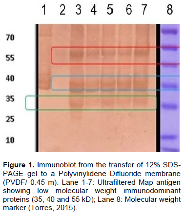
Vertical electrophoresis was done using 12% polyacrylamide gels (SDS-PAGE) placing 25 µL (15 µg) of ultra-centrifuged Map in each well. Gels were run at 80 V for 4.5 h at 4ºC. For immunoblot, the 12% SDS-PAGE gel was transferred to a Poly-vinylidene Difluoride membrane (PVDF/045 m). Bovine serum confirmed as positive by PCR and culture to Johne’s disease was used as first antibody at a 1:100 dilution. Reaction blocking was done with 3% milk and incubated for 1 h at ambient temperature with shaking. Afterwards, four washes for 5 min were carried out with Tween 20-Phosphate solution at pH 7.2, and a final wash with phosphate buffer solution at pH 7.2. A horseradish peroxidase marked (HRP) bovine anti-IgG conjugate was used as second antibody at a 1:10,000 dilution incubated for 30 min at ambient temperature. This was followed by four washes for 5 min with Tween 20-Phosphate solution at pH 7.2, a final wash with Phosphate buffer at pH 7.2 and revealed with diaminobenzidine (DAB) (Torres, 2015).
Sample size
460 serum and fecal samples, were worked coming from bovines, ovines and goats from the States of Morelos (33 ovines, 32 bovines), Jalisco (28 ovines, 30 bovines and 34 goats), Puebla (27 ovines), State of Mexico (21 ovines, 71 bovines), Guanajuato (35 ovines, 36 bovines and 17 goats), Colima (12 bovines), Hidalgo (10 bovines), Coahuila (20 bovines), Aguascalientes (10 bovines), Zacatecas (20 bovines), and Yucatan (24 bovines). Samples were collected during 2018 and 2019 from animals of farms that accepted to be part of the study. Sample size was estimated using Levy’s proportional formula (Levy’s and Lemeshow, 1980) using a prevalence for Johne’s disease of 10%, 1% error, and 95% confidence level. All the herds and flocks included in the study had a previous clinical history of Johne´s disease (>25% prevalence).
Blood samples from bovines were obtained from the coccygeal vein, while in ovines and goats from the jugular vein, using vacutainer tubes without anticoagulant. Samples were centrifuged at 3500 rpm for five min; serum transferred to 1.5 ml tubes and stored at -20°C until used. Feces were taken directly from the rectum using rectal long gloves and stored at -20°C.
Enzyme-linked immunoassay (ELISA)
Once the low molecular weight proteins (<100 kD) were obtained from the Map strain 3065 antigen, they were fixed to wells of ELISA microplates following the protocol described in Martinez et al. (2012). Serums were evaluated using in-house ELISA containing ultrafiltered protoplasmic antigen from Map strain 3065 with the following concentrations, antigen 8.653 µg/100 µl, serum 1:160 and HRP-labeled bovine anti-IgG 1:10,000. Commercial positive and negative control serums were used. The cut-off point for the in-house ELISAMap 3065 to differentiate between positive and negative animals ones was set at 0.20 OD (optical densities). The commercial anti-Map ELISA was processed according to manufacturer’s instructions.
DNA extraction and IS900 nested polymerase chain reaction (PCR-N IS900)
DNA extraction from feces and the PCR-N IS900 were carried out following the protocol described in Jaimes et al. (2008).
Statistical analysis
Results were analyzed using a Kappa test to measure the concordance between each ELISA and the PCR-N IS900. Pearson’s X2 test was used to determine differences between the diagnostic methods. All tests were carried out using the STATA 7®software.
RESULTS
Immunoblot
Immunoblot of the ultrafiltered Map 3065 antigen showed a profile composed of immunodominant proteins of 35, 40 and 55 kD (Figure 1).
Enzyme-linked immunoassay (ELISA) and PCR-N IS900
The in-house ELISA 3065 made from low molecular weight Map protein detected 142 (30%) of serums as positives, while the commercial ELISA detected 94 (20%). Using PCR-N IS900, a 210 bp amplicon of the Map-specific IS900 region was detected in a total of 151 samples (Table 1).

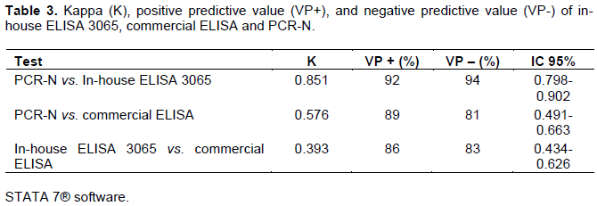
The PCR-N IS900 test was used as confirmatory to determine the concordance with the two ELISAs. The in-house ELISA 3065 had a greater sensitivity (87%) than the commercial ELISA (55%), while the specificity was relatively similar (96% versus 97%, respectively). The Kappa concordance value between PCR-N IS900 and in-house ELISA 3065 was considered good (0.850), while that of the commercial ELISA was considered moderate (0.576). When comparing between the ELISAs, sensitivity decreased to 50%, albeit specificity remained high (97%), and Kappa reached 0.393 (Tables 2 and 3).

DISCUSSION
The results obtained with this study show that an in-house ELISA made from low molecular weight proteins of the Map 3065 strain has 87% sensitivity and 96% specificity, while a commercial ELISA had 55 and 97%, respectively. This agrees with previous studies by some authors that have evaluated antigens for their use in ELISAs for the diagnosis of Johne’s disease and reported sensitivity ranging from 30 to 80% and specificity between 80 and 98% (Mathevon et al., 2017; Fry et al., 2008). When comparing both ELISAs, sensitivity decreased to 50% although specificity remained high at 97%, and both positive and negative predictive values remained above 80%.
It is important to point out that the predictive value is associated with the sensitivity and specificity of the test, as well as being highly dependent on the disease prevalence in the population where the test is applied (Martínez et al., 2012, Clark et al., 2008, Kokuina et al., 2006) The positive and negative predictive values, obtained with the in-house ELISA Map 3065 in this study, are considered high, these contribute to a good sensitivity and high specificity, which allows detecting animals that are infected with Map.
The commercial ELISA had a sensitivity and specificity within the expected ranges. It must be noted that the some ELISA test systems used to detect anti-Map antibodies have different cutoff points depending on the country of manufacturing and region where the test was standardized; therefore it varies from 0.25 to 0.7 optical densities (OD). Since this affects sensitivity, whenever a commercial anti-Map antibody ELISA test system is to be implemented, it is important to be aware of the cutoff value and adjust it according to the prevalence of Johne’s disease in the region (Clark et al., 2008; Fajardo et al., 1994). The cut-off point determined for the in-house ELISA Map 3065 was 0.20 OD, which allowed detecting greater quantity of infected animals that were also positive for nested PCR; with this, the sensitivity of the test was greater in animals that were beginning to show clinical signs of the disease; by using a cut-off point greater than 0.25 OD, there is the possibility to obtain a false-negative result, keeping infected animals in the herd that will eventually develop the disease.
It is thought that the early immune response against Map infection in primarily based on cellular immunity that is characterized by the production of interferon gamma, followed by antibody production as disease progresses. A study by Waters et al. (2003) showed that antibodies do appear in early stages of Map infection; therefore ELISA could be used as an early diagnostic tool.
To improve the sensitivity and specificity of serological tests well-defined antigens must be identified. Mon et al. (2012) evaluated a Map antigen panel for selecting candidates for Johne’s disease diagnosis, detecting a total of 54 recombinant proteins of which seven (Map 2513, 1693, 2020, 0038, 1272, 0209c and 0210c), with molecular weights between 25 and 790 kD, were evaluated in ELISA obtaining sensitivities and specificities above 75%. Moyano et al. (2021) and Chaubey et al. (2019) used Map recombinant and low molecular weight polyproteins in an ELISA to evaluate serums of goats, buffaloes and cattle obtaining sensitivity between 70 and 95%, and specificity between 80 and 97%, which are similar to the results in this study. The in-house ELISA made from ultrafiltered Map antigen from strain 3065 (<100 kD proteins) can be an option for the diagnosis of Johne’s disease since the sensitivity and specificity were of 87 and 93%, respectively. It is thought that ELISA can detect between 30 and 40% of the ruminants identified as infected by bacteriological culture of Map. Bacteriological culture is considered as the confirmatory test for the diagnosis of Johne’s disease but has many limiting factors such as a long incubation period (> 12 weeks), and contamination of cultures by fungi. As such, PCR can be used as the confirmatory tests since it allows the detection of a greater number of animals positive to the disease. PCR is capable of DNA detection of viable and unviable bacteria and the turnaround time for the results in this test is much quicker (Martínez et al., 2012). The IS900 marker is considered the standard for endpoint PCR and Q-PCR since there are between 14 and 20 copies of the sequence within the mycobacterium genome improving the sensitivity for detecting Map. To increase specificity when using primers for IS900, these should be designed so that they amplify regions that are close to the 5’ end of this inserted sequence since it is highly conserved (Cook and Britt, 2007). PCR-N IS900 was used in this study which detected a greater number of positive animals helping concordance with the low molecular weight Map proteins to be high and that positive and negative predictive values reach above 90%. Johne’s disease in domestic ruminants has a wide spectrum of immunological and pathological stages associated with the various phases of infection. Therefore, the sensitivity of ELISA tests for detecting anti-Map antibodies varies according to the disease stage: Low in early and subclinical stages; the level of bacterial shedding in feces and the age of the animals (Moyano et al., 2021). In this sense, no single antigen would be able to detect all infected animals which present an important challenge in the selection of antigens adequate for developing diagnostic techniques for the detection of Johne’s disease in livestock. Thus, the use of a mixture of low molecular weight antigenic proteins could be an interesting alternative for the development of serological tests because the increase of epitopes increases the possibility of detecting animals in differing stages of infection (Moyano et al., 2021; Chaubey et al., 2019; Mon et al., 2012; Shin et al., 2008). The immunoblot of the ultrafiltered Map strain 3065 antigen for determining immunodominant proteins, showed a profile containing proteins of 35, 40 and 55 kD. This shows that proteins used as antigens are weighing less than 100 kD (Torres, 2015). Current research on mycobacteriosis center on the evaluation of low molecular weight protein antigens. These antigens, since they are highly specific, are viable candidates for their use in diagnostic tests for detecting animals with Johne’s disease. Low molecular weight antigens have been previously used in serological diagnostic techniques such as ELISA and Fluorescence Polarized (FPA), showing high sensitivity and specificity for both techniques (Chaubey et al., 2016; Beck et al., 2005).
CONCLUSION
These results indicate that the established in-house ELISA Map 3065 detects antibodies specific to Map with high specificity and sensitivity and is a useful tool for the screening of Johne's disease.
Low molecular weight proteins (<100 kD) obtained from the ultrafiltered protoplasmic antigen of Map strain 3065 are a good candidate for their use as antigen in ELISA for diagnosing Johne’s disease and thus have a tool that can help in the implementation of health control monitoring program in herds. Considering that the diagnosis of the disease should be carried out with a serological test and a confirmatory one such as PCR or culture.
FUNDING
This Work was partially financed by FONSEC-SAGARPA-CONACYT SIGI 1281834685: “Development, production and validation of new-generation biologicals and diagnostic systems based on biotechnology to contribute to the prevention and control of diseases that affect livestock production in Mexico.”
CONFLICT OF INTERESTS
The authors have not declared any conflict of interests.
REFERENCES
|
Bates A, O'Brien R, Liggett S, Griffin F (2019). Control of Mycobacterium avium subsp. paratuberculosis infection on a New Zealand pastoral dairy farm. Veterinary Research. 29;15(1):266-279. |
|
|
Beck ST, Leite OM, Arruda RS, Ferreira AW (2005). Humoral response to low molecular weight antigens of Mycobacterium tuberculosis-by-tuberculosis patients and contacts. Brazilian Journal of Medical and Biological Research 38:587-596. |
|
|
Chaubey KK, Gangwar N, Pawaiya RS, Jatav GP, Sohal JS, Singh SV, Singh M, Gupta S, Kumaresan G, Kumar N, Jayaraman S (2019). Evaluation of newly developed six recombinant secretary proteins based cocktail ELISA and whole cell lysate based indigenous ELISA and tissue microscopy with Gold standard histopathology for the diagnosis of Johne's disease in slaughtered goats and buffaloes.Comparative Immunology Microbiology Infectious Diseases 66:101-110. |
|
|
Clark DL, Koziczkowski RP, Radcliff RA, Ellingson JLE (2008). Detection of Mycobacteriumavium subspecies paratuberculosis: Comparing fecal culture versus serumenzime-linked immunosorbentassay and direct fecal polymerasechainreaction. Journal Dairy Science 91(7):2620-2627 |
|
|
Cook KL, Britt JS (2007). Optimization of methods for detecting Mycobacterium avium subspecies paratuberculosis in environmental samples using quantitative, real-time PCR. Journal of Microbiological Methods 69:154-160. |
|
|
Eda S, Bannantine JP, Waters WR, Mori Y, Whitlock RH, Scott MC, Speer CA (2006). A Highly Sensitive and Subspecies-Specific Surface Antigen Enzyme-LikedImmunosorbent Assay for Diagnosis of Johne's Disease. Clinical and Vaccine Immunology 13(8):837-844. |
|
|
Fajardo-Gutiérrez A, Yamamoto-Kimura L, Yañez-Velasco L, Garduño EJ, Martínez GMC (1994). Utilidad de las curvas desensibilidad y especificidad conjunta en la aplicación de unaprueba de diagnóstico. Salud Pública Mexico 36(3):311-317. |
|
|
Fry MP, Kruze J, Collins MT (2008). Evaluation of four commercial enzyme-linked immunosorbent assays for the diagnosis of bovine paratuberculosis in Chilean dairy herds. Journal Veterinary Diagnostic Investigation 20(3):329-332. |
|
|
Gallaga MEP, Arellano RB, Santillán-Flores MA, Favila HLC, Córdova LD, Morales RJ, Díaz AE (2017). Situación Epidemiológica de la Paratuberculosis en las Principales Regiones caprinasdel Estado de Puebla, México. Quehacer Científico en Chiapas 12(1):36-45. |
|
|
Guzmán-Ruiz CC, Santillán-Flores MA, Córdova-López D (2016). Prevalence and possible risk factors for caprineparatuberculosis in intensive dairy production units in Guanajuato, Mexico. Journal of Veterinary Medicine and Animal Health 8(11):156-162. |
|
|
Jaimes NG, Santillán FMA, Hernández COA, Córdova LD, Guzmán RCC, Arellano RB, Díaz AE, Tenorio GVR, Cuellar OA (2008). Detección de Mycobacterium aviumsubespecieparatuberculosis, pormedio de PCR-anidada a partir de muestras de heces de ovino. Veterinaria México 39(4):377-386. |
|
|
Klausen J, Huda A, Ekeroth L, Ahrens P (2003). Evaluation of serum and milk ELISAs for paratuberculosis in Danish dairy cattle. Preventive Veterinary Medicine 58(3-4):171-178. |
|
|
Levy PS, Lemeshow S (1980). Sampling for Health Professionals Lifetime Learning Publications. Belmont, California. |
|
|
Kokuina E, Chico A, Estévez M, Pérez D, Gutiérrez A, CruzC (2006). Utilización y valor predictivo de la determinación deanticuerpos antinucleares en un Hospital de Referencia Nacionalde Salud. Revista Cubana de Medicina. 45(3):520-530. |
|
|
Martínez CAG, Santillán FMA, Guzmán RCC, Favila HLC, Córdova LD, Díaz AE, Hernández AL, Blanco OM (2012). Development of an enzyme-linked immunosorbent assay (ELISA) forthe diagnosis of bovine paratuberculosis. Revista Mexicanade Ciencias Pecurias 3(1):1-18. |
|
|
Mathevon Y, Foucras G, Falguières R, Corbiere F (2017). Estimation of the sensitivity and specificity of two serum ELISAs and one fecal qPCR for diagnosis of paratuberculosis in sub-clinically infected young-adult French sheep using latent class Bayesian modeling. Veterinary Research 13(1):230-241. |
|
|
Milián-Suazo F. Santillán-Flores MA, Zendejas-Martínez H, García-Casanova L, Hernández-Andrade L, Cantó-AlarcónG. (2015). Prevalence and Associated Risk Factors for Mycobacterium avium subsp. paratuberculosis in Dairy Cattle in Mexico. Journal of Veterinary Medicine and Animal Health 7(10)302-307. |
|
|
Mon ML, Viale M, Baschetti G, Alvarado-Pinedo F, Gioffre A, Trasvería G, Willemsen P, Bakker D, Romano MI (2012). Search for Mycobacterium avium Subspecies paratuberculosis Antigens for the Diagnosis of Paratuberculosis. Veterinary Medicine International 2012:1-9 ID 860362. |
|
|
Morón-Cedillo F J; Cortez-Romero C; Santillán-Flores MA; Figueroa-Sandoval B;Gallegos-Sánchez J (2015). Prácticas de ManejoAsociadas con la Seroepidemiología de Paratuberculosisovina en San Luis Potosí. Agroproductividad. 8(6):30-36. |
|
|
Moyano DR, Romero MA,Colombatti-Olivieri MA, Alvarado-Pinedo MF, Traveria G, Romano MI, Alonso MN (2021). Development and validation of a novel ELISA for the specific detection of antibodies against Mycobacterium avium subspecies paratuberculosis based on a chimeric polyprotein. Veterinary Medicine International 2021:1-10 |
|
|
Park KT, Ahn J, Davis WC, Koo HC, Kwon NH, Jung WK, Kim JM, Hong SK, Park YH (2006). Analysis of the seroprevalence of bovine paratuberculosis and the application of modified absorbed ELISA to field sample testing in Korea. Journal of Veterinary Science 7(4):349-354. |
|
|
Pinedo PJ, Rae DO, Williams JE, Donovan GA, Melendez P, and Buergelt CD (2008). Association among Results of Serum ELISA, Fecal Culture and Nested PCR on Milk, Blood and Faeces for the Detection of paratuberculosis in Dairy Cows. Transboundary and Emerging Diseases 55(2):125-133. |
|
|
Soto JP, Kruze J, Leiva S (2002). Comparación de tres métodos de diagnóstico de Paratuberculosis bovina en rebaños lecheros infectados. Archivos de Medicina Veterinaria. 24(2):265-273. |
|
|
Shin SJ, Cho D,Collins MT (2008). Diagnosis of Bovine Paratuberculosis by a Novel Enzyme-Linked Immunosorbent Assay Based on Early Secreted Antigens of Mycobacterium aviumsubsp. paratuberculosis. Clinical and Vaccine Immunology 15(8):1277-128. |
|
|
Speer CA, Scott MC, Bannantine JP, Waters WR, Mori Y, Whitlock RH, Eda S (2006). A Novel Enzyme-Linked Immunosorbent Assay for Diagnosis of Mycobacterium avium subsp. paratuberculosis Infections (Johne's Disease) in Cattle. Clinical and Vaccine Immunology 13(5):535-540. |
|
|
Torres VR (2015). Obtención y evaluación de proteínas de bajo peso molecular a partir de unextractocrudo de Mycobacteriumavium subsp. paratuberculosispara el diagnóstico de paratuberculosisporfluorescenciapolarizada. Tesis de Maestría. Posgrado-FMVZ UNAM, México. |
|
|
Valentin WP (2002). Johne's Disease: Pathogenesis and Problems Related to Diagnosis. In: Recent Developments and Perspectives in Bovine Medicine, XXII World Buiatrics Congress, Hanover, German pp. 18-23. |
|
|
Waters WR, Miller JM, Palmer MV (2003). Earlyinduction of humoral and cellular immune responses duringexperimental Mycobacterium aviumsubsp. paratuberculosisinfection of calves. Infection and Immunity 71(9):5130-5138. |
|
Copyright © 2024 Author(s) retain the copyright of this article.
This article is published under the terms of the Creative Commons Attribution License 4.0


