Full Length Research Paper

ABSTRACT
In recent years, Nattrassia mangiferae has received tremendous attention as a destructive plant pathogen that may potentially infect humans and animals. The pathogen was isolated from 61 species belonging to 47 genera and 30 family plants but was most frequently isolated from Cactaceae, Rosaceae, Euphorbiaceae, Anacardiaceae, and Rutaceae hosts. To systematically and comprehensively describe the progress, trends, and hotspots of N. mangiferae research, the 376 related publications from 1966 to 2022 were collected from the Scopus database. The bibliometric characteristics including publication output, countries/region of focus, author’s productivity, most proli?c authors, authorship pattern, citation frequency, institutes, most proli?c journals, and research focus were evaluated by using Excel 2013 and VOSviewer. Out of 376 original articles, there are 139 plant and 237 human studies. The leading countries based on the number of publications were the United States, the United Kingdom, and Brazil. A sharp increase in the number of studies related to the pathogenicity of N. mangiferae during 2018 to 2021 was observed, coinciding with an increase in the number of short reports and outbreak reports worldwide. The journal and subject categories with the most significant publications are Plant Disease and Medical Mycology, respectively. The most common document types were article, note, review, and letter. N. mangiferae is a thermophilic fungus, and warming temperatures may weaken trees and affect their susceptibility to this pathogen. The increase in reports of the pathogenicity of this pathogen on various plants and humans from 70 different countries in recent years shows the importance of this pathogen as a sever threat to the world. Our bibliometric analysis reveals the current research hotspots and topic trends on N. mangiferae, thus offering potential clues for further examination.
Key words: Neoscytalidium dimidiatum, plant pathogen, Onychomycosis, VOSviewer, Iran.
INTRODUCTION
The genus Nattrassia is a member of the Botryosphaeriaceae family (Dothideomycetes, Pezizomycotina, Ascomycota) that produces multiple morphologically distinct anamorphs (synanamorphs). The synanamorph of this genus is Neoscytalidium that contains four species, including Neoscytalidium dimidiatum, Neoscytalidium novaehollandiae, Neoscytalidium oculus, and Neoscytalidium orchidacearum (Crous et al., 2006; Pavlic et al., 2008; Huang et al., 2016; Calvillo-Medina et al., 2019). This genus may potentially infect plants (Türkölmez et al., 2019b; Goudarzi and Moslehi, 2020; Taguiam et al., 2021), humans, and animals (Bakhshizadeh et al., 2014; Dionne et al., 2015). The distribution of this genus has extended to all continents. Neoscytalidium genus can influence the different parts of plants by causing diseases that show various symptoms on aerial and underground parts of hosts, including shoot blight canker and gummosis (Polizzi et al., 2009), dieback (Ray et al., 2010), collar and root rot (Machado et al., 2012), brown spot (Lan et al., 2012), death of graft (Chen et al., 2013), internal black rot (Ezea et al., 2013), shoot and needle blight (Türkölmez et al., 2019a), tuber rot (Dervi? et al., 2020a), root and stem rot (Oksal and Ozer, 2021; Kuruppu et al., 2021), stem-end rot (Li et al., 2021), sooty canker (Yeganeh and Mohammadi, 2022), and leaf scorch (Güney et al., 2022) (Table 1). Members in the genus Neoscytalidium have been reported as pathogenic agents on various crops and trees, such as almond (Prunus dulcis), Avicennia marina Forssk., baobab (Adansonia gibbosa), bardi bush (Acacia synchronica), blue grevillea (Grevillia agrifolia), rattlepod (Crotalaria medicaginea), olive (Olea europaea L.), etc. (Table 1), causing economic yield losses (Pavlic et al., 2008; Ray et al., 2010; Sakalidis et al., 2011; Rahmani, 2018; Chakusary et al., 2019; Sabernasab et al., 2019; Dervi? et al., 2019; Mello et al., 2019; Türkölmez et al., 2019; Brito et al., 2020; Alananbeh et al., 2020; Oksal and Ozer, 2021; Kuruppu et al., 2021; Oren et al., 2022; Yeganeh and Mohammadi, 2022; Güney et al., 2022). Though, they have also been reported as opportunistic human pathogens causing chronic superficial infections of skin, nails, and nose, cutaneous, invasive cutaneous, deep cutaneous infection, onychomycosis, dermatomycosis, fingernail onychomycosis, superficial black onychomycosis, rhinosinusitis, brain abscess, and pulmonary disease (Bakhshizadeh et al., 2014; Dionne et al., 2015; Yang et al., 2019; Jo et al., 2020; González Cortés et al., 2021; Raiesi et al., 2022). Despite the issues in its identification, e.g., by using morphological and molecular techniques, the number of first-case and outbreak reports is increasing, revealing the virulence of the organism, which has already been detected in 70 countries.


Neoscytalidium spp. produces two different asexual states; arthric and pycnidia synanamorphs (Pavlic et al., 2008; Phillips et al., 2013). While Nattrass (1933) first described the pycnidial synanamorph as Hendersonula toruloidea, the production of two asexual morphs has led to some confusion in the taxonomy of this species (Crous et al., 2006). Other arthric and pycnidial existing morphs for H. toruloidea were Torula dimidiate (arthric morph), Scytalidium dimidiatum (arthric morph), and Fusicoccum dimidiatum (pycnidial morph) (Penzig, 1882; Sutton and Dyko, 1989; Farr et al., 2005). Crous et al. (2006), based on a DNA phylogeny of the family Botryosphaeriaceae revealed that the genus Scytalidium was polyphyletic, and the new genus Neoscytalidium was introduced to accommodate Fusicoccum-like with arthric morph (Crous et al., 2006).
To my knowledge, only a few bibliometric studies on plant disease have been done recently (Mayuri and Dayanithi, 2020; Avwerosuo 2021). Due to the increasing reports of pathogenicity in humans and plants and the associated public health concerns, no review has been done about this pathogen, and bibliometric analysis is still missing. In this regard, bibliometric analysis constitutes a systematic tool for monitoring this pathogen, offering veterans and new scientists an overview of the scientific panorama concerning this emerging pathogen. In this contribution, a bibliometric analysis of the studies on Nattrassia mangiferae published in the timespan from 1966 to 2022 is presented.
MATERIALS AND METHODS
Scopus (http://www.scopus.com), the most extensive database with abstracts and citations, journals, books, conference proceedings, and peer-reviewed literature, was chosen as the data source for the period 1966-2022 (Elsevier, 2017, 2019). To identify publications, the following keywords were used in the combined fields of title, abstract and keywords (per publication): “Hendersonula toruloidea” OR “Nattrassia mangiferae” OR “Scytalidium dimidiatum” OR “Scytalidium hyalinum” OR “Neoscytalidium dimidiatum” OR “Neoscytalidium novahollandiae” OR “Fusicoccum dimidiatum” OR “Neoscytalidium hyalinum” OR “Torula dimidiata” OR “Neoscytalidium orchidacearum” OR “Neoscytalidium oculus”. The downloaded search results contained full literature data such as document type (research articles, reviews, letters, book chapters, and books), abstract, keywords, author, year of publication, references, title, citations, and funding agency. All the retrieved documents were downloaded in the format of BibTeX, for easy recognition by VOSviewer (Version 1.6.18; 2020; Leiden University, Leiden, the Netherlands) (van Eck and Waltman, 2010) for further citation analysis. Then, the VOSviewer visualization software was used for citation analysis by co-occurrence and clusters of related publications, author collaboration (co-authorship), co-citation, country input, research hotspot, and frontier trend. Default parameter values of the VOSviewer were usually used in the analysis. Each term was represented by a label and a circle whose diameter and label size were directly proportional to their frequency (to avoid overlapping labels, only a subset of all labels is displayed in the maps). The colors in the network visualization represented clusters of similar terms as calculated by the program. The distance between the terms indicated the strength of the relationships.
RESULTS
Articles distributions by publication years
From 1966 to 2022, the total number of published manuscripts in Scopus was 376. Between 1966 and 1983 (17 years), the publication number per year was less than 5. The number of publications showed an increasing trend from 1984 (Figure 1) onwards, and the highest number of documents were published in 2021 (n=32). From 1966 onward, the average number of publications per year was 6.6, ranging from 0 (in 1967, 1968, 1971, 2006) to 32 (in 2021). Fifty-six percent of the articles were published between 2009 and 2022.

Document types and language of publication
The majority of articles retrieved were research articles (316; 84.04%) followed by notes (19; 5.03%), reviews (15; 3.98%), letters (13; 3.45%), conference papers (9; 2.39%), and short survey (3; 0.79%) (Figure 2). The ?rst article titled “microbial rotting of stored yams (Dioscorea species) in Nigeria” (Okafor, 1966) was published in Experimental Agriculture (2:179-182). The first report of the pathogenicity of this species in humans was made by Gentles and Evans (1970) as “infection of the feet and nails with H. toruloidea”. Almost all of the articles were published in English (343; 91.22%); others were published in French (14; 3.72%), Spanish (12; 3.19%), German (3; 0.79%), Portuguese (2; 0.53%), Chinese (1; 0.26%), and Italian (1; 0.26%) (Figure 3). Most of the published documents were in the form of original research articles, and English was the most common language used (Khan et al., 2020).


Journal analysis
The studies were published in 60 different journals, with the large majority belonging to the subject areas of medicine (208; 41.76%), agriculture and biological science (139; 27.91%), immunology and microbiology (49; 9.83%), biochemistry, genetics and molecular biology (37; 7.42%), environmental science (16; 3.21%), and veterinary (16; 3.21%) (Figure 4). Thirty two percent of the articles were published in one of these eight journals: Plant Disease (n=29), Medical Mycology (n=25), Mycoses (n=16), British Journal of Dermatology (n=13), Mycopathologia (n=13), Journal of Clinical Microbiology (n=10), Journal of Plant Pathology (8), and Journal de Mycologie Medicale (n=7) (Figure 5). The maximum number of articles was published in the Plant Disease followed by Medical Mycology (Figure 5). Among 10 journals, 7 of them belonged to medicine, and the rest are 3 related to plant diseases. The most cited article in the Plant Disease was by Chen et al. (2014) and their study reported N. dimidiatum as the cause of cankers and blights of English walnut in California.


Citation analysis
It is understood that highly cited articles have a significant impact on the concerned subject worldwide. The citation analysis of retrieved documents showed 3798 citations with an average of 10.10 citations per document. The most cited (240 times) article was published in 1989 titled “Onychomycosis, tinea pedis and tinea manuum caused by non?dermatophytic filamentous fungi nicht? dermatophyten?fadenpilze als erreger von onychomykosen, tinea pedis und tinea manuum” in the journal Mycoses. Three of the most cited articles were research articles. These articles discussed the diagnosis, epidemiology, clinical manifestations, and therapy of infections caused by this fungus.
Country, author, and institution analysis
The selected group of studies included 160 unique authors representing 70 countries across six continents. The five countries that contributed the most significant number of authors were the United States (50 articles), the United Kingdom (39 articles), Brazil (32 articles), France (25 articles), and Iran (25 articles), which took up 45.47% of the total (Figure 6). The identification of the most frequent authors helps to identify potential mentors and reviewers. A total of 160 authors were involved in publications retrieved during the defined period; 3 contributed more than ten manuscripts. In Table 2, we recorded top ten most productive and most cited authors, along with the institutions and countries they belonged. The authors listed came from Thailand, the United Kingdom, India, Nigeria, Turkey, and Spain. Four scholars appeared in both most productive and the most cited list: Moore MK (12 papers, 380 cites), Gugnani HC (11 articles, 206 cites), Hay RJ (7 articles, 332 cites), and Guarro J (6 articles, 166 cites). The leading organizations were the St John's Institute of Dermatology, UK, Mahidol University, Thailand, and the Mardin Artuklu University, Turkey, with 13, 12, and 11 publications.


Article title text mining
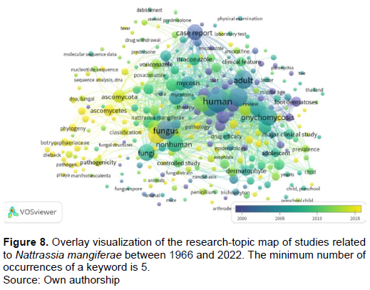
A text analysis of the words in the title and abstract of all publications (n=376) identified 2855 words (305 meet the threshold), after excluding words that carry very little information, which was ordered by frequency and displayed in a word cloud map. Figures 7 and 8 show the research-topic map of N. mangiferae studies between 1966 and 2022. Frequent terms were mapped into five clusters (Figure 7). Here, it is clear the particular interest in mycoses (onychomycosis, dermatophyte) caused by N. mangiferae. N. mangiferae has been reported as human pathogen causing chronic superficial infections of the skin, nails, and nose, onychomycosis, dermatomycosis, and pulmonary disease. Cluster 1 (red colored; n=86 terms) had keywords such as onychomycosis (highest with 103 occurrences), female, aged, middle-aged, dermatophyte, major clinical study, Trichophyton rubrum, tinea pedis, and skin disease. The second cluster (green color, n=83 terms) with a research theme highlighting isolation and purification, fungus identification, pathogenicity, phylogeny, plant disease, and classification. The third cluster (blue cluster, n = 60 terms) included humans, mycosis, antifungal agent, amphotericin b, itraconazole, and voriconazole. The fourth cluster (yellow color, n=42 terms) included human, male, adult, dermatomycoses, food dermatoses, nail diseases, and mitosporic fungi. The fifth cluster (violet color, n=32 terms) included nonhuman, controlled study, antifungal agents, in vitro study, and drug efficacy.

Bibliometric analysis of co-authorship
Authors from 70 countries showing at least two documents and having a minimum of 4 citations each were selected. Forty-six met the threshold, and these formed 6 clusters. The United States had the highest number of documents (n=49); 1737 citations with a total link strength of 139. The main collaborating partners of the United States were Canada and Italy. The United Kingdom, with 39 documents and 1314 citations, was in second place. The link strength for the UK was 18. The main collaborators of the UK were India, Nigeria and Spain. Brazil was at third place with 32 documents, having 451 citations, and eight total link strengths. Of the 851 organizations that were involved in research, collaborations were seen between 3 (with a minimum threshold of three documents and three citations).
DISCUSSION
Bibliometric analysis is a valuable tool for tracing the intellectual structure of a specific field of research. It allows a more structured literature review, including information and detection patterns (Vogel and Güttel, 2013). In this study, we utilized bibliometrics information visualization to analyze the literature set of N. mangiferae from 1966 to 2022 to gain a general view of the current research status, hot spots, and trends. Out of 376 published articles, 237 articles belonged to the subject area of medicine (237; 63.03%), and 139 articles were in the field of agriculture and biological science (139; 36.96%). Today, reports of the pathogenicity of this species on plants and humans are increasing.
Sabernasab et al. (2019) reported that minimum, optimum and maximum temperatures for growth of the N. mangiferae were, respectively, 10, 35, and 40°C. Similarly, Jamali and Banihashemi (2010) reported that the optimum temperature for growth of N. mangiferae was 35°C. Also, many researchers have shown that N. mangiferae could progressively grow and make symptoms in host plants in high temperatures and low relative humidity (Elshafie and Ba-Omar, 2002; Hassan et al., 2011; Mayorquin et al., 2016). Calavan and Wallace (1954) reported that N. mangiferae is aggressive on drought-stressed hosts. Drought stress leads to the reduced mechanical strength of the bark-wood bonds, and may result in bark cracks that can be invaded by N. mangiferae (Bettucci et al., 1999; Sabernasab et al., 2019). During the past decade, warming temperatures occurred in the world. This climate change may predispose trees to infection by this thermophilic fungus. N. mangiferae represent some of the most critical threats to urban and forest health where climate change is occurring. This study shows that the pathogen was isolated from 61 species belonging to 47 genera and 30 families of plants but was most frequently isolated from Cactaceae, Rosaceae, Euphorbiaceae, Anacardiaceae, and Rutaceae hosts. The pathogen produces high populations of black powdery spores under the bark of trees that are easily transmitted by wind and insects (Sabernasab et al., 2019), infecting other susceptible, injured, or weak hosts. Also, the broad host range of this pathogen and the high populations of black powdery spores can be a threat to human health, especially in people with defective immune systems. Among the top 10 most co-cited references from 1966 to 2022, seven of them concern onychomycosis, infection of the feet and nails, and superficial fungal infections caused by H. toruloidea, and then the rest of three papers are revisions of Hendersonula and its pathogenicity on citrus. Overlay
visualization of the research-topic map of studies related to N. mangiferae between 1966 and 2022 shows how the research topics moved from case report/foot dermatoses/dermatomycosis/hand dermatoses/ ketoconazole/miconazole/griseofulvin/clotrimazole (end of 2000), passing by human/male/adult/onychomycosis/ nonhuman (beginning of 2015), to plant disease/ pathogenicity/classification/phylogeny/sequence analysis (end of 2022) (Figure 8). Future studies should be focused on control measures for future outbreaks of N. mangiferae. Based on our results, the importance of this field is the interest of some countries like the USA, UK, Brazil, France, and Iran. The most productive organizations were the St John's Institute of Dermatology, UK, Mahidol University, Thailand, and the Mardin Artuklu University, Turkey. The most frequent author’s keywords are N. dimidiatum, onychomycosis, S. dimidiatum, N. mangiferae, dermatophytes, epidemiology, and dermatomycosis, which are all related to the pathogenicity of this fungus in humans. Foot and nail infection are one of the first diseases caused by N. mangiferae. After that, various diseases caused by this species have been reported in humans. Considering the optimal growth temperature of this pathogen, which is 37°C, and the growth potential in the human body, more investigation is required to characterize factors promoting diseases caused by this fungus. This can notify the strategies to prevent and manage diseases caused by this pathogen in woody plants, crops, and humans throughout the world.
CONCLUSION
In recent years, N. mangiferae has received tremendous attention as a destructive plant pathogen that may potentially infect humans, and animals. Based on 376 documents retrieved from the Scopus database, a bibliometric analysis of the N. mangiferae was carried out to fully understand the most productive countries, publications, journals, authors, institutions, and research categories, as well as research hotspots and future research directions. These results are helpful to researchers to analyze the existing publications, thereby helping them improve their research direction and keep up with the research frontier. It must be pointed out that due to the incomplete search items, some irrelevant articles may be collected, while some relevant articles may have been missed. Also, self-citation removing and non-English publications are not considered in this study, which will lead to an incomplete analysis. In future research, these deficiencies will be corrected to obtain more detailed and accurate research conclusions.
CONFLICT OF INTERESTS
The authors have not declared any conflict of interests.
REFERENCES
|
Al Raish SM, Saeed EE, Sham A, Alblooshi K, El-Tarabily KA, Abuqamar SF (2020). Molecular characterization and disease control of stem canker on royal poinciana Delonix regia caused by Neoscytalidium dimidiatum in the United Arab Emirates. International Journal of Molecular Sciences 21(3):1033. |
|
|
Alananbeh KM, Al-Qasim M, Gharaibeh A, Al-Hiary HA (2020). First report of shoot blight caused by Neoscytalidium dimidiatum on citrus in Jordan. Plant Disease 104 p. |
|
|
Al-Bedak OA, Mohamed RA, Seddek NH. (2018). First detection of Neoscytalidium dimidiatum associated with canker disease in Egyptian Ficus trees. Forest Pathology 48(2):e12411. |
|
|
Alidadi A, Kowsari M, Javan-Nikkhah M, Jouzani GRS, Rastaghi ME (2019). New pathogenic and endophytic fungal species associated with Persian oak in Iran. European Journal of Plant Pathology 155(3):1017-1032. |
|
|
Alkan M, Özer G, Ko?ar ?, Güney ?G, Dervi? S (2022). First report of leaf blight of Turkish oregano Origanum onites caused by Neoscytalidium dimidiatum in Turkey. Journal of Plant Pathology 104:471. |
|
|
Al-Sadi AM, Al-Ghaithi AG, Al-Fahdi N, Al-Yahyai R. (2014). Characterization and pathogenicity of fungal pathogens associated with root diseases of citrus in Oman. International Journal of Agriculture and Biology 16(2):371-376. |
|
|
Anwar SA, Mckenry MV, Ahmad HA (2012). Nematode and fungal communities associated with mango decline of southern Punjab. Pakistan Journal of Zoology 44(4):915-922. |
|
|
Arkam M, Alves A, Lopes A, ?echová J, Pokluda R, Eichmeier A, Zitouni A, Mahamedi AE, Berraf-Tebbal A (2021). Diversity of Botryosphaeriaceae causing grapevine trunk diseases and their spatial distribution under different climatic conditions in Algeria. European Journal of Plant Pathology 161(4):933-952. |
|
|
Arrieta-Guerra JJ, Díaz-Cabadiaz AT, Pérez-Pazos JV, Cadena-Torres J, Sánchez-López DB (2021). Fungi associated with dry rot disease of yam Dioscorea rotundata Poir. tubers in Cordoba, Colombia. Agronomy Mesoamerican 32(3):790-807. |
|
|
Avwerosuo E (2021). A Review on Research Trend on Sigatoka Diseases from 1965-2018: Bibliometric Approach. Turkish Journal of Computer and Mathematics Education (TURCOMAT) 12(12):3952-3965. |
|
|
Baban B, Choolaei A, Emami M, Shidfar M, Rezaei S (1995). The First Survey of Hendersonula toruloidea as a Human Pathogen in Iran. Journal of International Medical Research 23(2):123-125. |
|
|
Bahmani Z, Abdollahzadeh J, Amini J, Evidente A (2021). Biscogniauxia rosacearum the charcoal canker agent as a pathogen associated with grapevine trunk diseases in Zagros region of Iran. Scientific Reports 11(1):1-7. |
|
|
Bakhshizadeh M, Hashemian HR, Najafzadeh MJ, Dolatabadi S, Zarrinfar H (2014). First report of rhinosinusitis caused by Neoscytalidium dimidiatum in Iran. Journal of Medical Microbiology 63(7):1017-1019. |
|
|
Brito AC, de Mello JF, Câmara MP, Vieira JC, Michereff SJ, Souza-Motta CM, Machado AR (2020). Diversity and pathogenicity of Botryosphaeriaceae species associated with black root rot and stem cutting dry rot in Manihot esculenta in Brazil. European Journal of Plant Pathology 157(3):583-598. |
|
|
Calvillo-Medina RP, Martínez?Neria M, Mena?Portales J, Barba?Escoto L, Raymundo T, Campos?Guillén J, Jones GH, Reyes?Grajeda JP, González?y?Merchand JA, Bautista?de Lucio VM (2019). Identification and biofilm development by a new fungal keratitis aetiologic agent. Mycoses 62:62-72. |
|
|
Chang CW, Chen CY, Wang CL, Chung WH (2020). First report of leaf blight on Cattleya × hybrid caused by Neoscytalidium dimidiatum in Taiwan. Journal of Plant Pathology 102(3):921-921. |
|
|
Chuang M, Ni H, Yang H, Shu S, Lai S, Jiang YJPD (2012). First report of stem canker disease of pitaya Hylocereus undatus and H. polyrhizus caused by Neoscytalidium dimidiatum in Taiwan. Plant Disease 96(6):906-906. |
|
|
Correia KC, Silva MA, Netto MSB, Vieira WaS, Câmara MPS, Michereff SJ (2016). First report of grapevine dieback caused by Neoscytalidium hyalinum in Brazil. Plant Disease 100(1):213-213. |
|
|
Coutinho IBL, Cardoso JE, Lima CS, et al. (2018). An emended description of Neofusicoccum brasiliense and characterization of Neoscytalidium and Pseudofusicoccum species associated with tropical fruit plants in northeastern Brazil. Phytotaxa 358:251-264. |
|
|
Crous PW, Slippers B, Wingfield MJ, Rheeder J, Marasas WF, Philips AJ, ... & Groenewald JZ (2006). Phylogenetic lineages in the Botryosphaeriaceae. Studies in mycology 55(1): 235-253. |
|
|
De Mello JF, Brito ACQ, Vieira JCB, et al. (2021). Identification and pathogenicity of Botryosphaeriaceae species associated with root and stem rot of sweet potato in Brazil. Plant Pathology 70:1601-1615. |
|
|
Dervi? S, Özer G, Türkölmez ? (2020). First report of Neoscytalidium dimidiatum causing tuber rot of potato in Turkey. Journal of Plant Pathology 102:1295-1296. |
|
|
Dervis S, Türkölmez S, Çiftçi O, Serçe ÇU, Dikilitas M (2019). First report of Neoscytalidium dimidiatum causing canker, shoot blight, and root rot of pistachio in Turkey. Plant Disease 103. |
|
|
Dionne B, Neff L, Lee SA, Sutton DA, Wiederhold NP, Lindner J, Fan H, Jakeman B (2015). Pulmonary fungal infection caused by Neoscytalidium dimidiatum. Journal of clinical microbiology 53(7):2381-2384. |
|
|
Dy KS, Wonglom P, Pornsuriya C, Sunpapao A (2022). Morphological, molecular identification and pathogenicity of Neoscytalidium dimidiatum causing stem canker of Hylocereus polyrhizus in Southern Thailand. Plants 11(4):504. |
|
|
Elshafie AE, Ba-Omar T (2002). First report of Albizia lebbeck dieback caused by Scytalidium dimidiatum in Oman. Mycopathologia 154(1): 37-40. |
|
|
Esparham N, Mohammadi H, Gramaje D (2020). A survey of trunk disease pathogens within citrus trees in Iran. Plants 9(6): 754. |
|
|
Ezra D, Liarzi O, Gat T, Hershcovich M, Dudai M (2013). First report of internal black rot caused by Neoscytalidium dimidiatum on Hylocereus undatus Pitahaya fruit in Israel. Plant Disease 97(11):1513-1513. |
|
|
Farr DF, Elliott M, Rossman AY, Edmonds RL (2005). Fusicoccum arbuti sp. nov. causing cankers on Pacific madrone in western North America with notes on Fusicoccum dimidiatum, the correct name for Scytalidium dimidiatum and Nattrassia mangiferae. Mycologia 97(3):730-741. |
|
|
Feijo FM, Silva MJ, Nascimento AD, Infante NB, Ramos-Sobrinho R, Assunção IP, Lima GS (2019). Botryosphaeriaceae species associated with the pickly pear cactus, Nopalea cochenillifera. Tropical Plant Pathology 44(5):452-459. |
|
|
Ghasemi-Sardareh R, Mohammadi H (2020). Characterization and pathogenicity of fungal trunk pathogens associated with declining of neem Azadirachta indica A. Juss trees in Iran. Journal of Plant Pathology 102(4):1159-1171. |
|
|
González Cortés LF, Prada L, Bonilla JD, Gómez Lopez MT, Rueda LJ, Ibañez E (2021). Onychoscopy in a Colombian population with a diagnosis of toenail onychomycosis: an evaluation study for this diagnostic test. Clinical and Experimental Dermatology 46(8):1427-1433. |
|
|
Goudarzi A, Moslehi M (2020). Distribution of a devastating fungal pathogen in mangrove forests of southern Iran. Crop Protection 128. |
|
|
Güney ?G, Bozo?lu T, Özer G, Türkölmez ?, Dervi? S (2022). First report of Neoscytalidium dimidiatum associated with dieback and canker of common fig Ficus carica L. in Turkey. Journal of Plant Diseases and Protectio pp. 1-5. |
|
|
Güney ?G, Özer G, Turan ?, Ko?ar ?, Dervi? S (2021). First report of Neoscytalidium dimidiatum causing foliar and stem blight of lavender in Turkey. Journal of Plant Pathology 103(4):1347-1348. |
|
|
Gusella G, Morgan DP, Michailides TJ (2021). Further investigation on limb dieback of fig Ficus carica caused by Neoscytalidium dimidiatum in California. Plant Disease 105(2):324-330. |
|
|
Hashemi H, Mohammadi H (2016). Identification and characterization of fungi associated with internal wood lesions and decline disease of willow and poplar trees in Iran. Forest Pathology 46(4):341-352. |
|
|
Hashemi H, Mohammadi H, Abdollahzadeh J (2017). Symptoms and fungi associated with elm trees decline in Iran. European Journal of Forest Research 136(5):857-879. |
|
|
Hohenfeld CS, Santana MP, Junior LRC, De Oliveira EJ, De Oliveira S (2018). Modelling growth characteristics and aggressiveness of Neoscytalidium hyalinum and Fusarium solani associated with black and dry root rot diseases on cassava. Tropical Plant Pathology 43(5):422-432. |
|
|
Holland LA, Trouillas FP, Nouri MT, Lawrence DP, Crespo M, Doll DA, Duncan RA, Holtz BA, Culumber CM, Yaghmour MA, Niederholzer FJ (2021). Fungal pathogens associated with canker diseases of almond in California. Plant Disease 105)2):346-360. |
|
|
Hong CF, Gazis R, Crane JH, Zhang S (2020). Prevalence and epidemics of Neoscytalidium stem and fruit canker on pitahaya Hylocereus spp. in South Florida. Plant Disease 104(5):1433-1438. |
|
|
Huang SK, Tangthirasunun N, Phillips AJ, Dai DQ, Wanasinghe DN, Wen TC, Bahkali AH, Hyde KD, Kang JC (2016). Morphology and Phylogeny of Neoscytalidium orchidacearum sp. nov. (Botryosphaeriaceae). Mycobiology 44(2):79-84. |
|
|
Ismail SI, Ahmad Dahlan K, Abdullah S, Zulperi D (2021). First report of Neoscytalidium dimidiatum causing fruit rot on guava Psidium guajava in Malaysia. Plant Disease 105(1):220. |
|
|
Jamali S, Banihashemi Z (2010). The pathological and physiological study of Nattrassia mangiferae the cause of shade trees decline in Shiraz city. Iranian Journal of Plant Pathology 46:105-109. |
|
|
Jayasinghe CK, Silva WPK (1994). Foot canker and sudden wilt of Hevea brasiliensis associated with Nattrassia mangiferae. Plant Pathology 43(5):938-940. |
|
|
Jo SY, Lee S, Kim KH, Yi J (2021). A case of brain abscess caused by the dematiaceous mold Neoscytalidium dimidiatum in a Korean Man. Annals of Laboratory Medicine. 41(2):247-9. |
|
|
Kee YJ, Suhaimi NN, Zakaria L, Mohd MH (2017). Characterisation of Neoscytalidium dimidiatum causing leaf blight on Sansevieria trifasciata in Malaysia. Australasian Plant Disease Notes 12(1):1-4. |
|
|
Kuruppu M, Siddiqui Y, Kong LL, Ahmed K, Ali A (2021). First report of postharvest stem end rot disease on MD2 pineapple fruits caused by Neoscytalidium dimidiatum in Malaysia. Plant Disease 105(05):1564. |
|
|
Lin CH, Chen YX, Liu WB, Wu WQ, Miao WG, Zheng FC (2017). First report of Dioscorea esculenta dieback caused by Neoscytalidium dimidiatum in China. Plant Disease 101(7):1320. |
|
|
Machado AR, Pinho DB, De Oliveira SaS, Pereira OL (2014). New occurrences of Botryosphaeriaceae causing black root rot of cassava in Brazil. Tropical Plant Pathology 39:464-770. |
|
|
Machado AR, Pinho DB, Dutra DC, Pereira OL (2012). First report of collar and root rot of physic nut Jatropha curcas caused by Neoscytalidium dimidiatum in Brazil. Plant Disease 96:1697. |
|
|
Mayorquin JS, Wang DH, Twizeyimana M, Eskalen A (2016). Identification, distribution, and pathogenicity of diatrypaceae and botryosphaeriaceae associated with citrus branch canker in the Southern California desert. Plant Disease 100:2402-2413. |
|
|
Mayuri VR, Dayanithi BS (2020). Analysis and Visualisation of Research Trends in Aphanomyces Root Rot: A General Review. European Journal of Molecular and Clinical Medicine 7(7):2020. |
|
|
Mello JF, Brito ACQ, Motta CMS, Vieira JCB, Michereff SJ, Machado AR (2019). First report of Neoscytalidium dimidiatum causing root rot in sweet potato in Brazil. Plant Disease 103:373. |
|
|
Mirtalebi M, Sabahi F, Banihashemi Z (2019). Fruit rot caused by Neoscytalidium hyalinum on melon in Iran. Australasian Plant Disease Notes 14(1):1-4. |
|
|
Mirzâee MR, Mohammadi M, Rahimian H (2002). Nattrassia mangiferae, the cause of die-back and trunk cankers of Ficus religiosa and branch wilt of Psidium guajava in Iran. Journal of Phytopathology 150(4?5):244-247. |
|
|
Mohd MH, Salleh B, Zakaria L (2013). Identification and molecular characterizations of Neoscytalidium dimidiatum causing stem canker of red-fleshed dragon fruit Hylocereus polyrhizus in Malaysia. Journal of Phytopathology 161(11-12):841-849. |
|
|
Monteles RP, Sousa ES, Da Silva Matos K, De Brito VST, De Melo MP, Beserra JEA, Jr (2020). Neoscytalidium dimidiatum causes leaf blight on Sansevieria trifasciata in Brazil. Australasian Plant Disease Notes 15(1):1-4. |
|
|
Msikita W, Bissang B, James BD, Baimey H, Wilkinson HT, Ahounou M, Fagbemissi R (2005). Prevalence and severity of Nattrassia mangiferae root and stem rot pathogen of cassava in Bénin. Plant Disease 89(1):12-16. |
|
|
Msikita W, James B, Ahounou M, Baimey H, Facho BG, Fagbemissi R (1998). Discovery of new diseases of cassava in West Africa. Tropical Agriculture 75(1/2):58-63. |
|
|
Nouri MT, Lawrence DP, Yaghmour MA, Michailides TJ, Trouillas FP (2018). Neoscytalidium dimidiatum causing canker, shoot blight and fruit rot of almond in California. Plant Disease 102(8):1638-1647. |
|
|
Nourian A, Salehi M, Safaie N, Khelghatibana F, Abdollahzadeh J (2021). Fungal canker agents in apple production hubs of Iran. Scientific Reports 11(1):1-16. |
|
|
Oksal E, Çelik Y, Özer G (2019). Neoscytalidium dimidiatum causes canker and dieback on grapevine in Turkey. Australasian Plant Disease Notes 14(1):1-3. |
|
|
Oksal E, Yi?it T, Özer G (2020). First report of Neoscytalidium dimidiatum causing shoot blight, dieback and canker of apricot in Turkey. Journal of Plant Pathology 102(2):579-580. |
|
|
Oren E, Palac?o?lu G, Koca G, Ozan GN, Bayraktar H (2022). First report of Neoscytalidium dimidiatum causing branch dieback and canker on apple in Turkey. Journal of Plant Pathology 104(1):429-429. |
|
|
Ozer G, Günen TU, Ko?ar ?, Güney ?G, Dervi? S (2022). First report of Neoscytalidium dimidiatum causing blight of Melissa officinalis in Turkey. Journal of Plant Diseases and Protection 129(1):197-199. |
|
|
Panahandeh S, Mohammadi H, Gramaje D (2019). Trunk disease fungi associated with Syzygium cumini in Iran. Plant Disease 103(4):711-720. |
|
|
Pavlic D, Wingfield MJ, Barber P, Slippers B, Hardy GESJ, Burgess TI (2008). Seven new species of the Botryosphaeriaceae from baobab and other native trees in Western Australia. Mycologia 100(6):851-866. |
|
|
Penzig OA (1882). Funghi agrumicoli: contribuzione allo studio dei funghi parassiti degli agrumi. P. Fracanzani 1882. |
|
|
Phillips AJL, Alves A, Abdollahzadeh J, Slippers B, Wingfield MJ, Groenewald JZ, Crous PW (2013). The Botryosphaeriaceae: genera and species known from culture. Studies in mycology 76(1):51-167. |
|
|
Polizzi G, Aiello D, Castello I, Vitale A, Groenewald JZ, Crous PW (2011). Occurrence, molecular characterisation, and pathogenicity of Neoscytalidium dimidiatum on citrus in Italy. In. Acta Horticulturae. International Society for Horticultural Science 892:237-243. |
|
|
Raiesi O, Hashemi SJ, Yarahmadi M, Getso MI, Raissi V, Amiri S, Boroujeni ZB (2022). Allergic fungal rhinosinusitis caused by Neoscytalidium dimidiatum: A case report: Allergic fungal rhinosinusitis due to Neoscytalidium dimidiatum. Journal of Medical Mycology 32(1):101212. |
|
|
Ratanaprom S, Nakkanong K, Nualsri C, Jiwanit P, Rongsawat T, Woraathakorn N (2021). Overcoming encouragement of dragon fruit plant Hylocereus undatus against stem brown spot disease caused by Neoscytalidium dimidiatum using bacillus subtilis combined with sodium bicarbonate. Plant Pathology Journal 37(3):205-214. |
|
|
Ray JD, Burgess T, Lanoiselet VM (2010). First record of Neoscytalidium dimidiatum and N. novaehollandiae on Mangifera indica and N. dimidiatum on Ficus carica in Australia. Australasian Plant Disease Notes 5(1):48-50. |
|
|
Rolshausen PE, Akgül DS, Perez R, Eskalen A, Gispert C (2013). First report of wood canker caused by Neoscytalidium dimidiatum on grapevine in California. Plant Disease 97(11):1511. |
|
|
Rotem Y, Shoseyov O, Sztejnberg A (1995). The Role of Cellulase Endo?1,4?β?Glucanase in Gummosis Diseases in Apricot. Journal of Phytopathology 143(1):7-10. |
|
|
Sabernasab M, Jamali S, Marefat A, Abbasi S (2019). Morphological and molecular characterization of Neoscytalidium novaehollandiae, the cause of Quercus brantii dieback in Iran. Phytopathologia Mediterranea 58(2). |
|
|
Sakalidis ML, Ray JD, Lanoiselet V, Hardy GES, Burgess TI (2011). Pathogenic Botryosphaeriaceae associated with Mangifera indica in the Kimberley Region of Western Australia. European Journal of Plant Pathology 130(3):379-91. |
|
|
Sutton BC, Dyko BJ (1989). Revision of Hendersonula. Mycological Research 93(4):466-88. |
|
|
Suwannarach N, Kumla J, Lumyong S (2018). Leaf spot on cattleya orchid caused by Neoscytalidium orchidacearum in Thailand. Canadian Journal of Plant Pathology 40(1):109-114. |
|
|
Taguiam JD, Evallo E, Balendres MA (2021). Reduction of Selenicereus stem cuttings weight by fungal plant pathogens during storage. Journal of Phytopathology 169(9):577-580. |
|
|
Trakunyingcharoen T, Cheewangkoon R, To-Anun C, Crous P, Van Niekerk J, Lombard L (2014). Botryosphaeriaceae associated with diseases of mango Mangifera indica 43(4):425-438. |
|
|
Tsahouridou P, Thanassoulopoulos CJPD (2000). First report of Hendersonula toruloidea as a foliar pathogen of strawberry-tree Arbutus unedo in Europe. Plant Disease 84(4):487-487. |
|
|
Türkölmez S, Dervis S, Çiftçi O, Dikilitas M (2019a). First report of Neoscytalidium dimidiatum causing shoot and needle blight of pines Pinus spp. in Turkey. Plant Disease 103:2960-2961. |
|
|
Türkölmez ?, Dervi? S, Çiftçi O, Serçe ÇU, Türkölmez CG, Dikilitas M (2019b). First report of Neoscytalidium dimidiatum causing dieback, shoot blight, and branch canker of willow trees in turkey. Plant Disease 103(8):2139 |
|
|
Türkölmez ?, Dervi? S, Çiftçi O, Uluba? Serçe Ç, Dikilitas M (2019c). New disease caused by Neoscytalidium dimidiatum devastates tomatoes Solanum lycopersicum in Turkey. Crop Protection 118:21-30. |
|
|
Van Eck N, Waltman L (2010). Software survey: VOSviewer, a computer program for bibliometric mapping. scientometrics 84(2):523-538. |
|
|
Vogel R, Güttel WH (2013). The dynamic capability view in strategic management: A bibliometric review. International Journal of Management Reviews 15(4):426-446. |
|
|
Wang F, Zhang R, Yuan Z, Chen P (2021). Biological prevention and control of pitaya fruit canker disease using endophytic fungi isolated from papaya. Archives of Microbiology 203(7):4033-4040. |
|
|
Xie HH, Long LY, Huang S, Mao LY, Huang QW, Wang LP, Li JX (2021). First report of black spot caused by Neoscytalidium dimidiatum on sisal in Guangxi, China. Plant Disease 105(3):701-701. |
|
|
Xu M, Liu CL, Fu Y, Liao ZW, Guo PY, Xiong R, Cheng Y, Wei SS, Huang JQ, Tang H (2020). Molecular characterization and expression analysis of pitaya Hylocereus polyrhizus HpLRR genes in response to Neoscytalidium dimidiatum infection. BMC Plant Biology 20(1):1-20. |
|
|
Yang SJ, Ng CY, Wu TS, Huang PY, Wu YM, Sun PL (2019). Deep cutaneous Neoscytalidium dimidiatum infection: Successful outcome with amphotericin B therapy. Mycopathologia. 184(1):169-76. |
|
|
Yeganeh S, Mohammadi H (2022) Sooty canker, a destructive disease of banyan (Ficus benghalensis L.) trees in landscapes of Kish Island (Iran). Urban Forestry & Urban Greening 72:127573. |
|
|
Yi RH, Ling Lin Q, Mo JJ, Wu FF, Chen J (2015). Fruit internal brown rot caused by Neoscytalidium dimidiatum on pitahaya in Guangdong province, China. Australasian Plant Disease Notes 10(1):1-4. |
|
|
Zinsou V, Afouda L, Ahohuendo B, Wydra KJPJB (2017). Importance of fungal root rot pathogens of cassava in Benin. Pakistan Journal of Botany 49(5):2023-2028. |
|
Copyright © 2024 Author(s) retain the copyright of this article.
This article is published under the terms of the Creative Commons Attribution License 4.0


