African Journal of
Biotechnology

  • Abbreviation: Afr. J. Biotechnol.
  • Language: English
  • ISSN: 1684-5315
  • DOI: 10.5897/AJB
  • Start Year: 2002
  • Published Articles: 12486

Full Length Research Paper

A survey of the aflatoxin level and molecular identification of fungal contaminants in poultry feed mills from different geopolitical zones of Nigeria

Anthony C. Mgbeahuruike
  • Anthony C. Mgbeahuruike
  • Department of Veterinary Pathology and Microbiology, Faculty of Veterinary Medicine, University of Nigeria, Nsukka, Nigeria.
  • Google Scholar
Emmanuela I. Nwoko
  • Emmanuela I. Nwoko
  • Department of Human Kinetics and Health Education, Public Health Unit, Faculty of Education, University of Nigeria, Nsukka, Nigeria.
  • Google Scholar
Onwumere O. S. Idolor
  • Onwumere O. S. Idolor
  • Department of Agriculture Technology, Delta State Polytechnic, Ozoro, Delta State, Nigeria.
  • Google Scholar


  •  Received: 24 December 2019
  •  Accepted: 21 February 2020
  •  Published: 31 August 2020

 ABSTRACT

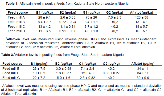
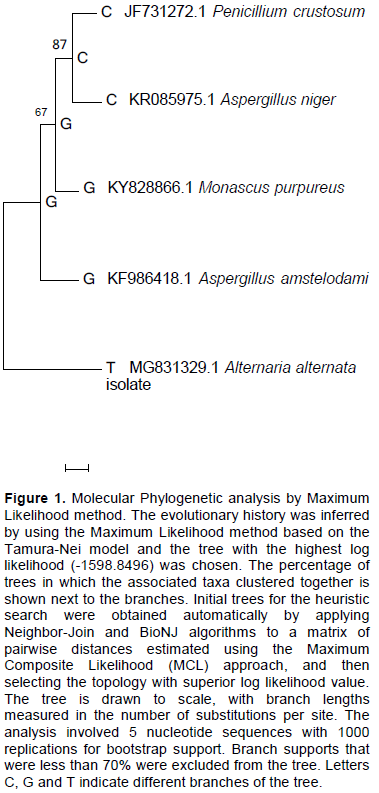
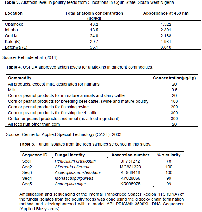
Aflatoxin contamination of poultry feeds in Nigeria is a common problem in most feed mills. A survey on the distribution of aflatoxin in feed mills from different parts of Nigeria was carried out. The aflatoxin concentration in most feed mills from the North-western part of Nigeria was low compared to the concentration in the feed mills from the South-eastern part. The total aflatoxin level in three feed mills (B, C and D) out of the four feed mills sampled in North-West ranged from 8.4 ± 2.7-13 ± 4.2 μg/kg. However, feed mill A, had higher concentration of aflatoxin 120 ± 38 μg/kg compared to the others. Three of the North-west feed mills with low aflatoxin concentration were below the 20 μg/kg recommended by United States Food and Drug Administration (USFDA) but the concentrations were higher than the 10 μg/kg recommended by the European commission (EU).  The total aflatoxin in the three feed mills sampled from the South-eastern part of Nigeria ranged from 30 ± 9.6 μg/kg in feed mill G to 34 ± 11 μg/kg in feed mill E. The results from the South-eastern part are comparable with the data from the South-western part of the country. AFG2 was very low in the feed mills sampled in the studied geopolitical regions while AFB1 appeared to be relatively high in all the feed mills in these regions. Screening of the contaminated feeds for aflatoxigenic fungi showed that Aspergillus species were the most common fungal contaminants, with Aspergillus amstelodami and Aspergillus niger being the most isolated fungi. The study has provided a comparative data on aflatoxin distribution in poultry feeds across some geopolitical zones in Nigeria. The obtained data could be useful in aflatoxin mapping in the studied geopolitical zones. Major fungal contaminants of the feed samples from all the geopolitical zones were also identified in this study.


Key words: Aflatoxin, Aspergillus, fungi, mycotoxin, Nigeria, feed mills, poultry feed.


 INTRODUCTION

A typical poultry ration is made up of several grain sources, each of which may be contaminated with one or several mycotoxins. Contamination of forages and cereals by mycotoxigenic fungi often occur in the field, or during processing, transportation and storage when conditions for growth such as temperature and relative humidity are available. Temperature and relative humidity of above 30°C and 80-100% respectively are favorable for fungal growth (Blaha and Lohnisky, 1990). Other conditions that favor fungal growth on feed or feed materials include nutrient availability (Njobeh et al., 2003) and oxygen supply (Filtenborg et al., 2000). Occurrence of mycotoxins in animal feed is most likely to occur in countries where the environment is hot and humid (Paterson and Lima, 2011). Mycotoxigenic fungi invade only a minor fraction of feed particles with appropriate condition for growth. Several mycotoxins occur in poultry feed and their ingredients, and these include aflatoxins, ochratoxins, fumonisins, cyclopiazonic acid, deoxynivalenol (DON), zearalenone (ZEN), nivalenol and ergot alkaloids (Zollner et al., 1999; Dalcero et al., 2002; Biselli and Hummert, 2005; Oliviera et al., 2008; Spanjer et al., 2008). Among these mycotoxins, aflatoxins are the most spread, most dangerous and most studied in the tropics. Aflatoxins are structurally related hepatocarcinogenic fungal metabolites produced on nuts and cereals by fungi of the Aspergillus species like A. flavus, A. parasiticus and A. nomius (Bressac et al., 1991; Groopman and Kensler, 1996). Aflatoxin contamination of animal feeds in the tropics is common due to the ubiquitous nature of the aflatoxin producing fungi. The concern for food safety due to aflatoxin contamination is more common in Sub-Saharan Africa, as compared to the temperate regions, where staple foods like maize and groundnut are less prone to aflatoxin contamination (Bankole and Adebanjo, 2003; Bankole et al., 2006). Aflatoxin-contamination of poultry feeds results in increased mortality of birds, decreased blood cell count, lower egg production, lower feed consumption rate, impaired resistance to infectious diseases, reduced vaccination efficiency and induced pathological damage to the liver and other organs (Mgbeahuruieke et al., 2018; Kamalavenkatesh et al., 2005). Approaches for reducing aflatoxin concentration in poultry feeds include, drying grains before storage to reduce the moisture content of the feed; and biological methods (Schaller, 2009). The use of microbial products which absorb mycotoxins from contaminated feeds has also been practiced (Xiao et al., 1991). Additionally, feed additives such as adsorbents have also been recommended as good detoxifying agents of contaminated feeds (Oguz et al., 2000; Kana et al., 2006; Mgbeahuruike et al., 2018). Studies have shown that the most frequently isolated fungal genera in both private and commercial feed mills in Nigeria is Aspergillus spp which is about 40% of mould, Penicillium spp, which is 20% in private feed mills and 13% in commercial feed mills (Adeniran et al., 2013). Commercial feed companies in Nigeria depend on feed ingredients which are produced locally for livestock feed production. Such companies purchase large quantities of grains and other feed materials during the production seasons; and  these  feed stuffs are stored and used gradually for feed production throughout the year. Additionally, the long storage of cereals during post-harvest periods and improper storage conditions are known to favour fungal growth, resulting in aflatoxin production in feed. Most feed mills suffer huge losses due to diseases caused by poor processing of feeds and feed contamination with microbial metabolites (Chelkowski, 1991; Hussein and Basel, 2001). The poultry industry in Nigeria has an annual growth rate of 2.17% (Killebrew and Plotnick, 2010). It is an important agricultural subsector and it provides food, employment and other economic resources for the country (Killebrew and Plotnick, 2010). Some of the birds raised in Nigeria as poultry include chickens, turkeys, ducks, guinea fowls, pigeons and ostrich (Killebrew and Plotnick, 2010). In Nigeria, there is no regulatory framework guiding levels of concentration of aflatoxin in poultry feeds. As a result, aflatoxin contamination of most livestock feeds in Nigeria are on the increase from the delivery of grains from harvesting point, to storage for feed manufacturing and finally to poultry farms in the form of feed. Commercial poultry sector in Nigeria ranges from the small scale peri-urban or rural operations to very large farms with well-integrated facilities. About 90% of the meat from broilers is sold in frozen, fried or roasted forms to consumers through fast food companies, commercial distributors, supermarkets and hotels or other hospitality industry operators (Adene and Oguntade, 2006). Since the meat and eggs sourced from the birds are major protein sources for many Nigerians (USDA-FAS, 2010), there is a need for appropriate monitoring and legislation of all aspects of quality issues in this sector. However, in other countries, maximum concentration of aflatoxin, DON, ZEN and ochratoxin A (OTA) are regulated in poultry feeds at various levels (FAO, 2004). In Africa, poultry industry suffers annual losses to the tune of over USD 670 million due to diseases caused by poor processing of feeds and feed ingredients (Bankole and Adebanjo, 2003). Because of these prevailing challenges, there is need to understand the rate of occurrence, distribution and concentrations of regulatory aflatoxin in poultry feed from different sources in Nigeria. Presently, there is no baseline data on the distribution of aflatoxin in feed mills in different parts of Nigeria. Additionally, there is a limited information on the major fungal species causing aflatoxin contamination of poultry feeds in Nigerian feed mills. The present study was carried out to investigate the prevalence of fungi and aflatoxin in commercial poultry feed mills across 2 geopolitical zones in Nigeria. Hence, we can have a base line data on aflatoxin distribution and fungal contamination in poultry feeds across the different geopolitical regions in Nigeria. 


 MATERIALS AND METHODS

Trial feed
 
The poultry feeds used in this trial were purchased from commercial feed dealers in Ogige Market Nsukka. All the feeds were freshly prepared feeds of 2-3 weeks old and they were purchased from 7 different commercial feed dealers. From our inquiry, 4 of the dealers purchased their feeds from 4 different companies A, B, C and D in Kaduna State, North-western part of Nigeria while the remaining 3 dealers purchased their feeds from 3 different feed mills E, F and G in Enugu State, the South-eastern part of Nigeria. After purchasing the feeds, they were placed in dark, water/air proof bags and kept in a storage room with a humidity of 80-100% and temperature of 25-30°C. The storage period for all the studied feed was two weeks.
 
Aflatoxin measurement
 
Aflatoxin analysis was done using high-performance liquid chromatography system consisting of model 600 pump, 717 autosampler, an in-line degasser and model 470 scanning fluorescence detector. The Aflatoxin extraction was carried out following the procedure described in (Mgbeahuruike et al., 2018). Briefly, 10 g of each feed sample was ground with mortar and pestle and transferred to extraction tubes containing water and acetonitrile (20:80%, v/v). The solution was vigorously mixed by shaking in a rotary shaker for 45 min before it was subjected to the extraction process. About 1 g of sodium chloride and 20 ml of n-hexane were added to each tube and mixed thoroughly. The resulting extract was filtered through folded filter paper (Schleicher and Shuell, 597½) and the filtrate was centrifuged for 10 min at 4000 g. The upper hexane phase was discarded while the lower methanol phase was used for immune affinity cleanup. An aliquot (1 ml) was diluted with 40 mL of de-ionized water, mixed thoroughly and the resulting solution was purified on immuno affinity columns (VicamAflaTest, Waters Corp.). The purified extract was further analyzed using reverse phase High Performance Liquid Chromatography (HPLC) (Shimadzu Corp.) with isocratic elution and fluorescence detection after post column derivatization with bromine by KOBRA CELL® (Rhone Diagnostics, Glasgow UK). The four known fractions of aflatoxin (Aflatoxin B1, B2, G1 and G2) were analyzed.
 
Fungal isolation
 
The feed samples from the different sources were finely ground for fungal isolation. The fungi were isolated by the dilution plate technique as reported by Ezekiel et al. (2014). One gram of each sample was suspended in 9 ml of sterile distilled water and vortexed for 2 min by hand inversion. A 0.1 ml aliquot of the suspension was spread plated in triplicates on Potato Dextrose Agar (PDA) and plates were incubated at 28°C for 48 h after which the colonies in each plate were counted and recorded as the fungal load per sample and the Colony Forming Unit (CFU/g). Each fungal colony from the mixed culture in each plate was carefully picked with a sterile toothpick and transferred again into sterile solid PDA plates for final purification prior to DNA extraction (Adebajo and Diyaolu, 2003). The fungal colonies were isolated based on their morphological characteristics such as color of isolate and physical appearance on agar (Pitt and Hocking, 2009).
 
Molecular characterization of fungal isolates and phylogenetic analysis
 
The extraction of the genomic DNA of the fungal isolates was done with the Zymo Research kit (Zymo-Research fungal/Bacterial Soil Microbe DNA, D6005, USA) supplied by Bio lab, South Africa, according to the manufacturer’s instructions. Amplification of the Internal Transcribed Spacer Region (ITS rDNA) of the fungal isolates from  the  poultry  feeds  was  carried  out  with  PCR  using universal primers ITS 1 and ITS 4 using established protocols (Mgbeahuruike et al., 2012). Cycling parameters included an initial denaturation at 94°C for 5 min, followed by 30 cycles of 94°C for 30 s, annealing at 60°C for 30 s, and extension at 72 ÌŠC for 30 s, with final extension at 72 ÌŠC for 8 min. PCR products were analyzed in 1% agarose gel and electrophoresed after staining with ethidiumbromide, then visualized under ultraviolet (UV) lights. Single PCR products were cleaned using Sephadex G-50 Fine DNA Grade (GE Healthcare, Sweden) followed by sequencing at Inqaba Biotechnical Industrial (Pty) Ltd, Pretoria, South Africa with PRISM™ Ready Reaction Dye Terminator Cycle Sequencing Kit, using the dideoxy chain termination method and electrophoresed with a model ABI PRISM® 3500XL DNA Sequencer (Applied Biosystems, Foster City, CA, USA), following the manufacturer’s instructions. Sequenced amplicons were used for BLAST search at the National Center for Biotechnology Information (NCBI). The obtained sequences were aligned by ClustalW following the method by Thompson et al. (1994) as implemented in MEGA 7 (Kumar et al., 2016). The aligned sequences were used to reconstruct a phylogenetic tree using Maximum Likelihood method based on the Tamura-Nei model (Tamura and Nei, 1993).
 
Statistical analysis
 
The data from the different aflatoxin fractions were analyzed using descriptive statistics and the average from different aflatoxin fractions were compared and represented in tables as means and standard deviations. Phylogenetic data was analysed using bootstrap method with 1000 replications per run. Branch support was placed at 100% and values that were less than 70% were excluded from the analysis.


 RESULTS

Feed samples from North West
 
The feed samples from Kaduna State, North-western Nigeria had very low of aflatoxin concentration except the sample from feed mill A where the aflatoxin level was high (Table 1). The aflatoxin level in the samples from feed mill A ranged from 2.6 ± 0.83 μg/kg for aflatoxin B2 to 78 ± 26 μg/kg for aflatoxin G1. However, aflatoxin B1 was relatively high but not as high as AFG1, in this same feed mill. The total aflatoxin content (AFB1+ AFB2+AFG1+AFG2) of feed samples from Feed mill A was 120 ± 38 μg/kg. In feed mills B, C and D, the different aflatoxin fractions (AFB2, AFG1 and AFG2) were low except AFB1 that appeared to be relatively higher than the other fractions (Table 1). In all the sampled feed mills in this region, AFG2 was abysmally low.
 
Feed samples from south east                               
 
The feed samples from feed mills in Enugu State, the South-eastern part of Nigeria presented a similar trend in aflatoxin concentration. AFB1 was relatively higher in the three feed mills sampled compared to the other aflatoxin fractions (Table 2). The total  aflatoxin  in  the  three  feed mills from South-eastern Nigeria ranged from 30 ± 9.6 μg/kg in Feed mill G – 34 ± 11 μg/kg in Feed mill E. AFG2 was very low in all the feed mills sampled in this region (Table 2). In the three feed mills from this region, AFB1 was relatively higher than the other fractions aflatoxin analyzed. The feed samples from South-eastern Nigeria showed a slightly higher level of aflatoxin concentration compared to the samples from some feed mills in the North-western part of the country.
 
 
Molecular identification and evolutionary relationship of the isolated fungi
 
The initial fungal culture on PDA produced several colonies of fungi with mixed morphology. Purification of the mixed fungal morphologies on fresh PDA produced pure cultures of each fungal isolate. Sequencing of the fungal ITS regions and subsequent BLAST searches in NCBI database showed that the major fungal contaminants in the feed samples were Penicillium crustosum, Alternaria alternate, Aspergillus amstelodami, Monascus purpureus, Aspergillus niger (Table 5). The sequences of the identified fungal species were deposited in GenBank with accession numbers JF731272, MG831329, KF986418, KY828866, KR085975 respectively. The percentage similarity of the identified fungal isolates with the fungal sequences in the NCBI database ranged from 78-100%. The identified fungal species belong to four major fungal genera. Aspergillus species, A. amstelodami and A. niger were the most common fungal species found in the feed samples. From the phelogenetic analysis, one major clade was observed and this cluster was made up of the following fungi, P. crustosum, A. niger, M. purpureus and A. amstelodami while A. alternate, formed the root  of  the tree showing its distant relationship from the other fungal species. In the order of evolutionary relationship, P. crustosum and A. niger appeared to be more closely related in this study, while M. purpureus and A. amstelodami appeared to be closer sisters in the evolutionary tree (Figure 1).


 DISCUSSION

The total aflatoxin concentrations in feeds from three  (B, C and D)  out of the four feed mills in Kaduna state were below the 20 μg/kg recommended by the United States Food and Drug Administration (USFDA) (Table 4). However, aflatoxin in these three feed mills including feed mill A were higher than the 10 μg/kg threshold recommended by the European Union (Mgbeahuruike et al., 2018). Feed mill A from the same state (Kaduna) had a reasonably high level of aflatoxin. The probable reason for this high level of aflatoxin could be due to poor method of preservation of feed ingredients. Due to high level of demands and supply, production of cheap low-quality feeds with the main aim of fast profit making becomes a common practice in some feed mills. In all the feed mills from the North-western state, AFB1 was considerably higher than the other aflatoxin fractions, except in feed mill A.  Analysis of aflatoxin concentration in all feed types in Nigeria has shown that AFB1 contaminated all chick feed, while layers mash, grower mash, finisher feed and starter feed had 46.4, 53.6, 62.5 and 72.7% contamination respectively (Ezekiel et al., 2012). AFB1 is a very potent fraction of the known aflatoxins (Mgbeahuruike et al., 2018). AFB1 has been implicated in increased mortality of birds in poultry farms, decreased blood cell count, lower  egg  production,  lower feed consumption rate, impaired resistance to infectious diseases, reduced vaccination efficiency and induced pathological damage to the liver and other organs (Mgbeahuruiek et al., 2018; Kamalavenkatesh et al., 2005). The high concentration of this aflatoxin fraction in this region could be attributed to the ubiquitous nature of the producing fungi. For the South eastern part of Nigeria, the AFB1 concentration in the feed samples was high in the three feed mills sampled when compared with the other fractions.  
 
The total aflatoxin (AFB1+AFB2+AFG1+AFG2) in the three feed mills sampled in Enugu state, South-eastern Nigeria were relatively higher than the total aflatoxin level in three out of the four feed mills (B, C and D) sampled in Kaduna state, North western part of Nigeria. The reason for this could be due to differences in weather conditions between the two regions. While the weather in the North is hot, dry and sunny, the South-eastern part of the country has a hot and humid tropical weather condition which provides the toxigenic fungal species with more favorable conditions for growth. In Ogun State, the South-western part of Nigeria, an independent study by Kehinde et al. (2014) showed a similar trend in the total aflatoxin level in poultry feed samples obtained from 5 different locations in the state (Table 3). Although the authors did not report the concentration of the different aflatoxin fractions, the total aflatoxin concentration in the feed mills studied were comparable to the results obtained from this study in the other two geopolitical regions  (South-eastern and North-western Nigeria). In the survey from the South-west region, the highest level of aflatoxin concentration, 95 μg/kg was found in a feed mill from Lafenwa town, this was followed by Obantoko town with aflatoxin concentration of 43.2 95 μg/kg (Table 3). Feed samples obtained from the other towns Idi-aba, Omida and Kuto had different concentrations of aflatoxin respectively (Kehinde et al., 2014).  Molecular identification of the fungal contaminants in the feeds showed five different fungal species P. crustosum, A. alternate, A. amstelodami, M. purpureus, A. niger belonging to four major genera of fungi (Table 5). Although four fungal genera were observed in the feeds, only two (Penicillium and Aspergillius) are known for mycotoxin production and may have contributed to the different fractions of aflatoxin observed in the feeds. However, Aspergillius species are known as the major producers of aflatoxin in the tropics (Mgbeahurike et al., 2018). Aspergillus species were the most abundant fungal species isolated from the feed samples. Aspergillus species such as A. tamarii, A. nominus, A. flavus, A. parasiticus and A. tubingensis have been implicated in many studies as the primary source of aflatoxin contamination of poultry feeds (Mgbeahuruike et al., 2018; Peterson et al., 2001; Klich et al., 2000). However, the two major Aspergillus species A. flavus and A. parasiticus known for heavy aflatoxin production in poultry feeds (Donner et al., 2009) were not found in this study. Heavy contamination of poultry feeds by  A.  niger  has  also  been   reported   in  other  studies  (Adeniran et al., 2013). Penicillium species are known to produce the mycotoxin, Trichothecenes, including DON (Rodrigues and Naehrer, 2012; Tiemann and Dänicke, 2007). However, in this study, we did not screen for the presence of Trichothecenes and other mycotoxins in the feed. Alternaria species and M. purpureus are not known as aflatoxin producing fungi, they could be in the feed as contaminants but not for aflatoxin production. Phylogenetic analysis of the identified fungal species confirmed an evolutionary relationship between the fungal isolates.


 CONCLUSION

The study has been able to make a comparative analysis of aflatoxin distribution in major poultry feed mills across some geopolitical zones in Nigeria. The observed aflatoxin level in most of the feed mills was slightly above the recommended levels of aflatoxin in poultry feeds (20 μg/kg) by the United States Food and Drug Administration (USFDA). The data from this study will act as a baseline data for aflatoxin mapping in the study areas and it will also act as a guide for poultry feed producers in the country. From the study also, we have succeeded in isolating and identifying some of the major fungal contaminants of poultry feeds in some feed mills in Nigeria.


 CONFLICT OF INTERESTS

The authors have not declared any conflict of interests.


 ACKNOWLEDGEMENTS

The authors appreciate the technical staff of the Department of Veterinary Microbiology and Pathology, Faculty of Veterinary Medicine, University of Nigeria Nsukka and also grateful to the technical staff of the Department of Chemistry, Environment and Feed Hygiene, National Veterinary Institute, Uppsala, Sweden for their support with the aflatoxin analysis.


 REFERENCES

Adebajo LO, Diyaolu SA (2003). Mycology and spoilage of retail cashew nuts. African Journal of Biotechnology 2(10):369-373
Crossref


Adene DF, Oguntade AE (2006). Nigeria: poultry sector country review.


Adeniran AL, Anthony MH, Lami MH (2013). Survey of mycotoxigenic fungi in concentrated poultry feed in Niger State, Nigeria. Journal of Food Research 2(2):128-135.
Crossref


Bankole SA, Adebanjo A (2003). Mycotoxins in food in West Africa: current situation and possibilities of controlling it. African Journal of Biotechnology 2(9):254-263.
Crossref


Bankole SA, Schollenberger M, Drochner W (2006). Mycotoxins in food systems in sub-Saharan Africa: a review. Mycotoxin Research 22:163-169.


Biselli S, Hummert C (2005). Development of a multicomponent method for Fusarium toxins using LC-MS/MS and its application during a survey for the content of T-2 toxin and deoxynivalenol in various feed and food samples. Food Additive and Contaminants 22(8):752-760.
Crossref


Blaha J, Lohnisky J (1990). Aflatoxin production by Aspergillus flavus strains isolated from Vietnamese feeds. Tropical Science 30:33-40.


Bressac B, Kew M, Wands J, Ozturk M (1991). Selective G to T mutations of p53 gene in hepatocellular carcinoma from Southern Africa. Nature 350(6317):429-431.
Crossref


Chelkowski J (1991). Cereal Grain: Mycotoxins, Fungi and Quality in Drying and Storage. Elsevier: Amsterdam.


Dalcero A, Magnoli C, Hallak C, Chiacchiera SM, Palacio G, Rosa CAR


(2002). Detection of ochratoxin A in animal feeds and capacity to produce this mycotoxin by Aspergillus section Nigri in Argentina. Food Additives and Contaminants 19(11):1065-1072.
Crossref


Donner M, Atehnkeng J, Sikora RA, Bandyopadhyay R, Cotty PJ (2009). Distribution of Aspergillus section Flavi in soils of maize fields in three agroecological zones of Nigeria. Soil Biology and Biochemistry 41(1):37-44.
Crossref


Ezekiel CN, Bandyopadhyay R, Sulyok M, Warth B, Krska R (2012). Fungal and bacterial metabolites in commercial poultry feeds from Nigeria. Food Additives and Contaminants. Part A, Chemistry, Analysis, Control, Exposure and Risk Assessment 29(8):1288-99.
Crossref


Ezekiel CN, Udom IE, Frisvad JC, Adetunji MC, Houbraken J, Fapohunda SO, Samson RA, Atanda OO, Agi-Otto MC, Onashile OA (2014). Assessment of aflatoxigenic Aspergillus and other fungi in millet and sesame from Plateau State, Nigeria. Mycology 5(1):16-22 
Crossref


Filtenborg O, Frisvad TC, Samson RA (2000). Specific association of fungi in foods and influence of physical environmental factors. In Samson RA, Hoekstra ES, Frisvad JC, Filtenborg O (Eds.), Introduction to Food-and Airborne Fungi (6th Ed., pp. 306-320). Utrecht: Central bureauvoor Schimmel culture.


Food and Agriculture Organization (2004). Worldwide regulations for mycotoxins in foodand feed in 2003. Food and Nutrition Papers No. 81. Rome: FAO.


Groopman JD, Kensler W (1996). Temporal patterns of aflatoxin-albumin adduct in hepatitis B surface antigenpositiveand antigen-negative residents of Daxin, Qidong County, and People's Republic of China. Cancer Epidemiology, Biomarkers and Prevention 5(4):253-261.


Hussein HS, Basel JM (2001). Toxicity, metabolism, and impact of mycotoxins on humans and animals. Toxicology 167(2):101-134.
Crossref


Kamalavenkatesh P, Vairamuthu S, Balachandran C, Muralimanohar B, Dhinakarray G (2005). Immunopathological effect of the mycotoxins cyclopiazonic acid and T- 2 toxin on broiler chicken. Mycopathologia 159:273-279.
Crossref


Kana JR, Teguia A, Tchoumboue J (2006). Effect of dietary plant charcoal from Canariumschweinfurthii Engl. and maize cob on aflatoxin B1 toxicosis in broiler chickens. Livestock Research and Rural Development 22. 

View. Accessed March 12, 2019.


Kehinde MT, Oluwafemi F, Itoandon EE, Orji FA, Ajayi OI (2014). Fungal Profile and Aflatoxin Contamination in Poultry Feeds Sold in Abeokuta, Ogun State, Nigeria. Nigerian Food Journal 32(1):73-79.
Crossref


Killebrew K, Plotnick R (2010). Poultry market in West Africa: Nigeria. EPAR Brief No. 84. Seattle (WA): Evans School of Public Affairs, Washington State University.


Klich MA, Mullaney EM, Daly CB, Cary JW (2000). Molecular and physiological aspects of aflatoxin and sterigmatocystin biosynthesis by Aspergillus tamari and A. ochraceoroseus. Applied Microbiology and Biotechnology 53:605-609.
Crossref


Kumar S, Stecher G, Tamura K (2016). MEGA7: Molecular Evolutionary Genetics Analysis version 7.0 for bigger datasets. Molecular Biology and Evolution 33(7):1870-1874.
Crossref


Mgbeahuruike AC, Ejioffor TE, Obasi CC, Shoyinka VC, Karlsson M, Nordkvist E (2018). Detoxification of Aflatoxin-Contaminated Poultry Feeds by 3 Adsorbents, Bentonite, Activated Charcoal, and Fuller's Earth. Journal of Applied Poultry Research 27(4):461-471.
Crossref


Mgbeahuruike AC, Karlsson M, Asiegbu FO (2012). Differential expression of two hydrophobin genes (Pgh1 and Pgh2) from the biological control agent Phlebiopsis gigantea. Fungal Biology 116(5):620-629.
Crossref


Njobeh BP, Dutton MF, Koch SH, Chuturgoon A (2003). Contamination with storage fungi of human foods. Camerounian International Journal of Food Microbiology 135:193-198.
Crossref


Oguz H, Kececi T, Birdane YO, Onder F, Kurtoglu V (2000). Effect of clinoptilolite on serum biochemical and haematological characters of broiler chickens during aflatoxicosis. Research in Veterinary Science 69(1):89-93.
Crossref


Oliviera CAF, Sebastiao LS, Fagundes H, Rosim RE, Fernandes AM (2008). Aflatoxins and cyclopiazonic acid in feed and milk from dairy farms in Sao Paulo, Brazil. Food Additives and Contaminants B 1(2):147-152.
Crossref


Paterson R, Lima N (2011). Further mycotoxin effects from climate change. Food Research International 44(9):2555-2566.
Crossref


Peterson SW, Ito Y, Horn BW, Goto T (2001). Aspergillus bombycis, a new aflatoxigenic species and genetic variation in its sibling species, A. nomius. Mycologia 93(4):689-703.
Crossref


Pitt JI, Hocking AD (2009). Fungi and Food Spoilage, 3rd edition, Springer Science plus Business Media, LLC, 233 Spring Street, New York, NY 10013, USA, 524 pp.


Rodrigues I, Naehrer K (2012). A three-year survey on the worldwide occurrence of mycotoxins in feedstuffs and feed. Toxins 4(9):663-675.
Crossref


Schaller JL (2009). Clinical Research of Mycotoxicosis on Animals www.personalconsult.com Accessed July 29, 2019.


Spanjer MC, Rensen PM. Scholten JM (2008). LC-MS/MS multi-method for mycotoxins after single extraction, with validation data for peanut, pistachio, wheat, maize, cornflakes, raisins and figs. Food Additives and Contaminants - Part A Chemistry, Analysis, Control, Exposure and Risk Assessment 25(4):472-89.
Crossref


Tamura K, Nei M (1993). Estimation of the number of nucleotide substitutions in the control region of mitochondrial DNA in humans and chimpanzees. Molecular Biology and Evolution 10(3):512-26.


Thompson JD, Higgins DG, Gibson TJ (1994). CLUSTAL W: improving the sensitivity of progressive multiple sequence alignment through sequence weighting, position-specific gap penalties and weight matrix choice. Nucleic Acids Research 22(22):4673-4680.
Crossref


Tiemann U, Dänicke S (2007). In vivo and in vitro effects on the mycotoxins zearalenone and deoxynivalenol on different non-reproductive and reproductive organs in female pigs: A review. Food Additives and Contaminants 24(3):306-314.
Crossref


USDA Foreign Agricultural Service (USDA-FAS) (2010). GAIN report: Nigeria grain and feed. Annual Report. Washington (DC): Department of Agriculture.


Xiao H, Marquardt RR, Frohlich AA, Phillips GD (1991). Effect of a hay and a grain diet on the bioavailability of ochratoxin A in the rumen of sheep. Journal of Animal Science 69(9):3715-3723.
Crossref


Zollner P, Jodlbauer J, Lindner W (1999). Determination of zearalenone in grains by high-performance liquid chromatography-tandem mass spectrometry after solid-phase extraction with RP-18 columns or immunoaffinity columns. Journal of Chromatography A 858(2):167-174.
Crossref


          */?>