ABSTRACT
This study is aimed at evaluating the antibacterial and anticandidal activity of coconut oil, fermented and unfermented coconut water and palm kernel oil (PKO) on Escherichia coli, Staphylococcus aureus, Pseudomonas aeruginosa, Corynebacterium sp. and Candida albicans. The coconut water was fermented with Saccharomyces cerevisiae and the pH, sugar and alcohol contents were determined before and after fermentation. The plant materials were screened for antimicrobial phytochemicals. The antimicrobial activity of the coconut water, coconut oil and palm kernel oil and the antibiotic susceptibility of the organisms were done on Muiller Hinton agar using disc diffusion method. The plasmids of the organisms were cured using 10% sodium dodecyl sulphate. The pH and ethanol content of the fermented coconut water were higher than the unfermented while the sugar content decreased after fermentation. There were no phytochemicals detected in the coconut water whereas all the phytochemicals were detected in the PKO while only reducing sugar and steroids were absent in the coconut oil. All the bacteria exhibited multi-drug resistance. After plasmid curing, there were reductions in the resistance indicating that the resistance is plasmid mediated. There was a reduction in the percentage antibiotic resistance of Corynebacterium sp. which was 60 and 0% before and after curing respectively. The PKO was more effective in inhibiting the bacteria and Candida sp. than coconut oil but were all resistant to the coconut water. PKO and coconut oil may thus be used as natural substitutes to control E. coli, S. aureus, P. aeruginosa and Candida sp.
Key words: Bacteria, antimicrobial, resistance, coconut water, coconut oil, palm kernel oil, phytochemicals.
The presence of microorganisms in food can bring about desirable and undesirable effects. Some of the adverse effects of their presence in food are spoilage of the food and/or food-borne diseases caused by organisms which
include Escherichia coli, Staphylococcus aureus and Pseudomonas aeruginosa (Pandey and Singh, 2011; Ashraf et al., 2018). Some of these diseases are life threatening and there is a growing concern of antibiotic resistance by many of these organisms (Sapkota et al., 2012; Wigmore et al., 2016; Collingnon and McEcwen, 2019). Repeated applications of chemical preservatives used in the prevention of spoilage (Shan et al., 2007) bring about bioaccumulation in food and feed chain. These result in microbial resistance to the applied chemicals, consequently having detrimental side effects on the health of the consumers (Blalonska, 2010; Akinyemi et al., 2006). About 90 to 95% of S. aureus strains isolated worldwide are penicillin and methicillin resistant (Borges et al., 2016). P. aeruginosa, S. aureus, Salmonella sp., Coagulase-negative Staphylococcus, Shigella, Enterococcus sp. and E. coli are among some of the bacteria with multidrug resistance (Fisher and Phillips, 2008). The prevalence and resistance of E. coli isolates to the third generation cephalosporins have been reported to be very high (Uribe-Beitrain et al., 2017).
The resistance of microorganisms to antimicrobials such as antibiotics can be caused by indiscriminate use of broad-spectrum antibiotics, immunosuppressive agents, organ transplants and intravenous catheters (Selvamohan et al., 2012). Due to these concerns, there are concerted efforts to develop potentially effective, safe and natural antimicrobials which can serve as food preservatives and substitutes for antibiotics in the treatment of diseases caused by multidrug- resistant organisms. The use of plants as an alternative is being studied globally since plants are considered nutritionally safe, biodegradable and possess antimicrobial phytochemicals (Berahou et al., 2007; Chika et al., 2007). Moreso, majority of the population of underdeveloped and most developing countries like Nigeria still depend mainly on traditional medicine using plants (Pallant and Steenkamp, 2008). This is because the health facilities are not well equipped, far from rural populace with expensive, or no means of transportation, and are expensive, as well as due to religious and cultural beliefs. Apart from high dependence of these plants ethno-medically in the underdeveloped and developing countries, there is also a growth trend of health and food supplements in the form of plants and their derivatives in different parts of the world.
Plants have been reported to be excellent sources of secondary metabolites which can be used in the production of modern medicines (Chandra et al., 2017). Many of the secondary metabolites such as tannins, flavonoids, alkaloids have been reported to contribute to antimicrobial activity. These secondary metabolites are produced by plants to fight against microbial attacks. The antimicrobial activity exhibited by plants and their extracts have been demonstrated in many studies (Akinpelu et al., 2015; Verma et al., 2012). Akinyemi et al. (2006) demonstrated the high antibacterial activity of three medicinal plants (Trema guineensis, Phyllanthus discoideus and Acalypha wilkesiana) on S. enteriditis, E. coli and S. aureus. Verma et al. (2012) found the extracts of Punica aranatum and Allium sp. to be effective against some food-borne spoilage bacteria (S. typhi, E. coli, B. cereus and S. aureus). Sarid-Saklani et al. (2019) reported the presence of flavonoids, terpenoid and tannins in Terminalia alata which were inhibitory to K. pneumoniae and S. pneumoniae.
Essential oil which are mainly plant derivatives were reported to possess antifungal, antibacterial and antiviral properties and are potential agents of food preservation and alternatives to treat infectious diseases (Astanin et al., 2011; Safaei-Ghonu and Ahd, 2010). Efficacy of essential oils has been reported in several studies against pathogens and food contaminants (Djenane et al., 2011). Plants from which essential oils have been derived include Cympbopogon citratus, Satureja montana, Eucalyptus sp., Allium sativum, Cinnamon, Cocos nucifera, Occimum gratissimum, Mentha longifolia and Piper nigrium.
Coconut is cultivated for its multi-purpose values (nutritional and medicinal). The products of C. nucifera include coconut water, copra, coconut oil, raw kernel, coconut cake and coconut milk. It is a unique source of various natural products for the development of drugs and industrial products and is effective against fungi, bacteria, viruses, parasites and dermatophytes (Floriana et al., 2015). It is also an antioxidant (Manisha and Shyamapada, 2011). Coconut water is a refreshing beverage believed to be rich in vitamins, minerals, amino acids, carbohydrates, enzymes, hormones and phytochemicals and sometimes fermented to increase its shelf life and probiotic content (Bandalam and Galvez, 2016). Coconut water and oil is claimed to cure some diseases by rural dwellers in Nigeria (Fowoyo and Alamu, 2018). Coconut oil contains a high amount of phytochemicals in form of alkaloids, flavonoids, saponins, tannins soluble carbohydrates, hydrogen cyanide, reducing sugar, terpenoids (Ojobor et al., 2018). Coconut oil was found to inhibit S. epidermidis, E. coli and Candida sp. (Rajan et al., 2016; Ogbalu, 2017). The anticandidal activity of this oil was comparable to that of fluconazole (Ogbalu, 2017). Shilling et al. (2013) reported that coconut oil inhibits Clostridium difficile.
Palm kernel oil is extracted from the kernel endosperm of Elaeis guineensis. It is used traditionally in the treatment of convulsion, dandruff, skin diseases, cold and cough (Ekwenye and Ijeomah, 2004). The inhibitory effect of palm kernel oil on some bacterial and fungal isolates was reported by Ugbogu et al. (2006). Floriana et al. (2015) found palm kernel oil to be effective against E. coli, S. aureus and Streptococcus sp while coconut oil was active against Listeria monocytogenes, S. aureus and Helicobacter sp.
This study is therefore aimed at evaluating the antibacterial and anticandidal activity of coconut oil, coconut water and palm kernel oil on some microorganisms associated with food spoilage and food-borne diseases.
Collection of samples
Plant materials
Mature coconuts, palm kernel oil and coconut oil were bought from Abraka Market, while the tender coconuts were harvested from farms in Abraka, Delta State, Nigeria. The oils were transported to Microbiology Laboratory, Delta State University, where they were kept in sterile, screw-capped bottles and kept in the refrigerator for analyses.
Collection of isolates
The bacterial isolates (S. aureus-9, E. coli-3, P. aeruginosa-1 and Corynebacterium sp-1) were collected from food-spoilage stock culture at Microbiology Laboratory, Delta State University, Abraka. They were cultured on Nutrient agar and Cysteine, Lactose and Electrolyte Deficient (CLED) agar, and incubated at 37°C for 24 h prior to usage.
Confirmation of isolates
The identity of each isolate was confirmed after incubation using morphological, cultural and biochemical characteristics which included Gram staining, motility test, catalase, oxidase, indole, citrate utilization and Triple Sugar Iron (TSI) agar tests.
Fermentation of coconut water
Coconut fruits were first washed with distilled water, then surface-sterilized using alcohol and opened at one of the eyes on the apical region using a sterile knife. The water was aseptically collected into a sterile glass measuring cylinder. The coconut water samples (mature and tender) were divided into two groups (A and B). The mature samples were labelled mA and mB while the tender ones were labelled tA and tB.
mA and tA were fermented using a modified method by Shiny et al. (2014). Dry baker’s yeast (90.5 g) was added to 450 ml of each coconut water sample in a measuring cylinder. The cylinder was covered with aluminium foil and incubated at 26±2°C for 24 h.
Determination of pH, sugar content and alcohol content
pH
The pH was determined using a pH meter.
Sugar content
A brix hydrometer was inserted into 450 mL of coconut water samples, fermented (mA and tA) and unfermented (mB and tB) and the readings were taken.
Alcohol content
This was done using the hydrometer to determine the specific gravity and the alcohol content calculated using the formula:
% Alcohol by volume = (FG-OG) x 131.25 (1)
Where FG = Final specific gravity, OG = Original specific gravity, and 131.25 is a constant brewery factor
Phytochemical screening of coconut water sample
Saponin, flavonoid, alkaloid, tannin, phenol, reducing sugar, terpenoid, steroid, glycoside and cardiac glycoside were screened for using the method of Hasbourne (1973) as follows:
1. Alkaloids: To 0.5 g of each extract, was added 5 ml of 2 N HCl and filtered. Dragndoff’s reagent was added and formation of a red precipitate was used to indicate the presence of alkaloids.
2. Phenols: About 3 to 4 drops of ferric chloride solution was added to the extract. The presence of phenol was indicated by the formation of bluish black colouration.
3. Terpenoids: 2 ml of chloroform and 3 ml concentrated H2SO4 was added to 5 ml of extract. This was done carefully to form a distinct layer. Positive result for the presence of Terpenoids was indicated by reddish brown colour formation.
4. Saponins: To a small quantity of the extract was added distilled water up to 20 ml and shaken vigorously. The formation of 1 cm layer of foam stable for 10 min indicates a positive result for saponin.
5. Flavonoids: Sodium hydroxide (10%) was added to the plant extract. The formation of yellow colouration indicated the presence of flavonoids.
6. Phenolics and Tannins: A 10% Lead acetate solution was added to little quantity of the extract already dissolved in distilled water. The presence of tannin was indicated by formation of white precipitate.
Preparation of standard inoculum
Suspension of each test organism was made by inoculating a loopful of the colony into peptone water and incubated at 37°C for 24 h. The overnight broth culture of organisms was diluted with sterile distilled water to an inoculum load of 1×10⁶ CFU/ml. It was standardized by comparing the turbidity with 0.5 McFarland turbidity standards (Murray et al., 2016).
Antibiotic susceptibility test
This was done using paper disc diffusion method. Sterile cotton swabs were dipped into each of the standardized inoculum suspension and used to inoculate freshly prepared Muller Hilton agar plates. The cotton swabs were pressed on the wall of the tubes to avoid flooding of the medium and the plates were completely covered for uniform growth. Appropriate antibiotic discs were aseptically placed on the inoculated agar plates which were incubated at 37°C for 24 h. They were examined for zones of inhibition (Murray et al., 2016).
Antimicrobial susceptibility test
Antimicrobial susceptibility was done using agar Kirby-Bauer disc diffusion method as described by Abbas et al. (2017). Different concentration (20, 40, 60, 80 and 100%) of fermented and unfermented coconut water samples were made in sterile universal containers. A sterile Whatmann No. 1 filter paper disc was impregnated with appropriate concentrations of the coconut water samples. The discs (equally spaced) were placed on a freshly and uniformly inoculated Muller-Hilton agar plates. The plates were incubated at 37°C for 24 h. After incubation, the plates were examined for zones.
Plasmid curing of bacterial isolates
Ten percent (10%) stock solution of sodium dodecyl sulphate (SDS) was prepared (Silhavey et al., 1984) by adding 10 g SDS to 90 mL of distilled water. This was heated with sterile magnetic heater for 15 min until homogeneous mixture was obtained. Bacterial isolates were inoculated into peptone water and incubated at 37°C for 24 h. Ten-fold serial dilution of the SDS solution was done and 0.5 mL of the standardized bacterial suspension were added to the dilutions in test tubes. The tubes with inoculated SDS were incubated at 37°C for 24 h. Solutions with growth were sub cultured by transferring 1 mL of the solutions and incubated at 37°C for 48 h to confirm their growth. Fresh peptone water solution was prepared and inoculated with 0.5 mL of cured tested cultures and incubated at 37°C for 24 h. Antibiotic susceptibility test was carried out on the organisms.
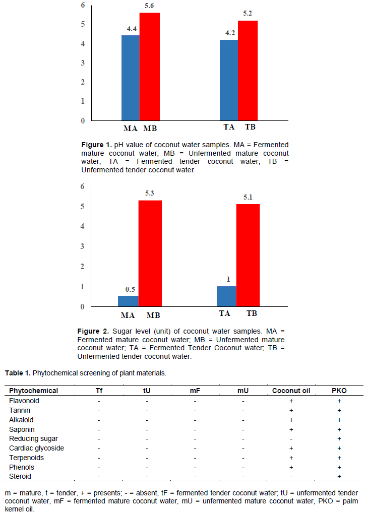
The pH values of fermented coconut milk (mA and tA) were found to be lower than the values of unfermented samples (mB and tB) (Figure 1). The ethanol content of the fermented tender coconut samples were higher than that of fermented mature coconut water samples, while the sugar content decreased significantly after fermentation (Figure 2). The phytochemical screening of the plant extracts indicated the presence of flavonoid, tannin, alkaloid, saponin, reducing sugar, cardiac glycoside and terpenoids. This was confirmed in this study (Table 1).

The antibiotic sensitivity profiles of the isolates showed that all the isolates exhibited multi drug resistance (Tables 2 to 3). All the isolates were resistant to tA while 81% were resistant to tB (only 2 of the 11 isolates were sensitive). There was 100 and 83% susceptibility to PKO and coconut oil respectively. The plasmid curing affected the antibiotic sensitivity profile of more than 90% of the gram negative bacteria and about 50% of the gram negative bacteria as shown in Table 4. The drug resistance of the affected isolates were reduced after the plasmid curing.
The fourteen isolates, that is, S. aureus (9), Corynebacterium sp. (1). E. coli (3) and P. aeruginosa (1) have been reported to be common pathogens which cause urinary tract infections (Odoki et al., 2019), nosocomial infections (Khan et al., 2019) among others. They have also been implicated in the spoilage of some foods.
The absence of phytochemicals in fermented and unfermented tender and mature coconut water (sap) may be attributed to the very high content of the water which may have diluted the chemicals if present, to undetected levels. Misra (2016) reported that the composition of coconut water was mainly water but after ultrafiltration contained total sugar (14.40 g/100 mL), reducing sugar (5.58 g/100 mL) and total protein (0.23 to 0.32 g/100 mL). This study used coconut water without processes used in concentrating the chemicals. The levels of the chemicals were undetected since they might have been diluted in the high-water content. Fowoyo and Alamu (2018) reported the moisture (%) of coconut water for both mature and immature coconut to be between 95.44 to 98.00%. The increase in acidity of the water after fermentation may be attributed to the presence of organic acids, free amino acids, fatty acids and dissolved CO2 arising from tissue respiration (Robert and Frim, 2009). The pH of tender coconut being more acidic than that of mature coconut water in this study corresponds with that of Fowoyo and Alamu (2018).
The detection of phytochemicals may be attributed to the natural ability of plants to produce secondary metabolites to combat microbial attacks. The lower sugar content in unfermented tender coconut water (0.5) than mature (1.0) and high sugar content in the unfermented mature coconut water (5.3 and 6.1) may be due to sugar present being converted to acid during metabolic processes in the plant and that the sugar being used by the kernel during development for the synthesis of fat (Fowoyo and Alamu, 2018). The alcohol content by volume was 1.3 and 2.0% for fermented mature and tender coconut water respectively. This showed a direct relationship with the sugar content. The tender coconut had a higher sugar content resulting in a higher alcohol content. Nakuwa et al. (2012) reported that on exposure, coconut water becomes vulnerable to oxidation and microbial contamination which brings about fermentation. Antibiotic sensitivity test showed all the gram positive organisms in the present study exhibiting multi drug resistance. Resistance may be due to prolonged use of the antibiotics, organisms putting up mechanisms to resist the antibiotics, as well as eating poultry and livestock with feeds treated with antibiotics. This is the practice in most of these countries where there are no checks and control resulting in indiscriminate use of antibiotics.
After curing, the gram positive isolates (apart from SA7) and EC1 and EC2 showed a reduction in the resistance level indicating that the resistance is plasmid mediated. This conforms with the result of Patward-han et al. (2017) that elimination of plasmids enables effective antibiotic therapy. The gene coding for the resistance is in the plasmid hence no resistance after the removal of the plasmid. This would have constituted a strategy for removal of the gene to make the organisms susceptible but other factors contributing to the resistance still have to be overcome, hence the need for a natural antimicrobial product.
The bacterial isolates exhibited resistance to all the tested concentrations of coconut water used (fermented and unfermented). This effect may be related to the absence of phytochemicals in coconut water. The result corresponds with the study of Rukimi et al. (2017) who stated that coconut water did not exhibit antimicrobial effect on microorganisms. However, the result disputed the report of Shiny et al. (2014) that fermented tender coconut water has antimicrobial effect on E. coli and the report of Fowoyo and Alamu (2018) that tender coconut water exhibited antimicrobial effect on S. aureus, E. coli, B. cereus and P. mirabilis.
All the isolates were susceptible to PKO and 86% to coconut oil. The high susceptibility of the organisms may be attributed to the many phytochemicals detected in the palm kernel and coconut oils. Coconut oil and palm kernel oil have been reported to contain lauric acid which is antibacterial, antifungal and antiviral (Manisha and Shyamapada, 2011). Contrary to the finding of this study, Abbas et al. (2017) reported a low susceptibility of E. coli to coconut oil extract. Ekwenye and Ijeomah (2004) also reported non-susceptibility of E. coli to PKO but there was susceptibility to virgin coconut oil when the PKO was mixed with coconut oil. The report of Floriana et al. (2015) that VCO and PKO were effective against E. coli and S. aureus conforms with that of this study. The difference in the effectiveness of the plant materials may be attributed to the fact that levels of the phytochemicals which invariably affect the microbial activity vary from plant to plant depending on factors such as method of extraction, solvent used in extraction and growing condition.
Food spoilage and food borne diseases are attributed to the presence of diverse microorganisms in food. Many of these organisms which are pathogenic are exhibiting alarming increase of multidrug resistance, in addition to the adverse effects of the synthetic chemicals in the drugs and the food preservatives. This study showed that the coconut water was not effective against any of the organisms so cannot serve as a potential antimicrobial. However, coconut oil and palm kernel oil may be used as natural alternatives in the control of some microorganisms associated with food spoilage and food borne diseases.
They may also serve as a substitute to synthetic antibiotics which are becoming less effective and have numerous side effects. Further work can still be done on the activity of concentrates of coconut water. Other species of coconut and E. guineensis from other geographical locations can also be researched into for their antimicrobial activity.
The authors have not declared any conflict of interests.
The authors acknowledge the laboratory staff of the Department of Microbiology, Delta State University for supply of culture as well as technical support.
REFERENCES
|
Abbas AA, Peter U, Akeh M, Adeola J, Ewenighi CO, Ishaku P (2017). Antibacterial activity of lauric acid on some selected clinical isolates. Annals of Clinical and Laboratory Research 10:2386-5780.
|
|
|
|
Akinpelu DA, Aiyegoro OA, Akinpelu OF, Okah AI (2015). Stem bark extract and fraction of Persia Americana (mill) exhibits bactericidal activities against strains of Bacillus cereus associated with food poisoning. Molecules 20:416-429.
Crossref
|
|
|
|
|
Akinyemi KO, Oluwa OK, Omomigbehin EO (2006). Antimicrobial activity of crude extracts of three medicinal plant used in South west Nigeria folk medicine on some food bacterial pathogens. African Journal of Traditional. Compounds and. Alternative Medicine 3(4):13-22.
Crossref
|
|
|
|
|
Ashraf AM, Abdulaziz AA, Khalid SA, Turki MD, Essain NS, Marwah MB (2018). Antimicrobial activity of some plant extracts against bacterial strains causing food poisoning diseases. Saudi Journal of Biological Sciences 25(2):361-368.
Crossref
|
|
|
|
|
Astanin A, Reichling J, Schnitizler P (2011). Comparative study on the antiviral activity of selected monoterpenes derived from essential oils. Phytotherapy Research 24:673-679.
|
|
|
|
|
Bandalam EB, Galvez LA (2016). Optimization of coconut water beverage fermented with Lactobacillus acidophilus. Annals of Tropical Research 38(1):196-202.
Crossref
|
|
|
|
|
Berahou A, Auhmani A, Fdil N, Benharref A, Jama M, Gadhi CA (2007). Antibacterial activity of Quercus ilex bark's extracts. Journal of Ethnopharmacology 112:426-429.
Crossref
|
|
|
|
|
Blalonska M (2010). Influence of pomegranate by-product and punicalaginus on selected groups of human intestinal microbiota. International Journal of Food Microbiology 140:175-182.
Crossref
|
|
|
|
|
Borges A, Abreu AC, Dias C, Saavedva MJ, Borges F, Simoes M (2016). New perspective on the use of phytochemicals as an emergent strategy to control bacterial infections including bioflum. Molecules 21(7):1-41.
Crossref
|
|
|
|
|
Chandra H, Bishnol P, Yodav A, Mishra AP, Nautiyal AK (2017). Antimicrobial resistance and the alternative resources with special emphasis on plant- based antimicrobials-A Review. Plants 6(2):16.
Crossref
|
|
|
|
|
Chika CO, Jude NO, Ifeanyi CO, Anyanwu NB (2007). Antibacterial activities and toxicological potentials of crude ethanolic extracts of Euphorbia hirta. Journal of American Sciences 3:11-16.
|
|
|
|
|
Collingnon PJ, McEwen SA (2019). One health: Its importance in helping to better control antimicrobial resistance. Tropical Medicine and Infectious Diseases 22:1-21.
Crossref
|
|
|
|
|
Djenane D, Yangueela J, Gomez D, Roncales P (2011). Perspectives on the use of essential oils as antimicrobials against Campylobacter jejuni CECT 7272 in retail chicken meats packaged in microaerobic atmosphere. Journal of Food Science 32:37-47.
Crossref
|
|
|
|
|
Ekwenye UN, Ijeomah CA (2004). Antimicrobial effects of palm kernel oil and palm oil. KMITL Science Journal 5(2).
|
|
|
|
|
Fisher K, Phillips C (2008). Potential antimicrobial uses of essential oils in food. Is citrus the answer? Trends in Food Science Technology 19:156-164.
Crossref
|
|
|
|
|
Floriana SL, Jansen S, Duri S (2015). Antibacterial activity of enzymatic hydrolysed virgin coconut oil and palm kernel oil against Staphylococcus aureus, Salmonella typhi and Escherichia coli. International Journal of Pharmaceutical Technology and Research 6(2):628-633.
|
|
|
|
|
Fowoyo P, Alamu J (2018). Nutritional composition and antimicrobial acidity of coconut water against selected gastrointestinal pathogens. International Journal of Microbiology and Application 5(1):1-8.
|
|
|
|
|
Hasbourne JB (1973). Phytochemical Methods: A Guide to modern technique of plant analysis (2nd edition) London: Chapman and Hall Ltd. 279.
|
|
|
|
|
Khan HA, Baig FK, Mchboob R (2019). Nosocomical infections: Epidemiology, Prevention, Control and Surveillance. Asian Pacific Journal of Tropical Bio-medicine 7(5):478-482.
Crossref
|
|
|
|
|
Manisha D, Shyamapada M (2011). Coconut (Cocos nucifera L.: Arecaceae): In health promotion and disease prevention. Asian Pacific Journal of Tropical Medicine 4(3):241-247.
Crossref
|
|
|
|
|
Misra P (2016). Meera: The coconut sap: A review. International Journal of Food Science and Nutrition 1(4):35-38.
|
|
|
|
|
Murray PR, Rosenthal KS, Pfaller MA (2016). Medical Microbiology 8th edition. Elsevier Int., Canada
|
|
|
|
|
Nakuwa LA, Leal WF, Freitasb DG, Cabralb LM, Penhub EM, Pentea dob AL (2012). Coconut water processing using ultrafiltration and pasteurization. In proceeding of International Congress on Engineering and Food National Technical University of Athens 2011:5.
|
|
|
|
|
Odoki M, Aleiro AA, Tibyangye J, Maurija JN, Wampande E, Kato CD, Agwu E, Bazira J (2019). Prevalence of bacterial urinary tract infections and associated factors among patients attending hospitals in Bushenyi district. Uganda International Journal of Microbiology 1-8.
Crossref
|
|
|
|
|
Ogbalu DO (2017). Antimicrobial properties of coconut oil on Candida sp. in Ibadan, Nigeria. Journal of Medicinal Food 2:384-387.
Crossref
|
|
|
|
|
Ojobor CC, Aorosike CA, Ezeanyika L (2018). Evaluation of phytochemical, proximate and nutritive potential of Cocos nucifera seeds. Journal of Experimental Research 6(2).
|
|
|
|
|
Pallant C, Steenkamp V (2008). In vitro bio activity of venda medicinal plants used in the treatment of respiratory conditions. Human Experimental Toxicology 27(11):859-866.
Crossref
|
|
|
|
|
Pandey A, Singh P (2011). Antibacterial activity of Syzygium aromaticum (clove) with metal ion effect against food borne pathogen. Asian Journal of Plant Science Research 1(2):69-80.
|
|
|
|
|
Patward-Han RB, Dhakephalkar PK, Chopade BA, Dhavale DD, Richardson LA (2017). Understanding and overcoming antibiotic resistance. PloS Biology 15(8):1-5.
Crossref
|
|
|
|
|
Rajan D, Samanthi G, Nasimuddin S (2016). A study on un-vitro antimicrobial activity of coconut water and coconut oil on Candida sp. World Journal of Pharmaceutical Sciences 4(12):266-268
|
|
|
|
|
Robert HS, Frim J (2009). Auxin and other signals on the move in plants. National Chemical Biology 5:325-332.
Crossref
|
|
|
|
|
Rukimi JN, Manasa S, Rohimi C, Siveesha LP, Tutu S, Umashankar GK (2017). Antibacterial efficacy of tender coconut water on streptococcus nutans. An in vitro study. Journal of International Society of Preventive and Community Dentistry 7:1-15.
Crossref
|
|
|
|
|
Safaei-Ghonu J, Ahd AA (2010). Antimicrobial and antifungal properties of essential oil and methanol extracts of Eucalyptus largiflorens and E. intertexta. Pharmacognosy Magazine 6:172:175.
Crossref
|
|
|
|
|
Sapkota R, Dasgupta R, Rawat DS (2012). Antibacterial effects of plants extracts on human microbial pathogens and microbial limit tests. International Journal of Research in Pharmaceutical Chemistry 2(4): 926-936.
|
|
|
|
|
Sarid-Saklani S, Mishra AP, Chandra H, Attanassova MS, Stankovic M, Sati B, Shariati MA, Nigan M, Khan MU, Plygmi S, Elmesellem H, Suleria HA (2019). Evaluation of polyphenol contents and antioxidant activities between ethanol extracts of Vitex negundo and V. trifolia leaves by different methods. Plants 6(4):45.
Crossref
|
|
|
|
|
Selvamohan T, Ramadas V, Kishore SSS (2012). Antimicrobial activity of selected medicinal plants against some selected human pathogens bacteria. Advances in Applied Science Research 3(5):3374-3381.
|
|
|
|
|
Shan B, Cai Y, Brooks JD, Corke H (2007). The in-vitro antibacterial activity of dietary spice and medicinal herb extracts. International Journal of Food Microbiology 117:112-119.
Crossref
|
|
|
|
|
Shilling M, Matt L, Visitacion MD, Haller NN, Grey SF, Woolverton CJ (2013). Antimicrobial effects of virgin coconut oil and its medium-chain fatty acids on Clostridium difficile. Journal of Medicinal Food 16(12):1079-1085.
Crossref
|
|
|
|
|
Shiny EA, Joseph MM, Iyer P (2014). Isolation, Characterization and identification of potential probiotics from fermented tender coconut water. BMR Microbiology 1(2):1-10.
|
|
|
|
|
Silhavey TJ, Berman ML, Enquist LW (1984). Experiments with gene fusions. Cold Spring Harbor, NY:Cold Spring Harbor Laboratory press.
|
|
|
|
|
Ugbogu OC, Onyeagba RA, Dam Chigbu OA (2006). Lauric acid content and inhibitory effect of palm kernel oil on two bacteria and Candida albicans. African Journal of Biotechnology 5(11):1045-1047.
|
|
|
|
|
Uribe-Beitrain MJ, Ahumada-Santos TP, Diaz-Camacho SP, Eslava-Campos CA, Osuna-Ramirez IO, Delgado-Vargas F (2017). High prevalence of multi-drug resistant Escherichia coli isolates from children with and without diarrhoea and their susceptibility to the antimicrobial activities of extracts of fruits nature to Mexico. Journal of Medical Microbiology 66:972-980.
Crossref
|
|
|
|
|
Verma V, Singh RM, Tiwari RK, Srivastava SV (2012). Antibacterial activity of extracts of citrus, Allium and Punica against food borne spoilage. Asian Journal of Plant Science Research 2(4):503-509.
|
|
|
|
|
Wigmore SM, Naiker M, Bean DC (2016). Antimicrobial activity of extracts from native plants of temperate Australia. Pharmacognosy Communications 6:80-84.
Crossref
|
|