Full Length Research Paper
ABSTRACT
The present research task is aimed at evaluating the influence of alpha lipoic acid (ALA) against phenytoin induced hepatotoxicity. The rats were divided into five groups of six animals each. Group 1 received 0.2% carboxy methyl cellulose (CMC, p.o), group 2 received 20 mg/kg phenytoin (p.o), groups 3, 4 and 5 received 50, 100 and 200 mg/kg (p.o) of ALA in 0.2% CMC, respectively 1 h prior to phenytoin for 45 days. On the 45th day, blood samples were collected and subjected to analysis of liver function test. Animals were sacrificed, antioxidant status and lipid peroxidation were estimated in the liver samples along with histopathological investigations. Phenytoin treatment was observed to induce liver injury, which was apparent from increased serum transaminases, alkaline phosphatase (ALP) and bilirubin in blood, and lipid peroxidation in liver. Phenytoin decreased the levels of albumin, total protein, and endogenous antioxidants along with reduction in body weight. Histopathological investigation revealed phenytoin induced periportal congestion and hepatic necrosis. ALA (100 and 200 mg/kg) significantly (P < 0.001) reduced the phenytoin elevated serum enzymes, ALP, bilirubin, lipid peroxidation, liver weight and significantly increased the levels of albumin, total protein, antioxidant levels and body weight reduced by phenytoin. ALA effectively reversed the phenytoin induced histopathological changes. ALA was found to be effective against phenytoin induced hepatotoxicity.
Key words: Phenytoin, alpha lipoic acid (ALA), hepatotoxicity, oxidative stress, antiepileptics, antioxidant.
INTRODUCTION
Aromatic antiepileptic drug (AAED) therapy has been expanded to a broad spectrum of psychiatric and neuro-logical disorders. However, the clinical use of these drugs is limited by several adverse effects, mainly hepatotoxicity. Metabolites of AAEDs are proven to be responsible for the occurrence of oxidative stress resulting in hepatic damage (Santos et al., 2008). Reactive metabolites from AAED lead to direct cytotoxicity and liver cell necrosis and liver cell necrosis (Björnsson, 2008).
Phenytoin is one of the most commonly used AAEDs in the treatment of generalized as well as secondarily gene-ralized tonic clonic seizures (Walker, 2005). Phenytoin induced hepatotoxicity is one of the most recurrently reported adverse effects induced by the drug (Walia et al., 2004). 10 to 38% of the patients were observed to show fatal outcome subsequent to phenytoin induced liver damage (Dreifuss and Langer, 1987). It was observed that there was an increase in hepatic enzymes such as transaminases, lactic dehydrogenase, alkaline phosphatase and gamma glutamyl transferase along with serum bilirubin in patients receiving phenytoin (Aldenhovel, 1988; Kazamatsuri, 1970; Smythe and Umstead, 1989). The drug also brought about morphologic and pathologic abnormalities such as primary hepatocellular degeneration and necrosis (Harden, 2000). 95% of phenytoin is metabolized in the liver and less than 5% is eliminated unchanged in the urine (Bajpai et al., 1996). AAED induced hepatotoxicity was considered to be due to defect in epoxide hydrolase detoxification process resulting in accumulation of arene oxides (Bavdekar et al., 2004; Kass, 2006). It was reported that the metabolites of phenytoin produced severe oxidative stress on the rat hepatic mitochondria resulting in mitochondrial dysfunction (Santos et al., 2008). The aforementioned studies suggested oxidative stress mediated via reactive oxygen species to be one of the contributing factors of phenytoin induced liver damage. Our previous study also confirmed that phenytoin induced liver damage had an etiological background of oxidative stress (Saraswathy et al., 2010).
Alpha lipoic acid (ALA) is a powerful antioxidant often termed as "universal antioxidant" as it neutralizes free radicals in both aqueous and lipid media of cells. ALA functions as both fat and water soluble antioxidant that easily crosses cell membranes, thereby it confers free radical protection to both interior and exterior cellular structures. The antioxidant capacity of ALA is retained in both its reduced and oxidised forms (Packer et al., 1995). ALA was used to treat liver poisoning induced by alcohol, mushroom and heavy metals. The antioxidant abilities of ALA and its role in glutathione recycling have encouraged its use in liver damage. ALA was also reported to exhibit a very signi?cant hepatoprotective effect against chloroquine induced hepatotoxicity than silymarin, a reference drug (Pari and Murugavel, 2004).
As phenytoin induced hepatic damage was induced by oxidative stress; the present study was undertaken to investigate the intervention of an antioxidant ALA on phenytoin induced hepatotoxicity.
MATERIALS AND METHODS
Animals
Adult male albino rats weighing 150 to 200 g were selected and housed in propylene cages at room temperature (25 ± 3°C). All through the study, they were fed ad libitum on standard pellet feed and freely provided drinking water. The study protocol was approved by the Institutional Animal Ethical Committee of M.S. Ramaiah College of Pharmacy, Reference number 220/abc/CPCSEA.
Study protocol
The rats were divided into five groups of six animals each. Group 1 served as control and received 0.2% carboxy methyl cellulose (CMC) (orally) for 45 days. Group 2 received 20 mg/kg phenytoin (orally) for 45 days. Groups 3, 4, and 5 received 50, 100 and 200 mg/kg (orally) of ALA in 0.2% CMC, respectively 1 h prior to administration of 20 mg/kg phenytoin for 45 days. On the 45th day of the drug administration, the animals were anaesthetized under ether anaesthesia and the blood samples were collected from retro orbital plexus for estimation of serum biochemical parameters such as total protein (Gomall et al., 1949; Lowry et al., 1951), albumin (Doumasa et al., 1971), serum glutamate oxaloacetate transaminase (SGOT) (Gella et al., 1985), serum glutamate pyruvate transaminase (SGPT) (Gella et al., 1985), alkaline phosphatase (ALP) (Rosalki et al., 1993) and total bilirubin (Pearlman and Lee, 1974) were analyzed by enzymatic kit (AGAPPE, India) and an autoanalyser (Chemistry Analyser (CA 2005), B4B Diagnostic Division, China). Animals were then sacrificed; liver tissues were dissected out and were rinsed with cold phosphate buffer (PB, 100 mM, pH 7.4), weighed, sliced for histopathological studies and stored at -40°C. The stored tissues were homogenized and the homogenate was centrifuged at 10,000 × g for 10 min at 4°C. The supernatant was stored at -40°C for estimation of lipid peroxidation (to measure the extent of oxidative stress) by malondialdehyde method (Chatterjee and Sil, 2006) and antioxidants such as superoxide dismutase (SOD) by pyrogallol auto oxidation method (Marklund and Marklund, 1974), catalase (Beer and Sizer, 1952) by hydrogen peroxide method and reduced glutathione (GSH) by Ellman’s method (Sedlak and Lindsay, 1968). The levels of endogenous antioxidants were estimated only in liver homogenates in order to assess the extent of liver damage.
Histopathological studies
Rats were anesthetized under ether anesthesia and sacrificed. The liver was fixed in 4% paraformaldehyde overnight. Block was prepared in block preparation unit (Shandon Histocenter-2) and sections (10 μm) were cut with the help of a microtome (Leica RM 2255, Lab India) and picked up on poly-l-lysine coated slides and were stained with hematoxylin and eosin (Li et al., 1998).
Statistical analysis
The results were expressed as mean ± standard error of mean (SEM; n=6). The statistical analysis was performed by means of analysis of variance (ANOVA) followed by Tukey-Kramer's Multiple Comparison Test. p value < 0.05 was considered as statistically significant. Data were processed with Graphpad Instat Software.
RESULTS
Effect of ALA on phenytoin induced alterations in hepatic parameters
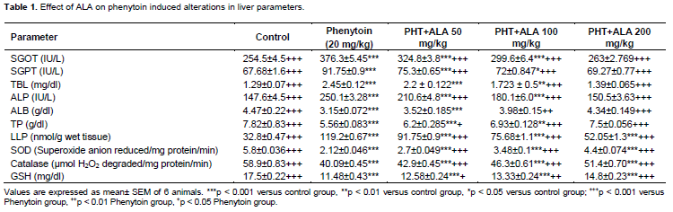
Administration of phenytoin 20 mg/kg for a period of 45 days significantly increased the levels of SGOT, SGPT, total bilirubin and ALP along with a significant decreasein the levels of albumin and total protein. ALA (50 and 100 mg/kg) significantly (p < 0.001) decreased the elevated SGOT levels when compared with phenytoin treated animals, but the values did not reach that of normal. ALA at its higher dose (200 mg/kg) dropped off the levels of SGOT near to that of normal control animals. ALA (50 and 100 mg/kg) significantly (p < 0.001) decreased the levels of SGPT when compared with phenytoin treated animals, but the values did not reach that of the normal. ALA (200 mg/kg) decreased the levels of SGPT near to that of normal control. ALA at 50 mg/kg showed no significant decrease in the levels of total bilirubin elevated by phenytoin, whereas at 100 mg/kg ALA significantly (p < 0.001) reduced the levels of total bilirubin and at 200 mg/kg the values were brought near that of normal values. ALA at the dose of 50 and 100 mg/kg significantly (p < 0.001) brought down the levels of ALP but not closer to the normal values, whereas the antioxidant at its higher dose (200 mg/kg) decreased the levels of ALP near to that of normal control. ALA at the dose of 50 mg/kg showed no significant increase in the levels of albumin, whereas at 100 and 200 mg/kg ALA augmented the levels of albumin in a dose dependent fashion and at the dose of 200 mg/kg, a significant (p < 0.001) increase in the levels of albumin near to that of normal control was observed. ALA at the dose of 50 mg/kg slightly increased the levels of total protein (p < 0.05), whereas at 100 and 200 mg/kg, significantly (p < 0.001) augmented the levels of total protein (Table 1).
Effect of ALA on phenytoin enhanced liver lipid peroxidation
Administration of phenytoin 20 mg/kg for a period of 45 days significantly increased the lipid peroxide contents in liver. ALA at all the three doses (50, 100 and 200 mg/kg) significantly (p < 0.001) reduced the liver lipid peroxidation in a dose dependent manner but the values did not reach the normal (Table 1).
Effect of ALA on phenytoin depleted endogenous enzymatic and non enzymatic antioxidants
Administration of phenytoin 20 mg/kg for a period of 45 days significantly decreased the endoge-nous enzymatic antioxidants such as SOD as well as catalase and non enzymatic antioxidant GSH in liver. ALA at all the three doses (50, 100 and 200 mg/kg) significantly increased the endoge-nous antioxidant levels decreased by phenytoin in a dose dependent manner but the values did not reach the normal (Table 1).
Effect of antioxidants on phenytoin induced alterations in body weight, absolute and relative liver weight
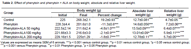
At the end of 45 days of treatment with phenytoin, there was a statistically significant decrease in body weight and an increase in the absolute and relative liver weights when compared with thecontrol group. ALA at the dose of 50 mg/kg showed no significant difference in body weight or absolute and relative liver weight. ALA at its higher doses (100 and 200 mg/kg) reversed the phenytoin induced weight loss and decreased the absolute and relative liver weights significantly when compared with phenytoin group (Table 2).
Effect of ALA on phenytoin induced alterations in liver histopathology
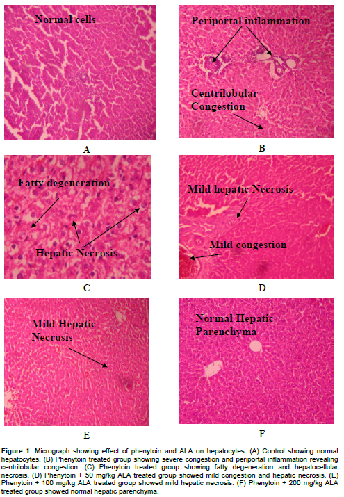
On histopathological examination, the livers of the control group revealed normal hepatic architecture (Figure 1A). Figure 1B and C represented the phenytoin group and showed severe congestion, periportal inflammation revealing centrilobular congestion, fatty degeneration and hepatocellular necrosis. Phenytoin + ALA (50 mg/kg) treated group showed mild hepatic necrosis and mild congestion in liver (Figure 1D), ALA (100 mg/kg) showed mild hepatic necrosis (Figure 1E). Thus ALA (50 and 100 mg/kg) decreased the extent of hepatic damage induced by phenytoin. ALA (200 mg/kg) treated group showed normal hepatic parenchyma (Figure 1F).
DISCUSSION
AAED induced hepatotoxicity correlated to the accumulation of arene oxides metabolites of phenytoin which are reported to be involved in the pathogenesis of hepatotoxicity (Bavdekar et al., 2004). Santos et al. (2008) elucidated the mechanism of phenytoin induced hepatic damage and revealed that oxidative stress to be one of the potential mechanisms responsible for phenytoin associated hepatotoxicity.
Oxidative stress induced by the metabolites of phenytoin have been suggested to be formed after its biotransformation both in humans and in rats (Bavdekar et al., 2004; Shear and Spielberg, 1988; Roy and Snodgrass, 1988; George and Farrell, 1994; Madden et al., 1996; Kalapos, 2002; Zaccara et al., 2007). Santos et al. (2008) demonstrated AAED induced depletion of the mitochondrial antioxidant defense in rat liver. These findings might explain the potential role of mitochondrial toxicity and oxidative stress in the hepatotoxicity in associated with AAED therapy. Also, in the present study, phenytoin increased lipid peroxidation and depleted the endogenous antioxidants such as SOD, catalase and GSH in liver revealing massive oxidative stress in liver.
SGOT, SGPT, ALP and bilirubin are markers used to assess hepatic damage (Sallie et al., 1991; Ncibi et al., 2008; Gokcimen et al., 2007; Eraslan et al., 2009). A low serum albumin indicates poor liver function, reductions in albumin levels shows the presence of underlying liver disease (Kalender et al., 2010). In this investigation phenytoin treated rats showed a significant increase in the levels of SGOT, SGPT, bilirubin and ALP and decrease in the levels of albumin and total protein which indicates the hepatotoxic nature of the drug phenytoin. Phenytoin was observed to alter protein and free amino acid metabolism and their synthesis in the liver. The body weight of phenytoin treated rats was decreased whereas the relative liver weight was increased.
Phenytoin exhibited periportal inflammation, hemorrhage, sinusoidal congestion and hepatic necrosis in rat liver which was revealed by histopathological investigation. These changes were online with the changes in various biochemical parameters investigated and liver damage was considered to arise from the toxic effects of phenytoin mediated via oxidative stress.
ALA was used to treat liver poisoning induced by alcohol, mushroom and heavy metals. The antioxidant abilities of ALA and its role in glutathione recycling have encouraged its use in liver damage. ALA (100 mg/kg/day) was reported to exhibit a signi?cant hepatoprotective against chloroquine induced hepatotoxicity. It was also observed that ALA had a better protective effect than silymarin, a reference drug (Pari and Murugavel, 2004). Hesham (2007) elucidated the effects of ALA against tamoxifen (TAM) induced liver damage, oxidative stress and DNA fragmentation. ALA was described to scavenge free radicals, prevent DNA fragmentation, reduce liver injury and protect oxidative stress induced by TAM intoxication. The study suggested the use of ALA in the prophylactic treatment of TAM induced liver injury than its use as curative agent (post-TAM administration) (Hesham, 2007). The effects of ALA and its reduced form dihydrolipoic acid (DHLA) was studied by Foo et al. (2011) against thioacetamide (TAA) induced liver fibrosis in rats and the possible underlying mechanisms in hepatic stellate cells in vitro. It was found that co- administration of ALA to rats chronically treated with TAA inhibited the development of liver cirrhosis, as indicated by reductions in cirrhosis incidence, hepatic fibrosis and AST, ALT activities. ALA exhibited beneficial role in the treatment of chronic liver diseases caused by ongoing hepatic damage (Foo et al., 2011). Liu et al. (2010) explored the effect of ALA (10 mg/kg/day) and vitamin C (25 mg/kg/day) on arsenic (50 mg/L water) induced oxidative stress. It was observed that the combination of both the antioxidants significantly decreased the TBARS level of the brain and liver and thereby attenuated oxidative stress, restored the δ-ALAD activity against arsenite induced toxicity (Liu et al., 2010). Investigation of influence of ALA treatment in malathion (100 mg/kg) induced toxicity revealed that the pretreatment with ALA significantly attenuated the physiological and histopathological alterations induced by malathion (Al-Attar, 2010). ALA was reported to exhibit protective effect against combination of Isoniazid and Rifampicin (INH-RIF) induced hepatotoxicity (Saad et al., 2010).
In the present study, supplementation with ALA (200 mg/kg) decreased the markers of hepatotoxicity such as SGOT, SGPT and bilirubin which were elevated by phenytoin. ALA supplementation also restored the levels of albumin and total protein decreased by phenytoin. In addition, ALA restored the total body weight of the rats and decreased the relative liver weight against phenytoin induced alterations. ALA also has improved the hepatic histopathological damages induced by phenytoin. ALA at the dose of 200 mg/kg exerted significant protection against phenytoin induced toxicity by its ability to ameliorate the lipid peroxidation and thus oxidative stress through its free radical scavenging activity, which improved the levels of antioxidant defense system.
CONCLUSION
The results of the present investigation revealed the protective effect of ALA against phenytoin induced oxidative stress and hepatotoxicity. ALA also reversed the histopathological damages induced by phenytoin in liver. ALA at a dose of 100 and 200 mg/kg was effective in reducing the oxidative stress and hepatic damage. The enzyme inducing property of phenytoin might possibly explain the relative inefficiency of ALA at 50 mg/kg. This investigation reports the beneficial ALA on phenytoin induced hepatotoxicity mediated via oxidative stress.
CONFLICT OF INTEREST
There is no conflict of interest as regard this study.
REFERENCES
|
Al-Attar AM (2010). Physiological and histopathological investigations on the effects of ALA in rats exposed to malathion. J. Biomed. Biotechnol. 1-8. crossref |
||||
|
Aldenhovel HG (1988). The influence of long-term anticonvulsant therapy with diphenylhydantoin and carbamazepine on serum gamma glutamyl transferase, aspartate aminotransferase, alanine aminotransferase and ALP. Eur. Arch. Psychiatry Neurol. Sci. 237(5):312-316. crossref |
||||
|
Bajpai M, Roskos LK, Shen DD, Levy RH (1996). Roles of cytochrome P4502C9 and cytochrome P4502C19 in the stereoselective metabolism of Phenytoin to its major metabolite. Drug Metab. Dispos. 24(12):1401-1403. Pubmed |
||||
|
Bavdekar SB, Muranjan MN, Gogtay NJ, Kantharia V, Kshirsagar NA (2004). Anticonvulsant hypersensitivity syndrome: lymphocyte toxicity assay for the confirmation of diagnosis and risk assessment. Ann. Pharmacother. 38(10):1648-1650. crossref |
||||
| Beer RF, Sizer IW (1952). A spectrophotometric method for measuring the breakdown hydrogen peroxide by catalase. J. Biol. Chem. 195(1):133-140. | ||||
|
Björnsson E (2008). Hepatotoxicity associated with antiepileptic drugs. Acta Neurol. Scand. 118(5):281-290. crossref |
||||
|
Chatterjee M, Sil PC (2006). Hepatoprotective effect of aqueous extract of Phyllanthus niruri on nimesulide induced oxidative stress in vivo. Indian J. Biochem. Biophys. 43(5):299-305. Pubmed |
||||
|
Doumasa BT, Watson WA, Biggs HG (1971). Albumin standards and the measurement of serum albumin with bromocresol green. Clin. Chim. Acta 31(1):87-96. crossref |
||||
|
Dreifuss FE, Langer DH (1987). Hepatic considerations in the use of antiepileptic drugs. Epilepsia 28(2):S23-S29. crossref |
||||
|
Eraslan G, Kanbur M, Silici S (2009). Effect of carbaryl on some biochemical changes in rats: the ameliorative effect of bee pollen. Food Chem. Toxicol. 47(1):86-91. crossref |
||||
|
Foo NP, Lin SH, Lee YH, Wu MJ, Wang YJ (2011). α-Lipoic acid inhibits liver fibrosis through the attenuation of ROS-triggered signaling in hepatic stellate cells activated by PDGF and TGF-β. Toxicology 282(1-2):39-46. crossref |
||||
|
Gella FJ, Olivella T, Cruz Pastor M, Arenas J, Moreno R, Dueban R, Gomez JA (1985). A simple procedure for routine determination of aspartate aminotrasferase and alanine aminotransferase with pyridoxal phosphate. Clin. Chim. Acta 153(3):241-247. crossref |
||||
| George J, Farrell GC (1994). Anticonvulsivant hepatotoxicity. In: Farrell GC (ed.), Drug-induced Liver Disease. Churchill Livingstone, Edinburgh, London. pp. 143-144. | ||||
|
Gokcimen A, Gulle K, Demirin H, Bayram D, Kocak A, Altuntas I (2007). Effects of diazinon at different doses on rat liver and pancreas tissues. Pestic. Biochem. Physiol. 87(2):103-108. crossref |
||||
| Gomall AG, Bardawill CJ, David MM (1949). Determination of serum proteins by means of biuret reaction. J. Biol. Chem. 177(2):751-766. | ||||
|
Harden CL (2000). Therapeutic safety monitoring: what to look for and when to look for it. Epilepsia 41(8):S37-S44. crossref |
||||
| Hesham AEB (2007). Lipoic acid attenuates DNA fragmentation, oxidative stress and liver injury induced by tamoxifen in rats. Asian J. Tradit. Med. 2:175-188. | ||||
|
Kalapos MP (2002). Carbamazepine provoked hepatotoxicity and possible etiopathological role of glutathione in the events. Retrospective review of old data and call for new investigation. Adv. Drug React. Toxicol. Rev. 21(3):123-141. crossref |
||||
|
Kass GE (2006). Mitochondrial involvement in drug-induced hepatic injury. Chem. Biol. Interact. 163(1), 145-159. crossref |
||||
|
Kazamatsuri H (1970). Elevated serum ALP levels in epilepsy during diphenylhydantoin therapy. N. Engl. J. Med. 283(25):1411-1412. crossref |
||||
|
Li Y, Powers C, Jiang N, Chopp M (1998). Intact, injured, necrotic and apoptotic cells after focal cerebral ischemia in the rat. J. Neurol. Sci. 156(2):119-132. crossref |
||||
|
Liu CB, Feng YH, Ye GH, Xiao M (2010). Effects of α-lipoic acid and vitamin C on oxidative stress in rat exposed to chronic arsenic toxicity. Zhonghua Lao Dong Wei Sheng Zhi Ye Bing Za Zhi 28(12):891-894. Pubmed |
||||
|
Lowry OH, Rosebrough NJ, Farr AL, Randall RJ (1951). Protein measurement with the Folin phenol reagent. J. Biol. Chem. 193(1):265-75. Pubmed |
||||
|
Madden S, Maggs JL, Park BK, (1996). Bioactivation of carbamazepine in the rat invivo. Evidence for the formation of reactive arene oxide(s). Drug Metab. Dispos. 24(4):469-479. Pubmed |
||||
|
Marklund S, Marklund G (1974). Involvement of the superoxide anion radical in the auto oxidation of pyragallol and a convenient assay for superoxide dismutase. Eur. J. Biochem. 47(3):469-474. crossref |
||||
|
Ncibi S, Othman MB, Akacha A, Krifi MN, Zourgui L (2008). Opuntia ficus indica extract protects against chlorpyrifos-induced damage on mice liver. Food Chem. Toxicol. 46(2):797-802. crossref |
||||
|
Packer L, Witt EH, Tritschler HJ (1995). Alpha-lipoic acid as a biological antioxidant. Free Radic. Biol. Med. 19(2):227-250. crossref |
||||
|
Pari L, Murugavel P (2004). Protective effect of alpha lipoic acid against chloroquine induced hepatotoxicity in rats. J. Appl. Toxicol. 24(1):21-26. crossref |
||||
|
Pearlman FC, Lee RTY (1974). Detection and measurement of total bilirubin in serum with use of surfactants as solubilizing agents. Clin. Chem. 20(4):447-453. Pubmed |
||||
|
Rosalki SB, Foo AY, Burlina A, Prellwitz W, Strieber P, Neumeier D, Klein G, Poppe WA, Bodenmuller H (1993). Multicentre evaluation of iso ALP test kit for measurement of bone alkaline phosphatase ativity in serum and plasma. Clin. Chem. 39(4):648-652. Pubmed |
||||
|
Roy D, Snodgrass WR (1988). Phenytoin metabolic activation: role of cytochrome P450, glutathione, age, and sex in rats and mice. Res. Commun. Chem. Pathol. Pharmacol. 59:173-190. Pubmed |
||||
|
Saad EI, El-Gowilly SM, Sherhaa MO, Bistawroos AE (2010). Role of oxidative stress and nitric oxide in the protective effects of alpha-lipoic acid and aminoguanidine against isoniazid rifampicin induced hepatotoxicity in rats. Food Chem. Toxicol. 48(7):1869-1875. crossref |
||||
|
Sallie R, Tredger JM, Willam R (1991). Drugs and the Liver. Biopharm. Drug Dispos. 12:251-259. crossref |
||||
|
Santos NA, Medina WS, Martins NM, Rodrigues MA, Curti C, Santos AC (2008). Involvement of oxidative stress in the hepatotoxicity induced by aromatic antiepileptic drugs. Toxicol. Vitro 22(8):1820-1824. crossref |
||||
| Saraswathy GR, Maheswari E, Santhrani T (2010). Effect of Vitamin C supplementation on phenytoin induced hepatotoxicity. Glob. J. Pharmacol. 4(3):127-135. | ||||
|
Sedlak J, Lindsay RH (1968). Estimation of total, protein bound & non-protein SH groups in tissue with Ellman's reagent. Anal. Biochem. 25(1):192-205. crossref |
||||
|
Shear NH, Spielberg SP (1988). Anticonvulsant hypersensitivity syndrome. In vitro assessment of risk. J. Clin. Investig. 82(6):1826-1832. crossref |
||||
|
Smythe MA, Umstead GS (1989). Phenytoin hepatotoxicity: a review of the literature. DICP 23(1):13-18. Pubmed |
||||
|
Kalender S, Gokce Uzun F, Durak D, Demir F, Kalender Y (2010). Malathion-induced hepatotoxicity in rats: The effects of vitamins C and E. Food Chem. Toxicol. 48(2):633-638. crossref |
||||
|
Walia KS, Khan EA, Ko DH, Raza SS, Khan YN (2004). Side effects of antiepileptics - a review. Pain Pract. 4(3):194-203. crossref |
||||
|
Walker M (2005). Status epilepticus: an evidence based guide. BMJ 331(7518):673–677. crossref |
||||
|
Zaccara G, Franciotta D, Perucca E (2007). Idiosyncratic adverse reactions to antiepileptic drugs. Epilepsia 48(7):1223-1244. crossref |
||||
Copyright © 2024 Author(s) retain the copyright of this article.
This article is published under the terms of the Creative Commons Attribution License 4.0


