Full Length Research Paper
ABSTRACT
The use of medicinal plants in traditional medicine is common practice in developing countries. However, such unregulated and unreasonable use may pose a risk in short or long-term toxicity to humans. The aim of this study was to investigate the phytochemical and toxicological characteristics of Annona senegalensis and Trichilia prieureana two plants from Benin, which are traditionally used for the treatment of colds, chest pain, fever, stomach problems, diabetes, malaria, bechias, venereal diseases and colic. The acute toxicity of ethanolic extracts of A. senegalensis and T. prieureana at a single dose of 5000 mg/kg according to OECD 423 protocol was studied. Biochemical and hematological examinations, as well as histological studies of the liver and kidneys were performed. Phytochemical screening of these plants revealed the presence of secondary metabolites, including coumarins, lignans, sterols and triterpenes, anthracene derivatives, tannins, flavonoids and alkaloids. In contrast, saponins and naphthoquinones were not detected in the T. prieureana extract. Acute toxicity data indicated that the liver and kidneys of treated animals were not affected and did not undergo structural nor functional changes. These observations demonstrate the safety of these plants and justify their use in traditional medicine to treat many diseases.
Key words: Annona senegalensis, Trichilia prieureana, acute oral toxicity.
INTRODUCTION
Herbs have existed since ancient times, through many civilizations. In Africa, herbal medicine is deeply rooted in the culture of the people and plays an important role in traditional medicine (Ali et al., 2008). Around the world, the use of medicinal plants has seen an unprecedented surge in interest since the 1980s (Lompo, 1998). The World Health Organization (WHO) estimates that traditional medicine, particularly herbal medicine, covers the health needs of 80% of the population in developing countries (WHO, 2002). In Benin, there is a high demand for herbal medicine among the rural population, especially far from medical centers; moreover, this use of plants is still partly based on the idea that plants are a natural remedy without any risk. However, plants are both useful and toxic (Sambo et al. 2005).
Toxicity caused by the use of medicinal plants is a medical and socio-economic problem; herbal preparations that are considered safe may contain contaminants such as micro- organisms that cause disease because of the way they are prepared.
Among these medicinal plants, our research focuses on the plant species Annona senegalensis and Trichilia prieureana from tropical Africa. They are widely used by traditional healers to treat a variety of ailments including: Colds, chest pain, fever, stomach problems, diabetes, malaria, bechias, venereal diseases and colic (Abou-Arab et al, 2000). The aim of this study is to evaluate the acute toxicity of ethanolic extracts of A. senegalensis and T. prieureana by evaluating level of some markers of renal and hepatic functions of hematological parameters in rats and to promote the understanding of certain plants with toxic effects, particularly those used in traditional medicine, and to raise awareness among the population of the risks of poisoning and possible abuses in the uncontrolled use of plants.
MATERIALS AND METHODS
Study material
Plant and extracts preparation
The plant material used consisted of A. senegalensis leaves and T. prieureana leaves. Plant organs were collected in July 2018 in the commune of Tori-Bossito (Atlantic, Southern Benin), Longitude: 2.145, Latitude: 6.50306 and certified at the Benin National Herbarium with voucher numbers YH237/HNB for A. senegalensis and YH238/HNB for T. prieureana.
Herbalists collected these leaf samples at well-defined times for use in the Pharmacopoeia.
Animal
Animals used in this experiment were male albino rats (Rattus norvegicus) of Wistar strain with an average weight of 150 g. They were acclimatized under normal conditions in the animal house of the Laboratory of Physiopathology / Molecular Pharmacology and Toxicology, Faculty of Science and Technology of the University of Abomey-Calavi. These rats had free access to food and water. The standard rations were in the form of pellets provided by the group Véto service SA (GVS - Benin) in charge of animal nutrition and were composed as follows: 16% protein, 60% ENA (non-nitrogenous extracts, that is, carbohydrates), 3% lipids, 5% minerals and vitamins, 12% moisture. The caloric intake of carbohydrate is 2900 kcal/kg.
Ethical approval
This research is part of a thesis. This study has been authorized by the Committee of the Doctoral School of Life and Earth Sciences (ED-SVT) of the University of Abomey-Calavi (UAC/Benin) under the number 168702.
Phytochemical analysis
Phytochemical analyses were performed on both plants. Phytochemical screening of plants for secondary metabolites was performed according to the methods described by Wagner and Blat (2001) and Bruneton et al. (2009). The chemical groups assayed were total polyphenols, tannins, coumarins, saponins, triterpenes, anthracene derivatives, steroids, flavonoids and alkaloids.
Acute toxicity test
The acute oral toxicity study of ethanolic extracts of A. senegalensis and T. prieureana was evaluated according to the Organisation for Economic Co-operation and Development (OECD) guideline 423 in Wistar strain albino rats (Rattus norvegicus), where the limit test dose of 5000 mg/kg was used.
The animals were deprived of food but not water overnight prior to the test. After fasting, the animals were weighed and the extracts were administered orally by gavage through an esophageal tube at 5000 mg/kg body weight. The amounts of extract administered were regularly adjusted to remain constant in relation to the weight of the animal. The control group received distilled water. After administration, the animals were again deprived of food for 3 to 4 h. The animals were observed individually at least once during the first 30 min and at least twice during the first 24 h after treatment. Close attention was paid to them daily for 14 days after treatment. All observations were systematically recorded. Particular attention was paid on various manifestations of tremors, convulsions, salivation, diarrhea, lethargy, sleep and coma. At the end of the treatment, the rats were deprived of food the last night before sampling. Blood samples (day 1 and day 14) were collected from all animals by retroorbital sinus puncture under ether anesthesia. The blood sample was collected in two types of tubes, one containing EDTA and the other without anticoagulant (dry tube). The samples in the EDTA tubes were used for hematological analysis. The dry tubes were centrifuged at 4000 min for 10 min, and the resulting serum was stored at -20°C for analysis of biochemical parameters. After collection, two animals per batch were randomly selected and sacrificed under ether anesthesia for removal of organs such as liver and kidney to see if the extracts caused damage to kidney and liver tissue (histological studies). These organs were flushed with 0.9% saline and fixed in 10% buffered formalin.
Body weights
The individual body weight of each animal was determined shortly before test substance administration, 24 h after administration, 7 days after administration, and at day 14.
Biochemical and hematological examinations
Biochemical and hematological examinations were performed in the Pathophysiology/Molecular Pharmacology and Toxicology Laboratory. Biochemical tests included aspartate aminotransferase (AST), alanine aminotransferase (ALT), urea, creatinine, total bilirubin. Hematological examinations included red and white blood cell count, hemoglobin level, hematocrit, mean corpuscular volume (MCV), mean corpuscular hemoglobin content (MCH), and mean corpuscular hemoglobin concentration (MCHC) determination.
Histopathological observations
Histological sections of the liver and kidneys were performed at the Histopathology Laboratory of the Institute of Applied Biomedical Sciences (ISBA) of the University of Abomey-Calavi. The pathomorphological study consisted of hematoxylin-eosin staining of 5 μm-thick thin slides. This is a routine staining after which the nuclei, stained by hematoxylin, appear dark blue and the cytoplasm, stained by eosin, appears pink. Microscopic observation of these sections was performed with the ZEISS camera microscope at different magnifications in order to select only the most representative photographs.
Data analysis
Data collected from biochemical and hematological analyses were expressed as mean ± SEM (n = 6). Statistical differences between the treated and control groups were determined by analysis of variance (ANOVA) followed by the Fisher LSD test using Systat 10.0. Differences between groups were considered significant at P <0.05.
RESULTS
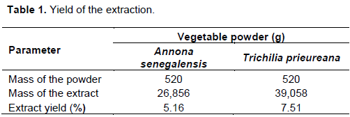
Yield of the extraction
The extraction yield obtained for ethanolic extraction for the studied plants is presented in Table 1. The extraction results show that T. prieureana has a better yield than A. senegalensis.
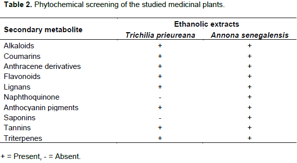
Phytochemical screening
Table 2 shows that the secondary metabolite content of plant extracts varies. Ethanolic extracts of A. senegalensis and T. prieureana show potent secondary metabolites including coumarins, lignans, sterols and triterpenes, anthracene derivatives, tannins, flavonoids and alkaloids. On the other hand, the presence of saponins and naphthoquinones has not been demonstrated in T. prieureana extract.
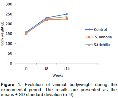
Evolution of animal weight
The graph below shows the evolution of the weight of the control animals as well as the treated ones as a function of time under the influence of the extracts. From the graph, we observe an increase in the weight of the animals from the 1st to the 8th day. The weight of animals treated with A. senegalensis is lower than those treated with T. prieureana. The weight of the control animals is higher than the treated ones. From the 8th to the 14th day, the weight of the control rats continues to increase while the treated ones begin to stabilize (Figure 1).
Acute toxicity
LD50 of the studied plant extracts
At the dose tested (5000 mg/kg), animals from different batches did not die. Again, no obvious signs of toxicity were observed. Thus, the LD50 of the studied plants is higher than 5000 mg/kg.
Effect of the studied plants extracts on biochemical parameters
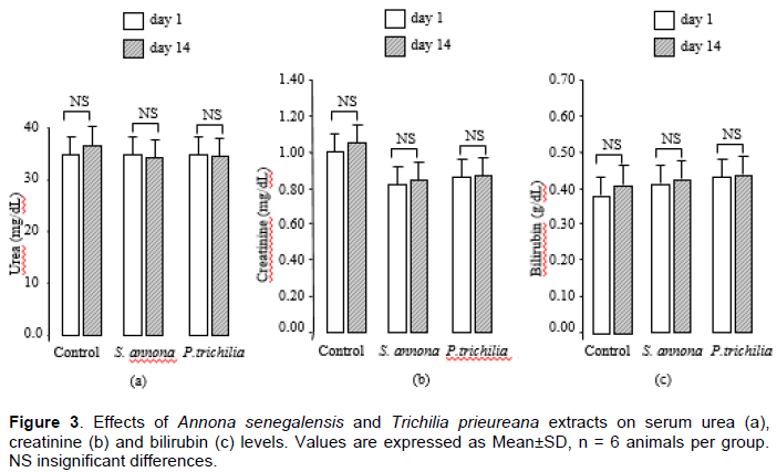
Aspartate aminotransferase (AST) and alanine aminotransferase (ALT) were considered for the exploration of liver function (Figure 2). There was no significant difference in mean between the treated and control groups (P>0.05). The renal parameters assessed for renal function were uremia and creatinine. Figure 3a and b shows the evolution of the serum urea, creatinine level between day 1 and day 14 in the different batches. In this study, treatment with the different plant extracts did not cause significant effects on these parameters P>0.05).

Effect of the studied plant extracts on hematological parameters
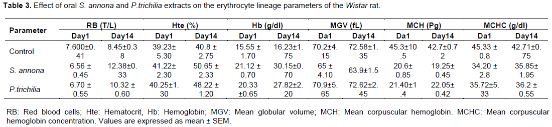
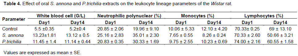
The results of the hematological tests are summarized in Tables 3 and 4. The mean red blood cells count of rats treated with the plants extracts had a significantly (P<0.05) higher red blood cells count and Day 14 compared to the untreated control group and Day 1 groups. The mean hematocrit of rats treated with the plants extracts had a significantly (P<0.05) higher hematocrit count and Day 14 compared to the untreated control group and Day 1 groups. The mean hemoglobin a significantly (P<0.05) higher concentration of rats treated with the plants extracts and Day 14 compared to the untreated control group and Day 1 groups. No significant changes were observed in levels of mean corpuscular hemoglobin (MCH), mean corpuscular hemoglobin concentration (MCHC), and mean globular volume (MGV) of variation among the groups.No significant changes were observed in the total white blood cell (WBC), however, neutrophils were significantly (P<0.05) elevated in the rats treated with the plants extracts and Day 14 compared to the untreated control group and Day 1 groups, while the lymphocyte count was significantly (P<0.05) lower in the rats treated with the plants extracts and Day 14 compared to the untreated control group and Day 1 groups.
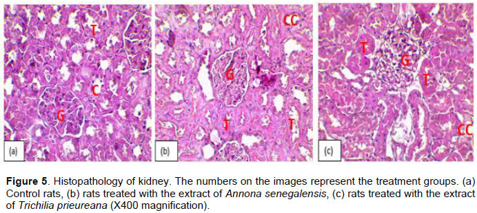
Histopathologic evaluations of the liver and kidneys
Histological examination of rat liver sections treated with 5000 mg/kg body weight extract showed normal architecture, normal appearance of central veins, and hepatic sinusoids lined with similar endothelial and Kupffer cells (Figure 4).
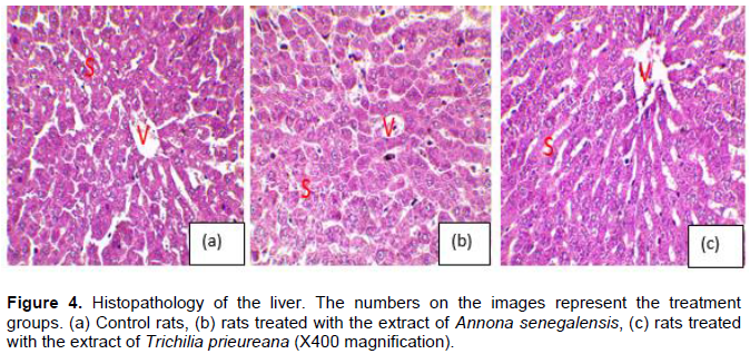
As can be seen in this figure, the liver parenchyma of treated rats has the same appearance as control rats. The hepatocytes (arrows) are normal in appearance, arranged in cords separated by sinusoidal waves. These sinusoids line up around the centrilobular vein (v). Tissue sections were made to confirm the biochemical data.
Figure 5 shows the renal histology of treated and control animals. Analysis of the data in this figure showed that the renal parenchyma of treated rats had a typical structure of control rats. The glomerulus (G), distal duct (T), and collecting duct (CC) were clearly discernible.Therefore, the extract did not affect the renal structure in any way.
DISCUSSION
The acute toxicity tests provide preliminary information on the toxic level of the extracts for which no other toxicological information was available according to the OECD recommended guidance document for acute oral toxicity tests based on oral LD50 values. Such information can be used to: (i) deal with cases of accidental ingestion of a large amount of the material. (ii) determine possible target organs that should be scrutinized and/or special tests that should be conducted in repeated-dose toxicity tests. (iii) select doses for short-term and sub-chronic toxicity tests when no other toxicology information is available (Gad and Chengelis, 1988). Furthermore, the majority of pharmaceutical companies only use acute toxicity studies to determine the minimum lethal or maximum non-lethal dose.
Such toxicity studies assess the hazard, namely the basic toxicity of the substance, and the risk is determined by considering the probability of exposure to a particular hazard at certain levels (Klaassen and Eaton, 1991).
In the present study, sufficient information was obtained on the acute toxicity of ethanolic extracts of A. senegalensis and T. prieureana from Benin flora, according to OECD guideline 423, for the traditional treatment of a variety of diseases to be classified as non-toxic and safe, as evidenced by its high LD50 > 5000 mg/kg body weight. Ethanolic extracts of A. senegalensis and T. prieureana content secondary metabolites including coumarins, lignans, sterols and triterpenes, anthracene derivatives, tannins, flavonoids and alkaloids. On the other hand, the presence of saponins and naphthoquinones has not been demonstrated in the T. prieureana extract. It must be recognized that different extraction methods from the same herb or plant part will yield end products with different chemical compositions (Williamson et al., 1996). The presence of tannin, as well as other identified bioactive molecules, is suggestive of the medicinal properties of the plant studied and its therapeutic use in several pharmacopoeias.
Flavonoids have hypoglycemic properties as they improve glycemic and oxidative metabolism in altered diabetic status. They also stimulate insulin secretion by modifying the Ca2++ concentration. (Hii et al., 1985). The liver is the main site of biotransformation and detoxification of xenobiotics (Gueguen et al., 2006). Therefore, the liver is particularly vulnerable to exogenous substances (Lee et al., 1993; Sturgill and Lambert, 1997). Similarly, the kidneys, the major organ of excretion of xenobiotics and their metabolites, are particularly sensitive to toxic effects (Gueguen et al., 2006). Numerous studies have confirmed that elevated serum levels of liver enzymes, ALT and AST transaminase are not directly related to liver injury, but elevated levels can lead to inflammation, cell leakage, and liver damage (Kausar et al., 2010). Therefore, the increase in liver enzymes ALT and AST after administration of ethanolic plant extracts may be due to the fact that some phytochemicals may be toxic to the liver and cause liver damage at increasing doses. Necrosis from hepatotoxic chemicals can occur within distinct zones in the liver, either distributed diffusely, or occur massively. Many chemicals produce zonal necrosis, that is, necrosis confined to a specific zone of the hepatic acinus (Roberts et al., 2003). However, the remarkable ability of the liver to regenerate itself makes it able to withstand moderate zonal or diffuse necrosis. Over a period of several days, necrotic cells are removed an replaced with new cells; and normal hepatic architecture and function are restored (Roberts et al., 2003).
In this study, no significant effect on the activity of these enzymes was found. This was confirmed by histopathological studies, which showed that no structural changes in the liver were observed. The renal parameters assessed were uremia and creatinine. Any increase in the levels of these markers indicates possible renal tissue damage (Yuliandra et al., 2015). In this study, it should be noted that treatment with the different plant extracts did not cause significant effects. These data, reflecting no functional changes in the kidneys, are supported by histological studies showing no structural changes in the organ. The hematopoietic system of animals is an important indicator reflecting physiological and pathological states in animals and humans (Redondo et al., 2014), especially in the bone marrow where the production of red blood cell occurs (Kifayatullah et al., 2015).
An increase in red blood cell count was observed in experimental rats treated with the plant extract. Erythrocyte parameters were increased in the test group compared to the untreated control group, implying that treatment with the plant extract resulted in increased erythropoiesis (Idoh et al., 2016).
The effects of A. senegalensis and T. prieureana on hematological parameters in rats was investigated in this study. The extracts caused an increase in the erythrocyte count. This was confirmed by the increased hematocrit and percentage hemoglobin in the test group. In normal circumstances, local tissue anoxia apparently leads to the formation of a glycoprotein called erythropoietin, which stimulates increased production of erythrocytes (Bowman et al., 1980) It is very likely that these plant extracts contains erythropoietin-like agent(s) which is/are responsible for the increased production of erythrocytes.
The total WBC (leucocyte) counts were not significantly altered following extracts administration. However, examination of the differential counts revealed that ethanolic extracts of A. senegalensis and T. prieureana at a single dose of 5000 mg/kg led to reduction in lymphocyte count but increased the neutrophil count in the rats; thus, the total WBC count remained largely unaltered. The reduction in lymphocyte count could probably be due to cell margination rather than destruction. It is also possible that the extracts contains agents that stimulate the bone marrow to produce neutrophils and release them into the blood. Neutrophils are the major granulocytes to be activated when the body is invaded by bacteria and they provide the first line of defense against invading microorganisms (Ganong et al., 2005). The granules of the neutrophil contains many enzymes, which makes it a powerful and effective killer machine and, hence, deficiency of neutrophils in the body leads to myriad defects, including conditions such as chronic granulomatous disease. To summarize, from this study we conclude that oral administration of Annona senegalensis and Trichilia prieureana at a single dose of 5000 mg/kg increases red blood cell count, hemoglobin level, and the proportion of neutrophils.
CONCLUSION
The results of this safety evaluation of A. senegalensis and T. prieureana showed that the tested extracts of these plants at dose 5000 mg/kg b.w/day had no serious adverse effect on body growth, hematological and biochemical parameters as well as on gross and histopathological appearance of liver and kidneys in rat. Moreover, higher dose 5000 mg/kg b.w/day of the extracts of A. senegalensis and T. prieureana The extracts of A. senegalensis and T. prieureana having a potential to increase the hematological indices that are red blood cells, hematocrit, hemoglobin count up on prolonged administration, it, however, may induce irritation of liver tissues as a side effect. Further investigation on other vital organs and nonrodent species, including human is recommended since mild hepatotoxicity was observed on prolonged administration. Nevertheless, absence of any morbidity as well as any significant sign of toxicity even at high dose up to 5000 mg/kg b.w appears to support the general safety of A. senegalensis and T. prieureana ethenolic extracts for treatment of type 2 diabetes in Benin and may contribute towards the development of new antidiabétique drug.
CONFLICT OF INTERESTS
The authors have declared no conflicts of interest.
REFERENCES
|
Abou-Arab AA, Abou Donia MA (2000). Heavy metals in Egyptian spices of medicinal plants and the effect of processing on their levels. Journal Agricultural Food Chemical 48(6) :2300-4000. |
|
|
Ahmad M, Lim CP, Akowuah GA (2013). Safety assessment of standardised methanol extract of Cinnamomum burmannii. Phytomedicine 20 (12):1124-1130. |
|
|
Ali BH, Blunden G, Tanira MO, Nemmar A (2008). Some phytochemical, pharmacological and toxicological properties of ginger (Zingiber officinale Roscoe): A review of recent research. Food and Chemical Toxical 46:409-420. |
|
|
Bowman WC, Rand MT(1980). Drugs affecting coagulation, fibrinolysis, haematopoiesis and the functioning of blood cells. Textbook of Pharmacology. Oxford: Blackwell Publishers 21(1):21-53. |
|
|
Bruneton (2009). Pharmacognosie: Phytochimie, Plantes Médicinales. Tec & Doc Lavoisier, P. 456. |
|
|
Gad SC, Chengelis CP (1988). Acute toxicity testing perspectives and horizons. The Telford Press. Caldwell, pp. 165-167. |
|
|
Ganong WF (2005). Review of Medical Physiology. 22nd ed. Singapore: McGraw Hill, pp. 515-517. |
|
|
Hii CS, Howell SL (1985). Effects of flavonoids on insulin secretion and Ca2+ handling in rat islets of Langerhans. Journal of Endocrinology 107:1-8. |
|
|
Idoh K, Agbonon A, Potchoo Y, and Gbeassor M (2016). Toxicologique assessment of the hydroethanolic leaf extract of Clerodendrum capitatum in Wistar rats. The Pan African Medical Journal 24:66. |
|
|
Kausar MW, Moeed K, Asif N, Rizwi F, Raza S (2010). Correlation of bilirubin with liver enzymes in patients of falciparum malaria. International Journal of Pathology 8(2):63-70. |
|
|
Kifayatullah M, Mustafa MS, Senguptha P, Sarker MMR, Das A, Das SK (2015). Evaluation of the acute and sub-acute toxicity of the ethanolic extract of Pericampylus glaucus (Lam.) Merr inBALB/c mice. Journal of Acute Disease 4(4):309-315. |
|
|
Klaassen CD, Eaton DL (1991). Principles of toxicology, in Casarett and Doull's Toxicology. The Basic Science of Poison (Amdur MO, Doull JD and Klaassen CD eds), pp. 32-33. |
|
|
Kneifel W, Czech E, Kopp B (2002). Microbial contamination of medicinal plants-a review. Planta Medica 68 (1):5-15. |
|
|
Lawal BAS, Aderibigbe AO, Essiet GA, Essien AD (2007). Hypotensive and Antihypertensive Effects of Aframomum melegueta Seeds in Humans. International Journal of Pharmacology 3(4):311-318. |
|
|
Lee WM (1993). Drug-induced hepatotoxicity. Alim Pharmacol Ther 7: 477-485. |
|
|
Pushpa Latha B, Rama Manohar Reddy L, Mannur Ismail S, Vijaya T (2010) Medicinal plants and their derivatives as potential source in treatment of obesity. Asian Journal of Biological Sciences 1(4):719-727. |
|
|
Roberts S, James RC, Franklin MR (2003). Hepatotoxicity: toxic effects on the liver. In: Williams PL, James RC, Roberts SM, editors. |
|
|
Principles of toxicology: environmental and industrial applications. 2nd ed. New York: John Wiley & Sons, Inc., pp. 111-28. |
|
|
Said O, Khalil K, Fulder S, Azaizeh H (2002). Ethnobotanical survey of medicinal herbs of the Middle East region. Journal of Ethnopharmacology 83:251-256. |
|
|
Smart DJ, Ahmedi KP, Harvey JS, Lynch AM (2011). Genotoxicity screening via the gH2AX by flow assay. Mutation Research 715(1-2):25-31. |
|
|
Sturgill MG, Lambert GH (1997). Xenobiotic-induced hepatotoxicity : mechanisms of Liver and methods of monitoring hepatic function. Clinical Chemistry 43:1512-1526. |
|
|
Thanaboripat D, Suvathi Y, Srilohasin P, Sripakdee S, Patthanawanitchai O, Charoensettasilp S (2007). Inhibitory effect of essential oils on the growth of Aspergillus flavus. KMITL Science and Technology Journal 7:1-7. |
|
|
Wagner H, Bladt S (2001). Plant Drug Analysis. 2nd ed. Springer. 384. |
|
|
WHO Strategies for Traditional Medicine 2002-2005.World Health Organization, Geneva, 65p. |
|
|
Williamson EM, Okpako DT, Evans FJ. (1996). Pharmacological methods in phytotherapy research. Selection, Preparation And Pharmacological Evaluation of Plant Material 1:131-154, John Wiley & Sons Ltd., Chichester, England. |
|
|
Yuliandra Y, Armenia A, Salaa AN, and Ismed F (2015). Uji toksisitas subkronis ekstrak ethanol tali putri (cassytha filiformis L.) terhadap fungsi ginjal tikus. Jurnal Sains Farmasi andKlinis 2(1):54-59. |
|
|
Sambo M H (2005). Etude du traitement traditionnel du diabète par une recette et les écorces de tronc de Manilkara multinervis Dub (Sapotaceae). Thèse De Pharmacie, Université De Bamako, P. 112. |
|
|
Lompo M, Ouedrago S, Sourabi S, Guissou IP (1998). Valorisation d'une plante médicinale anti-inflammatoire. Pharmaceuticals MM Trade Africa 10:68-79. |
|
|
Gueguen Y, Mouzat K, Ferrari L,Tissandie E, Lobaccaro JM, Batt AM, Paquet F,Voisin P, Aigueperse J, Gourmelon P, Souidi M. (2006). Cytochromes P450: xenobiotic metabolism, regulation and clinical importance. Annales de Biologie Clinique (Paris) 64:535-48 |
|
Copyright © 2024 Author(s) retain the copyright of this article.
This article is published under the terms of the Creative Commons Attribution License 4.0


