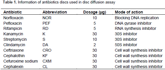
Full Length Research Paper

ABSTRACT
The current work aimed to determine the prevalence of Escherichia coli in fresh ground beef purchased from butchers' shops in Suez Governorate, Egypt, and the antibiotics susceptibility pattern of the isolated bacteria. E. coli was isolated and detected on tryptone bile glucuronide agar (TBGA) plates as chromogenic selective medium for this species. The sensitivity and resistance of the isolated bacteria to antibiotics were performed according to the National Committee for Clinical Laboratory Standards guidelines (NCCLS). A total of 299 bacterial isolates were recovered from 130 ground beef and E. coli had the highest frequency of occurrence (81.5%). The isolated enteric bacteria were identified phenotypically and genotypically as Serratia marcescens, E. coli, Enterobacter cloacae and Klebsiella pneumoniae and deposited in the GenBank nucleotide sequence database under accession numbers KU237235, KU237236, KU237237 and KU237238, respectively. Antibiotic susceptibility test showed that the four isolated species were susceptible to norfloxacin, pefloxacin, kanamycin and ceftriaxone, and resistant to clindamycin and the other tested antibiotics showed different susceptibility pattern with each tested species. Precautions and strict hygienic measures should be taken during the processing stages of ground beef in order to avoid contamination by enteric bacteria.
Key words: Contamination, Enterobacteriaceae, genotype, resistance, susceptibility, meat.
INTRODUCTION
MATERIALS AND METHODS
RESULTS AND DISCUSSION
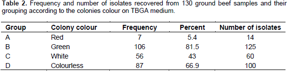
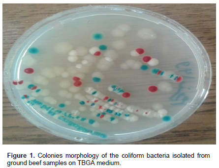
The appearance of these different coloured colonies besides the E. coli colonies might be due to different intracellular enzymes by these bacteria or different reactions between the components of the medium and intracellular metabolites. Generally, this chromogenic medium may be suitable for better detection of these four groups of isolated bacteria. Isolation and enumeration of E. coli is used as reliable indicator of fecal contamination and probability of toxigenic microorganisms’ presence in this food. Group B which represents E. coli had the highest frequency of occurrence and represented by 81.5% recovered from 106 samples out of 130 samples, while groups A, C and D is represented by 5.4, 43 and 66.9%, respectively (Table 1). The results are almost in line with that of Greeson et al. (2013) who studied the prevalence of Enterobacteriaceae in 36 samples of meat and reported that E. coli was the most frequent contaminant and its prevalence was 72.2%. E. coli is known as a fecal contamination indicator in foods due to its presence in the intestinal tract.
The gastrointestinal tract and the hands of personnel were recorded as major transferors of Klebsiella spp. and E. coli (Gundogan and Yakar, 2007). On the other hand, the current study results are in contrast with those of Mohammed et al. (2014) who recorded low frequency (15.89%) of E. coli isolated from 384 meat samples. Also, a similar study on isolation of E. coli from retailed meat was performed by Nossair et al. (2014) who isolated different members of Enterobacteriaceae from 50 samples of retailed meat collected from buffaloes and it was found that different species of bacteria were isolated at different rates, where E. coli is the highest isolated bacterium (40%) followed by K. pneumoniae. The obtained results showed that the examined ground beef of cow origin were highly contaminated with E. coli and other enteric bacteria which showed fecal contamination potential for severe hazard (Mohammed et al., 2014). The isolated bacteria were members of the intestinal flora of human and animals and many of them might lead to food deterioration and toxicities (Gundogan and Yakar, 2007; Haryani et al., 2007).
These results emphasized the role played by meat in transmission of E. coli that could constitute public hazard and food poisoning outbreaks (Reuben and Gyar, 2015; Kabiru et al., 2015). Contamination of both carcasses and the environment by E. coli from the intestinal contents of cattle during slaughter is one of the most significant risk factors in transmission to humans (Koohmaraie et al., 2005; Bosilevac et al., 2009). Moreover, Enterobacteriaceae contaminating ground beef in butchers' shops may originate from human carriers (workers) who handle and prepare the meat during cutting and grinding. Also, infected rodents that may be present in the butchers' shops or slaughterhouse could represent a neglected nsource of contamination by E. coli and other coliforms bacteria (Okonko et al., 2010). The four isolated coloured bacterial groups were characterized morphologically, biochemically and genotypically.
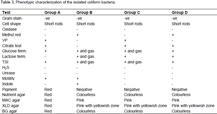
The morphological and biochemical characterization of the total recovered bacterial isolates belonging to the different four groups: A, B, C and D were performed by conventional methods. All the tested isolates belonging to the four groups were negative for Gram staining and have short rods shape. Also, the four groups were negative for oxidase and positive for glucose fermentation. On the other hand, the four groups differed in their result for lactose fermentation as the test was positive for the three groups: B, C and D, and was negative for group A. The results of the other biochemical tests showed more differences between the four bacterial groups in this investigation. Group A was characterized by red pigment production and was positive for VP, citrate and motility, whereas, was negative for methyl red, H2S, urease and indole. Group B was positive for methyl red, motility and indole but negative for VP, citrate, H2S and urease.
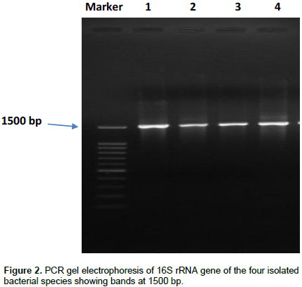
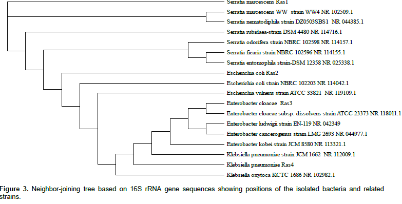
Group C was positive for VP and citrate but was negative for methyl red, H2S, urease, motility and indole. The fourth group showed positive result for each methyl red and citrate and gave result for VP, H2S, urease, motility and indole. The colonies colour differed on different culture media as presented in Table 3. Based on the morphological and biochemical characterizations, the isolated strains belonging to the four groups: A, B, C and D were related to Serratia marcescens, E. coli, Enterobacter cloacae, Klebsiella pneumoniae, respectively, according to the Bergey’s Manual of Determinative Bacteriology (Holt, 1994). The phenotypic-based identification was confirmed by genotypic identification. One isolate was selected from each of the four groups and their DNA were extracted, and a fragment of about 1500 bp from each one was amplified using the universal primers 16S F and 16S R (Figure 2). Comparison between 16S rRNA gene sequences of the tested isolates Ras1, Ras2, Ras3 and Ras4 belonging to groups A, B, C and D, respectively and 16S rRNA gene sequences on GenBank database as determined using Blast search analysis, were done.
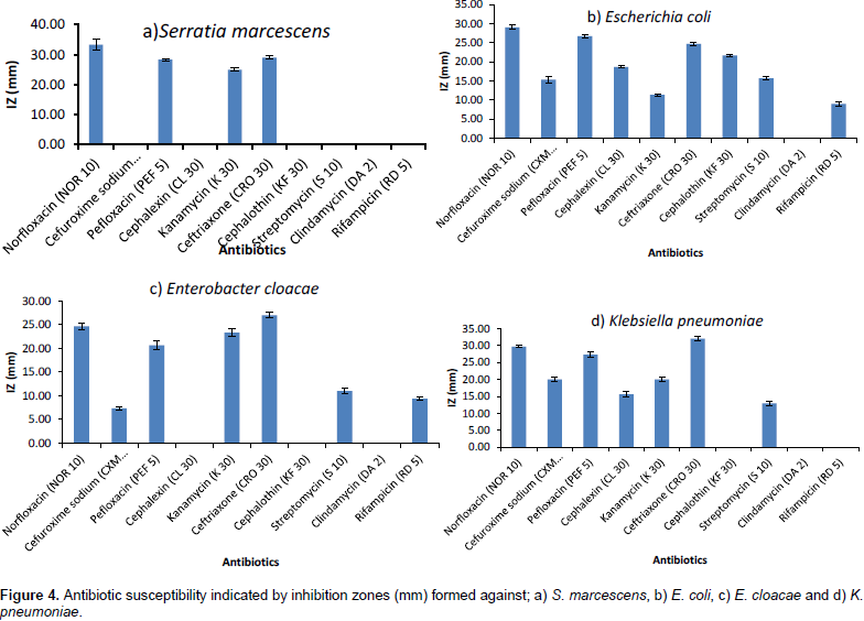
Other tested antibiotics showed differential susceptibility. All the tested isolates of the four tested species were resistant to clindamycin. The tested isolates of S. marcescens were also resistant to cefuroxime sodium, cephalexin, cephalothin, streptomycin and rifampicin, whereas, were highly sensitive to norfloxacin, pefloxacin, kanamycin and ceftriaxone. The tested isolates of E. coli were highly sensitive to norfloxacin, pefloxacin, ceftriaxone and cephalothin but with moderate sensitivity to cefuroxime sodium, cephalexin and streptomycin and had low sensitivity to the others antibiotics. E. cloacae isolates were resistant for cephalexin, cephalothin and clindamycin, however, were highly sensitive to norfloxacin, kanamycin and ceftriaxone. On the other hand, K. pneumoniae was resistant to cephalothin, rifampicin in addition to clindamycin and was highly sensitive to norfloxacin, pefloxacin and ceftriaxone. Shiga toxin-producing E. coli isolated from samples of meat were multi-resistant, exhibiting resistance to ampicillin, ciprofloxacin, tetracycline, sulfamethoxazole-trimethoprim, gentamycin and streptomycin (Li et al., 2011).
In a study carried out by Kalmus et al. (2011) to evaluate antibiotic resistance of E. coli, ampicillin, streptomycin and tetracycline resistance were observed in 24.3, 15.6 and 13.5%, respectively, among the E. coli isolates. While examining the hygienic and sanitary quality of pasteurized cow's milk, E. coli was identified in 77.05% of the samples and the highest rates of resistance to antimicrobial agents were obtained for ampicillin (19.2%), cephalothin (18.9%) and tetracycline (17.1%) (Zanella et al., 2010). E. coli and other coliforms recovered from humans and animals had antibiotic resistance and several species were resistant to many antimicrobial agents commonly used in human and veterinary medicine (Greeson et al., 2013). The treatment of cattle with common antibiotics leads to increase in resistance and transfer of these resistant strains to human hosts (Rinsky et al., 2013; Barnett and Linder 2014; Cordoba et al., 2015).CONCLUSION
CONFLICT OF INTERESTS
ACKNOWLEDGEMENTS
REFERENCES
|
Abd-Alla MH, Morsy FM, El-Enany AE, Ohyama T (2012). Isolation and characterization of a heavy-metal-resistant isolate of Rhizobium leguminosrum bv. viciae potentially applicable for biosorption of Cd2+ and Co2+. Int. Biodeter. Biodegr. 67:48-55. |
|
|
Ayhan K, ضzkan G, Noveir MR (2000). Determination of histidine decarboxylase activity produced by Enterobacteriaceae isolated from ground meat. J. Ciencia. 10:243-250. |
|
|
Barnett ML, Linder JA (2014). Antibiotic prescribing for adults with acute bronchitis in the United States, 1996-2010. Jama. 311(19):2020-2022. |
|
|
Bauer AW, Kirby MM, Sherris JC, Truck M (1966). Antibiotic susceptibility testing by a standardized single disk method. Am. J. Clin. Pathol. 45:493-496. |
|
|
Bosilevac JM, Guerini MN, Kalchayanand N, Koohmaraie M (2009). Prevalence and characterization of salmonellae in commercial ground beef in the United States. Appl. Environ. Microbiol. 75:1892-1900. |
|
|
Cordoba G, Siersma V, Lopez-Valcarcel B, Bjerrum L, Llor C, Aabenhus R, Makela M (2015). Prescribing style and variation in antibiotic prescriptions for sore throat: cross-sectional study across six countries. BMC family practice 16:1-8. |
|
|
Doucet F, Trieu-cuot P, Andremont A, Courvalin P (2001). Inducible transfert of conjugative transposon Tn1545 from Enterococcus faecalis to Listeria monocytogenes in the digestive tracts of gonobiotic mice. Antimicrob. Agents Chemother.,35:185-187. |
|
|
Feng PC, Hartmann PA (1982). Fluorogenic assays for immediate confirmation of Escherichia coli. Appl. Environ. Microbiol. 43:1320-1329. |
|
|
Greeson K, Suliman GM, Sami A, Alowaimer A, Koohmaraie M (2013). Frequency of antibiotic resistant Salmonella, Escherichia coli, Enterococcus, and Staphylococcus aureus in meat in Saudi Arabia. Afr. J. Microbiol. Res. 7(4):309-316. |
|
|
Gundogan N, Avci E (2013). Prevalence and antibiotic resistance of extended-spectrum beta-lactamase (ESBL) producing Escherichia coli and Klebsiella species isolated from foods of animal origin in Turkey. Afr. J. Microbiol. Res. 7(31):4059-4064. |
|
|
Gundogan N, Cıtak S, Yalcin E (2011). Virulence properties of extended spectrum beta-lactamase-producing Klebsiella species in meat samples. J. Food Prot. 74:559-564. |
|
|
Gundogan N, Yakar U (2007). Siderophore production, serum resistance, hemolytic activity and extended spectrum beta lactamase-producing Klebsiella species isolated from milk and milk products. J. Food Saf. 3:251-260. |
|
|
Hara-Kudo Y, Niizuma J, Goto I (2008). Surveillance of Shiga toxin-producing Escherichia coli in beef with effective procedures, independent of serotype. Foodborne Pathog. Dis. 5: 97-103. |
|
|
Haryani Y, Noorzaleha AS, Fatimah AB, Noorjahan BA, Patrick GB, Shamsinar AT, Laila RAS, Son R (2007). Incidence of Klebsiella pneumonia in street foods sold Malaysia and their characterization by antibiotic resistance, plasmid profiling, and RAPD-PCR analysis. Food Cont. 18:847-853. |
|
|
Holt JG, Krieg NR, Sneath PH (1994). Bergey's Manual of Determinative Bacteriology, Lippincott Williams and Wilkins 9th Ed, Baltomore, P 787. |
|
|
Jordan D, Phillips D, Sumner J, Morris S, Jenson I. (2007). Relationships between the density of different indicator organisms on sheep and beef carcasses and in frozen beef and sheep meat. J. Appl. Microbiol. 102(1):57-64. |
|
|
Kabiru KM, Bello M, Kabir J, Grande L, Morabito S (2015). Detection of pathogenic Escherichia coli in samples collected at an abattoir in Zaria, Nigeria and at different points in the surrounding environment. Int. J. of Env. Res. Public Health. 12:679- 691. |
|
|
Kalmus P, Birgit A, Age K, Toomas O, Kalle K (2011). Udder pathogens and their resistance to antimicrobial agents in dairy cows in Estonia. Acta Veterinaria Scandinavica 53:4. |
|
|
Kang DH, Koohmaraie M, Siragusa GR (2001). Application of multiple antimicrobial interventions for microbial decontamination of commercial beef trim. J. Food Prot. 64:168-171. |
|
|
Kodaka H, Armfield AY, Lombard GL, Dowell VR (1982). Practical procedure for demonstrating bacterial flagella. J. Clin. Microbiol. 16:948-952. |
|
|
Koohmaraie M, Arthur TM, Bosilevac JM, Guerini M, Shackelford SD, Wheeler TL (2005). Post-harvest interventions to reduce/eliminate pathogens in beef. Meat Sci. 71:79-91. |
|
|
Li MC, Wang F, Li F (2011). Identification and molecular characterization of antimicrobial-resistant shiga toxin–producing Escherichia coli isolated from retail meat products. Foodborne Pathog. Dis. 8(4):489-493. |
|
|
Mohammed O, Shimelis D, Admasu P, Feyera T (2014). Prevalence and antimicrobial susceptibility pattern of E. coli isolates from raw meat samples obtained from abattoirs in Dire Dawa City, Eastern Ethiopia. Int. J. Microbiol. Res. 5(1):35-39. |
|
|
Molina F, LÙŽpez-Acedo E, Tabla R, Roa I, GÙŽmez A, Rebollo JE (2015). Improved detection of Escherichia coli and coliform bacteria by multiplex PCR. BMC Biotechnol. 15:48. |
|
|
Nossair MA, Kamal K, Nahla AE, Samaha IA (2014). Detection of some enteric pathogens in retailed meat. Alexandria J. Veterinary Sci. 44:67-73. |
|
|
Okonko IO, Ikpoh IS, Nkang AO, Udeze AO, Babalola TA, Mejeha OK, Fajobi EA (2010). Assessment of bacteriological quality of fresh meats sold in Calabar Metropolis, Nigeria. EJEAFChe 9(1): 89-100. |
|
|
Paterson DL (2006). Resistance in Gram-negative bacteria (Enterobacteriaceae). Am. J. Med. 119: 520-528. |
|
|
Popovic NT, Skukan AB, Dzidara P, Coz-Rakovac R, Strunjak-Perovic I, Kozacinski L, Jadan M, Brlek-Gorski D (2010). Microbiological quality of marketed fresh and frozen seafood caught off the Adriatic coast of Croatia. Vet. Med. 55(5):233-241. |
|
|
Ray B, Bhunia A (2008). Spoilage of specific food groups. Fundamental food microbiology, CRC Press, Boca Raton, Florida: pp. 209-223. |
|
|
Reuben CR, Gyar SD (2015). Isolation and antibiogram of shiga toxin-producing Escherichia coli O157:H7 from diarrhoeic HIV/AIDS patients in Lafia, Central Nigeria. Int. Res. J. Microbiol. 6:20-26. |
|
|
Rinsky JL, Nadimpalli M, Wing S, Hall D, Baron D, Price LB, Larsen J, Stegger M, Stewart J, Heaney CD (2013). Livestock-associated methicillin and multidrug resistant Staphylococcus aureus is present among industrial, not Antibiotic-free livestock operation workers in North Carolina. PLoS ONE 8(7):1-11. |
|
|
Saidi R, Khelef D, Kaidi R (2014). Antibiotic susceptibility of enterobacteriaceae species isolated from mastitic milk in Algeria. Asi. Pacif. J. Rep. pp. 311-316. |
|
|
Sanger F, Nicklen S, Coulson AR (1977). DNA sequencing with chain-terminating inhibitors. Proc. Nat. Acad. Sci. 74:5463-5467. |
|
|
Schoeder CM, White DG, Meng J (2004). Retail meat and poultry as a reservoir of antimicrobial-resistant Escherichia coli. Food Microbiol. 21:244-255. |
|
|
Zanella GN, Mikcha JM. Bando E, Siqueira VL, Machinski M (2010). Occurrence and antibiotic resistance of coliform bacteria and antimicrobial residues in pasteurized cow's milk from Brazil. J. Food. Prot. 9:1684-1687. |
|
Copyright © 2024 Author(s) retain the copyright of this article.
This article is published under the terms of the Creative Commons Attribution License 4.0


