African Journal of
Biotechnology

  • Abbreviation: Afr. J. Biotechnol.
  • Language: English
  • ISSN: 1684-5315
  • DOI: 10.5897/AJB
  • Start Year: 2002
  • Published Articles: 12487

Full Length Research Paper

Molecular diagnosis of phytoplasma transmission from zygotic embryos to in vitro regenerated plants of coconut palm (Cocos nucifera L.)

DARAMCOUM Wentoin Alimata Marie Pierre
  • DARAMCOUM Wentoin Alimata Marie Pierre
  • Université Félix Houphouët Boigny, UFR Biosciences, Laboratoire de Génétique, 22 BP 582 Abidjan 22, Côte d’Ivoire.
  • Google Scholar
KONAN Konan Jean-Louis
  • KONAN Konan Jean-Louis
  • Centre National de Recherche Agronomique (CNRA), Station Marc Delorme of Port-Bouët, 07 BP 13 Abidjan 07, Côte d’Ivoire.
  • Google Scholar
YAO Saraka Didier Martial
  • YAO Saraka Didier Martial
  • Université Peleforo Gon Coulibaly, UFR des Sciences Biologiques, BP 1328 Korhogo, Côte d’Ivoire.
  • Google Scholar
YAIMA Arocha Rosete
  • YAIMA Arocha Rosete
  • Sporometrics, 219 Dufferin Street, Suite 20C, Toronto, ON M6K 3J1, Canada.
  • Google Scholar
KOFFI Eric-Blanchard Zadjéhi
  • KOFFI Eric-Blanchard Zadjéhi
  • Centre National de Recherche Agronomique (CNRA), Station Marc Delorme of Port-Bouët, 07 BP 13 Abidjan 07, Côte d’Ivoire.
  • Google Scholar
YOBOUE Koffi
  • YOBOUE Koffi
  • Centre National de Recherche Agronomique (CNRA), Station Marc Delorme of Port-Bouët, 07 BP 13 Abidjan 07, Côte d’Ivoire.
  • Google Scholar
KOUASSI Kan Modeste
  • KOUASSI Kan Modeste
  • Centre National de Recherche Agronomique (CNRA), Station Marc Delorme of Port-Bouët, 07 BP 13 Abidjan 07, Côte d’Ivoire.
  • Google Scholar
KOUADJO Claude Ghislaine
  • KOUADJO Claude Ghislaine
  • Centre National de Recherche Agronomique (CNRA), Station Marc Delorme of Port-Bouët, 07 BP 13 Abidjan 07, Côte d’Ivoire.
  • Google Scholar
KOFFI Edmond
  • KOFFI Edmond
  • Centre National de Recherche Agronomique (CNRA), Station Marc Delorme of Port-Bouët, 07 BP 13 Abidjan 07, Côte d’Ivoire.
  • Google Scholar
KOFFI Kouadio Kan Ghislain
  • KOFFI Kouadio Kan Ghislain
  • Centre National de Recherche Agronomique (CNRA), Station Marc Delorme of Port-Bouët, 07 BP 13 Abidjan 07, Côte d’Ivoire.
  • Google Scholar
N’GUETTA Assanvo Simon-Pierre
  • N’GUETTA Assanvo Simon-Pierre
  • Université Félix Houphouët Boigny, UFR Biosciences, Laboratoire de Génétique, 22 BP 582 Abidjan 22, Côte d’Ivoire.
  • Google Scholar


  •  Received: 24 April 2018
  •  Accepted: 05 June 2018
  •  Published: 04 July 2018

 ABSTRACT

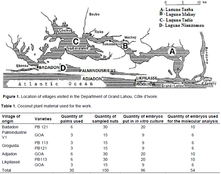
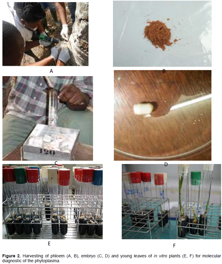
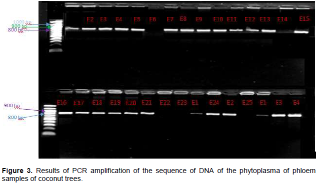
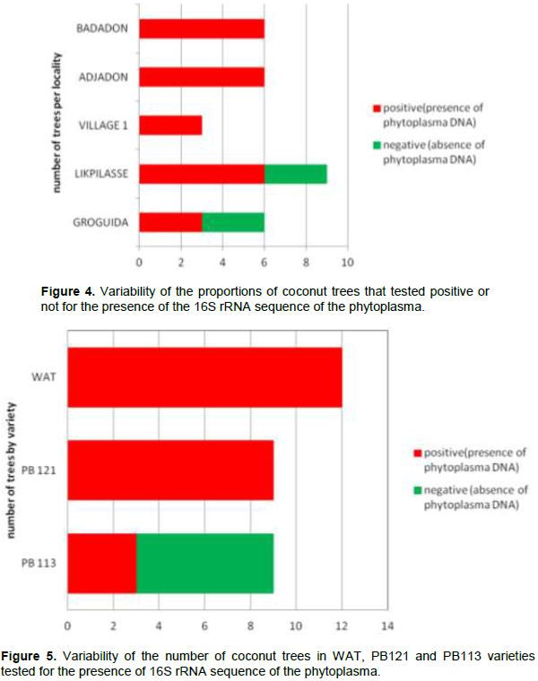
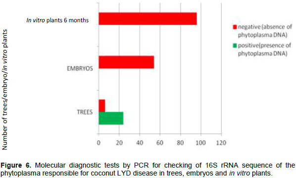
The aim of this study was to investigate the transmission of the lethal yellowing disease (LYD) of coconut tree caused by a phytoplasma from the zygotic embryo to the regenerated plantlet in vitro. From a total of 30 trees, 150 mature coconut nuts where harvested. These nuts were used to extract 150 zygotic embryos. From this package, 96 zygotic embryos were used to regenerate 96 young coconut seedlings in vitro and the 54 others were used to extract total DNA. From the stem of the 30 palms at the stage 1 of the LYD, phloem sample were also collected. From the regenerated in vitro-plantlets at 6 months age, leaf sample were collected. From the molecular diagnosis by PCR, 80% of the phloem samples carried the 16S rRNA gene of the phytoplasma responsible for LYD. All the zygotic embryos and in vitro-plantlets regenerated were healthy. So, coconut zygotic embryos can be used for the safe exchange of genetic material regarding lethal yellowing disease. The regenerated in vitro plantlet are free of disease.

Key words: Coconut, phytoplasma, transmission, in vitro.


 INTRODUCTION

The coconut tree originates from two geographical areas, namely, the islands of Southeast Asia and South India (Gunn et al., 2011). The ancient origins of the local coconut tree bordering the West African coast are probably India and Mozambique (De Nuce and Wuidart, 1979). From its center of origin, the coconut was disseminated by flotation of nuts at the mercy of marine currents and, later, by human travel and migration (Harries et al., 2004; Baudouin and Lebrun, 2009). Human migrations were those of Austronesians, Arabs and Europeans. It was introduced from Mozambique (East Africa) to Côte d'Ivoire (West Africa) by Portuguese navigators in the early 16th century (De Nuce and Wuidart, 1979).
 
 
Creation of the coconut collection for the purposes of research in the context of the varietal development passes through the exchange of the plant material. With regards to the coconut tree, the exchanges of the genetic material are done using nut in order to create the diversity from the family of brood stocks in a country's collection. However, these exchanges are not easily done because of the volume and mass of the nut, which is the organ usually used (Orozco-Segovia et al., 2003). These nuts that do not have dormancy often carry disease germs like lethal yellowing disease (LYD). Studies on the conservation and transfer of coconut material in the form of embryos have been investigated in several laboratories (Assy-Bah et al., 1989; Danso et al., 2009; Rillo and Paloma, 1991; Cueto et al., 2012; Yoboue et al., 2014).
 
The lethal yellowing disease threatens the entire world coconut grove. This disease has already destroyed more than thousands of hectares of coconut plantations in several regions of the world such as East Africa, the Caribbean and Central and West Africa (Been, 1981; Oropeza et al., 2005; Dollet et al., 2009; Konan et al., 2013). Phytoplasmas are transmitted to plants during food activity by their vectors, mainly leafhoppers, planthoppers and psyllids (Weintraub and Beanland, 2006).
 
The embryos contain a completely differentiated vascular system and they can be a source of propagation or transmission of the lethal yellowing disease (Harrison et al., 1995; Cordova et al., 2003). The exchange of germplasm of coconut, usually affected by the embryos, becomes difficult, especially when the embryos come from areas where the disease occurs (Jones et al., 1999).
 
The disease  is  known  by  different  names  in  various countries; in Ghana it is called Wilt disease of Cape Saint Paul (CSPW), in Togo, it is known as Kaïncopé disease, in Nigeria it is Awka and in Cameroon, it is named Kribi disease. The phytoplasmas that cause the diseases are variable from one country to another. Disease appeared in southern Côte d'Ivoire in the department of Grand-Lahou and threatens one of the world's largest coconut collections (Konan et al., 2013).
 
The presence of the disease in all parts of the world creates mistrust between the coconut producing countries and the exchange of plant material. This has a negative impact on research works and the culture of coconut. The objective of this work was to check the presence or absence of the phytoplasma responsible for lethal yellowing disease in zygotic embryos and regenerated plants obtained from trees affected by this disease. These results will serve as a guide and help to ensure the exchange of plant material between coconut producing countries using zygotic embryos.


 MATERIALS AND METHODS

The study focused on zygotic embryos from mature coconut nuts (10-12 months of age) harvested from trees that are visually affected by the LYD and are in stage 1 of the disease. The visual symptoms of LYD are: at stage 0 or apparently breast, the tree does not present symptoms; in stage 1, yellowing of the apical leaves; in stage 2, the fall of immature and mature nuts; in stage 3, leaf and crown leaf loss; at stage 4, only trunks and stems remain. Mature nuts were collected in coconut plantations which contained two types of hybrid (PB121 or MYD x WAT and PB113 or CRD x RIT) and the West African Tall (WAT) variety. The three types of coconut are sensitive to the disease. Sample were collected in five villages of Grand-Lahou Department in Côte D’Ivoire (5° 14'39'' North and 5° 00'11 '' West) that are Badadon, Palmindustrie V1, Groguida, Adjadon and Likpilassé (Figure 1). Sampling is also undertaken for phloem of stem of each palm. A total of 30 trees were sampled including 6 at Badadon, 3 at Village 1, 6 at Groguida, 6 at Adjadon and 9 at Likpilassé (Table 1).
 
 
Sampling of phloem, mature nuts, zygotic embryos and plantlets
 
To check the gene 16s RNAr of  the  phytoplasma  responsible  for the lethal yellowing disease, one phloem sample per tree was taken for the mother trees; the phloem (Figure 2) was taken with an electric drill. A total of 30 phloem samples from each of the mother trees was collected and stored at -4°C prior for extraction of the total DNA.
 
 
Mature nuts, 10-12 months old, recognizable by the brown colour of their epidermis (De Nuce and Wuidart, 1982) or noise of water inside were harvested from the bunches on the mother trees for 5 to 10 nuts. A total of 150 nuts were harvested from the 30 sampled mother trees. The cylinders of the endosperm or solid albumen that contain the zygotic embryos were carefully removed from the nuts and disinfected as recommended by N'Nan et al. (2012) and Yoboue et al. (2014). After disinfection of the endosperm cylinders, extraction and disinfection of the zygotic embryos were carried out in the laboratory under aseptic conditions for operation in air-flow cabinet (ASSY-Bah et al., 1989; Yoboue et al., 2014). The samplings were taken on seemingly healthy trees or in stage 1 trees because during the evolution of the disease, the mature and immature nuts fall early enough; therefore, for these stages (2, 3 and 4), there are no mature nuts on the trees. The zygotic embryos from the nuts (a zygotic embryo/whole nut) from the same mother tree were divided into two batches. For each sampled parent tree, one of the batches of zygotic embryos was used to run tests in order to detect the presence of the phytoplasma DNA. The embryos that compose the other batches were transferred directly to in vitro regeneration medium contained in the test tubes as one embryo per tube. The  composition  of  the  in vitro  regeneration  medium   was continuously modified to successively induce germination of the zygotic embryos and organogenesis (root, stem and leaf) in order to obtain, after 6 months, a complete seedling (Figure 3).
 
 
Total DNA extraction
 
The extractions of the total DNA of phloem from mother trees, zygotic embryos and leaves of regenerated plantlets in vitro were done in a buffer CTAB according to the protocol of Harrison et al. (2013). DNA concentrations in the various extracts were read through a spectrophotometer Nanodrop 2000 (thermo-scientific, USA).
 
PCR and detection of sequence 16S rRNA of the phytoplasma
 
The conventional PCR was performed with 25 ng of total DNA in a reaction volume of 25 μl containing 1 μM of each P1 universal primer (Deng and Hiruki, 1991) and P7 (Schneider et al., 1995). Concerning the Nested-PCR, the CSPWD G813F/AwkaSR primers (Tymon et al., 1998) were used to amplify the area between the 16s rRNA and 23S gene for detection of West African phytoplasma strain (Nigeria, Ghana and Cote d’Ivoire) of the phytoplasma (N’nan et al., 2014). The characteristics of these primers used are recorded in Table 2.


The analysis of the results of the PCR was carried out by electrophoresis on a 1% agarose gel prepared with a TBE buffer at 95 V for 45 min. The agarose gel was pre-saturated with "SYBR Safe  DNA"  during  preparation.  Fragment  sizes  were   measured using a 100 bp molecular weight marker. After migration, the gel was observed on a screen using a digital imaging system "digidoc-il 120 imaging system".


 RESULTS

The display on 1% agarose gel of PCR products obtained from the phloem DNA extracts revealed a total of 80% (24 trees out of 30 tested) positive (presence of the sequence between 800 and 900 bp characteristic of the DNA length of phytoplasma) within the trees (Figure 4). The distribution of trees infected with the lethal yellowing disease revealed that 100% of the individuals tested had the disease in the villages; Badadon, Palmindustrie V1 and Adjadon (Figure 5). A proportion of 20% of trees that tested negative were observed in the villages of Likpilassé (3 trees) and Groguida (3 trees). In the three varieties studied, all PB121 trees and WAT were positive while out of 9 trees of PB 113, 3 were positive and 6 negative (Figure 5).
 
 
Molecular diagnosis by PCR carried out for zygotic embryos and in vitro plants did not reveal the presence of phytoplasmic DNA on the embryos obtained from the nuts harvested from the trees that tested positive as well as the embryos obtained from the nuts harvested from trees that tested negative at Likpilassé and Groguida (Figure 6).


 DISCUSSION

From a total number of 30 sampled trees, 24 trees tested positive to the sequence 16S rRNA of the phytoplasma responsible for the lethal yellowing disease followed up by the molecular diagnosis by PCR. This indicates the presence of the  phytoplasma  responsible  for  the  lethal yellowing disease in these trees. It confirms the presence of the disease in the Department of Grand-Lahou as published by Konan et al. (2013) and Yaima et al. (2014). Contrary results were obtained by N'nan et al. (2014) in their works on the lethal yellowing disease in Ghana where the authors did not obtain any amplification for samples taken from trees at the onset of the disease.
 
When embryo was extracted, the PCR analysis revealed that not all embryos carry phytoplasma because there was no amplification. Similar results were obtained by N'nan et al. (2014) where all embryos tested did not have amplification of the AwkaSR gene. Indeed, during the evolution of the disease, nuts fall very early which could exempt them. Similarly, the seedlings obtained after 6 months of in vitro cultures are not carriers of the disease. These results confirm the absence of the phytoplasma in the embryo and the seedling obtained in vitro. 80% of the phytoplasma-bearing trees produced nuts with healthy in vitro embryos and seedlings, which indicates that the disease is not transmitted from the seed. The absence of DNA of the phytoplasma in embryo demonstrated by the authors also agrees with the work of McCoy et al. (1983) and Cousin (2001). According to the latter, the transmission of the phytoplasma by the embryo contradicts the biological principles which shows that the seeds do not transmit the phytoplasma. The works of Nipah et al. (2007) and Myrie et al. (2011) support this point of view. Indeed, the authors showed that in vitro culture of embryos from infected plants leads to healthy plants.
 
The current work confirms this hypothesis because all the embryos collected in endemic areas are healthy, not carrying the phytoplasma. The methods of extractions and detections were carried out under strict laboratory conditions. Embryos are not transmitters of lethal yellowing.
 
However, results in disagreement with those reported in this study were previously reported. Harrison et al. (1995) and Cordova et al. (2003) showed the possible presence of phytoplasma in the embryo. The works of Nipah et al. (2007) also reported the presence of phytoplasma from zygotic embryos while working on the Great West Africa (GWA). Cordova et al. (2003) studies on the amplification of 16S rRNA of the phytoplasma gene in the embryo by PCR. According to these authors, the DNA of the phytoplasma is available in the embryo at a very low concentration. Therefore, for the detection of the phytoplasma by PCR, a large number of embryos should be used in order to get a significant quantity of the DNA of the phytoplasma in the total DNA extracted from the embryo. Harrison et al. (1995) have already proposed that several cycles of ultracentrifugation may be useful for amplification of the 16S rRNA gene of the phytoplasma. 


 CONCLUSION

This study was conducted to check the phytoplasma responsible for the coconut lethal yellowing disease of coconut tree in embryos and seedlings. The results of the molecular diagnosis by PCR revealed that unlike affected trees, the zygotic embryos from the nuts harvested from these trees and seedlings regenerated from the in vitro culture of these zygotic embryos, do not carry the gene responsible for lethal yellowing disease. This is due to the fact that in vitro embryo regeneration generates healthy plants. Thus, the use of embryo for the exchange of plant material even from lethal yellowing disease areas is recommended.


 CONFLICT OF INTERESTS

The authors have not declared any conflict of interests.


 REFERENCES

Assy-Bah B, Durand-Gasselin T, Engelmann F, Pannetier C (1989). Culture in vitro d'embryons zygotiques de cocotiers (Cocos nucifera L.): Méthode révisée et simplifiée d'obtention de plants de cocotiers transférables au champ. Oléagineux 44(11):515-523.


Baudouin L, Lebrun P (2009). Coconut (Cocos nucifera L.) DNA studies support the hypothesis of ancient Austronesian migration from Southeast Asia to America. Genetic Resources Crop Evolution 56:257-262.
Crossref


Been BO (1981). Observation on field resistance to lethal yellowing in Jamaica. Oléagineu, 36(1):9-11.


Cordova I, Jones P, Harrison NA, Oropeza C (2003). In situ PCR detection of phytoplasma DNA in embryos from coconut palms with lethal yellowing disease. Molecular Plant Pathology 4:99-108.
Crossref


Cousin MT (2001). Phytoplasmes et phytoplasmoses: caractéristiques, symptômes et diagnostic. Cahiers d'Agriculture 10:361-376.


Cueto CA, Johnson VB, Bourdeix R, Engelmann F, Kembu A, Konan JL, Kouassi Kan M, Rivera RL, Vidhanaarachchi V, Wwise SF (2012). Technical guidelines for the safe movement and duplication of coconut (cocos nucifera l.) germplasm using embryo culture transfer protocols. COGENT; Bioversity International, Montpellier, France.


Danso K, Quaicoe R, Oduro V, Dery S, Owusu-Nipah J, Amiteye S, Malaurie B (2009). In vitro germination response and plantlets development of healthy and diseased zygotic embryo of coconut. International Journal of Integrative Biology 7(1):26-31.


De Nuce De Lamothe, Wuidart W (1982). L'observation des caracteristiques de developpement vegetatif, de floraison et de production chez le cocotier. Oléagineux 37(6):291-300.


De Nuce L, Wuidart W (1979). Les cocotiers grands de Port Bouët (Côte d'Ivoire). 1- Grand Ouest Africain, Grand de Mozambique, Grand de Polynésie, Grand de Malaisie. Oléagineux 34(7):339-349.


Deng S, Hiruki C (1991). Amplification of 16S rRNA genes from culturable and non-culturable mollicutes. Journal of Microbiological Methods 14:53-61.
Crossref


Dollet M, Jannot C, Baudouin L, Cirad JO (2009). Le cocotier en Afrique et la maladie du jaunissement mortel OCL 16(2):74-75.


Gunn BF, Baudouin L, Olsen KM (2011). Independent origins of cultivated coconut (Cocos nucifera L.) in the old world tropics. PLoS ONE 6, e21143.
Crossref


Harries H, Baudouin L, Cardena R (2004). Floating, boating and introgression: Molecular techniques and the ancestry of coconut palm populations on pacific islands. Ethnobotany Research and Applications 2:37-53.
Crossref


Harrison NA, Richardson PA, Tsaï JH (1995). Detection and diagnosis of Lethal Yellowing: conventional methods and molecular techniques. In: Oropeza C, Howard FW, Ashburner GR (eds.) Lethal Yellowing research and practical aspects, Developments in Plant Pathology, Springer Netherlands, pp. 79-91.
Crossref


Harrison NA, Davis RE, Helmick E (2013). DNA Extraction from Arborescent Monocots and How to Deal with Other Challenging Hosts. In Phytoplasmas: methods and protocols (eds. M. Dickinson and J. Hodgetts), Humana Press, Springer New York Heidelberg Dordrecht London, UK, ISSN 1064-3745, pp. 147-58.
Crossref


Jones P, Tymon AM, Mpunami AA (1999). Detection and diagnosis of African Lethal Yellowing like diseases. In: Oropeza C, Verdeil JL, Ashburner GR, Carde-a R, Santamaría J (eds) Current Advances in Coconut Biotechnology, Kluwer Academic Publishers (Dordrecht/ Boston/London) pp. 197-220.
Crossref


Konan Konan JL, Allou K, Atta Diallo H, Saraka Yao D, Koua B, Kouassi N, Benabid R, Michelutti R, Scott J, Arocha-Rosete Y (2013). First report on the molecular identification of the phytoplasma associated with a lethal Yellowing-type disease of coconut palms in Côte d'Ivoire. New Disease Reports 28:3.
Crossref


Konan Konan JL, Lekadou T, N'goran B, Kouassi N, Gbalou Y. (2013) Project FIRCA (Fonds Interprofessionnel pour la Recherche et le Conseil Agricoles). Cocotier CNRA – COC No. 588. Etude de la maladie du cocotier identifiée dans le Départment de Grand-Lahou, 35p.


Mccoy RE, Howard FW, Tsai JH, Donselman HM, Thomas D, Basham HG, Atilano RA, Eskafi FM, Britt L, Collins ME (1983). Lethal Yellowing of palms. University of Florida Agricultural Experiment Stations Bulletin 834, Gainesville, Florida, USA.


Myrie W, Oropeza C, Sáenz L, Harrison N, Roca MM (2011). Reliable improved molecular detection of coconut lethal yellowing phytoplasma and reduction of associated disease through field management strategies. Bulletin of Insectology 64:203-204.


N'Nan O, Borges M, Konan Konan JL, Hocher V, Verdeil J-L, Tregear J, N'Guetta ASP, Engelmann F, Malaurie B (2012). A simple protocol for cryopreservation of zygotic embryos of ten accessions of coconut (Cocos nucifera L.). In Vitro Cellular and Developmental Biology-Plant 48:160-166.
Crossref


Nipah JO, Jones P, Hodgetts J, Dickinson M (2007). Detection of phytoplasma DNA in embryos from coconut palms in Ghana, and kernels from maize in Peru. Bulletin of Insectology, 60:385-386.


N'Nan-Alla Oulo, Ernest Gonedele BI, Abou Bakari Kouassi, Auguste Kouassi, Rene Philippe, Bernard Malaurie, Michel Dollet (2014). Isolation of phytoplasma dna from the coconut palms (Cocos nucifera L.) collected from Ghana, Volume – 2(5) pp. 596-500.


Oropeza C, Escamilla JA, Mora G, Zizumbo D, Harrison NA (2005). Coconut lethal yellowing. In Coconut genetic resources, edited by Batugal p, Ramanatha Rao V, et Olivier J, IPGRI, Selangor Darul Ehsan (Malaisie), pp. 349-363.


Orozco-Segovia A, Batis Ana I, Rojas-Arechiga M, Mendoza A (2003). Seed biology of palms: a review. Palm 45(2):79-94.


Rillo EP, Paloma MBF (1991). Storage and transport of zygotic embryos of Cocos nucifera L. for in vitro culture. Plant Genetic Resource Newsletter 86:1-4


Schneider B, Seemüller E, Smart CD, Kirkpatrick BC. 1995. Phylogenetic classification of plant pathogenetic mycoplasmalike organisms or phytoplasmas. In: Razin S, Tully JG, eds. Molecular and Diagnostic Procedures in Mycoplasmology, Volume I. San Diego, CA, USA: Academic Press, 369-79.
Crossref


Tymon AM, Jones P, Harrison NA. 1998. Phylogenetic relationships of coconut phytoplasmas and the development of specific oligonucleotide PCR primers. Annals of Applied Biology 132:437-452.
Crossref


Weintraub PG, Beanland L (2006). Insect vectors of phytoplasmas. Annual Review of Entomology 51:91-111.
Crossref


Yaima Arocha-Rosete., Konan Konan JL, Diallo AH, Allou K, Scott JA. (2014) Identification and molecular characterization of the phytoplasma associated with a lethal yellowing-type disease of coconut in Côte d'Ivoire. Canadian Journal of Plant Pathology 36:141-150.
Crossref


Yoboue K, Alla-N'Nan O, Konan JL, Sie RS, Kouassi M, Yao SD, Koffi BE (2014). Méthode simple d'échange de germoplasme de cocotier (Cocos nucifera L.) par l'utilisation d'embryons zygotiques. Journal of Applied Biosciences 80:7048-7059.
Crossref


          */?>