African Journal of
Biotechnology

  • Abbreviation: Afr. J. Biotechnol.
  • Language: English
  • ISSN: 1684-5315
  • DOI: 10.5897/AJB
  • Start Year: 2002
  • Published Articles: 12487

Full Length Research Paper

In vitro selective cytotoxicity of activated parasporal proteins produced by Bacillus thuringiensis serovars kumamotoensis and tohokuensis against human cancer cell lines

Maher Obeidat
  • Maher Obeidat
  • Department of Biotechnology, Faculty of Agricultural Technology, Al-Balqa Applied University, Al-Salt 19117, Jordan.
  • Google Scholar


  •  Received: 08 August 2017
  •  Accepted: 16 October 2017
  •  Published: 22 November 2017

 ABSTRACT

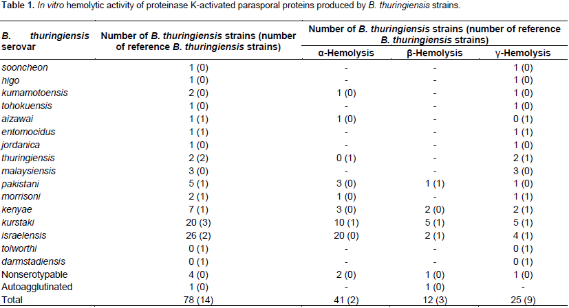
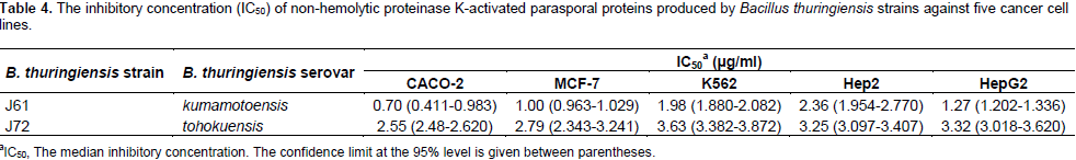
The anti-cancer activity of alkali-solubilized protease-activated parasporal proteins produced by 78 local Bacillus thuringiensis strains and 14 reference B. thuringiensis strains was screened against five human cancer cell lines (CACO-2, Hep2, HepG2, K562, and MCF-7). Activated parasporal proteins were tested for their hemolytic activity against human erythrocytes. It was found that activated parasporal proteins of 25 local B. thuringiensis strains and 9 reference strains were non-hemolytic. Non-hemolytic parasporal proteins produced by 9 local B. thuringiensis strains were found to exhibit no to low cytotoxicity against human non-cancerous Hs27 cells. Out of them, activated parasporal proteins of two local B. thuringiensis strains (J61; B. thuringiensis serovar kumamotoensis and J72; B. thuringiensis serovar tohokuensis) were found to produce high to very high in vitro selective cytotoxicities, preferentially toxic to cancerous cells, against all cancer cell lines used in this study. This is the first observation of the anti-cancer activity from B. thuringiensis serovar kumamotoensis. Based on IC50 values, activated parasporal proteins of J61 strain produced the most significant cytotoxicity against all cancer cell lines. Furthermore, CACO-2 and MCF-7 cells were found to be the most sensitive. Thus, parasporal proteins produced by B. thuringiensis serovar kumamotoensis strain J61 and/or B. thuringiensis serovar tohokuensis strain J72 may be used as alternative or improving means for current cancer therapy.

Key words: Bacillus thuringiensis, kumamotoensis, tohokuensis, parasporal, cancer.


 INTRODUCTION

Cancer is a major public health problem worldwide and is the second cause of death after cardiovascular diseases (coronary heart disease and hypertension) in Jordan as well as in the world. According to the Ministry of Health annual report (MOH, 2013), cancer of all types (in particular breast cancer, colorectal cancers, lymphomas, lung cancer, and leukemia) was responsible for the death of over 10,000 Jordanians from 1996 until now. A total of 8,744 new cancer cases were registered by Jordan Cancer Registry (JCR) in 2013. Out of new cases of cancer recorded amongst Jordanians in 2013, colorectal cancer (12.7%) followed by lung cancer (11%) were the most common among Jordanian males. Where as, breast cancer (36.5%) followed by colorectal cancer (9.4%) were the most common among Jordanian females. The spread of cancer is increasing over the world especially in developing countries (Plummer et al., 2016). The number of new cancer cases diagnosed among Jordanians has increased up to 51% in the past ten years with 0.9:1 male to female ratio according to JCR.
 
There is a great deal of controversy within the medical community over what kind of medical treatment is most efficacious in treating cancer. Most conventional medical treatments for cancer are of little help, so alternative techniques are required to be developed to target cancer. In this research, the possibility of using Bacillus thuringiensis (Bt) as a source of anti-cancer compounds that are usually too difficult to create synthetically will be investigated. The bacterium B. thuringiensis is a Gram-positive spore-forming soil bacterium. Several strains of this bacterium produce crystalline parasporal proteins during sporulation. These parasporal proteins often contain d-endotoxin proteins (known as insecticidal Cry proteins) that can exhibit strong insecticidal activity against agriculturally and medically important insect pests (Saraswathy and Kumar, 2004).
 
It has long been believed that B. thuringiensis is characterized by insecticidal activity associated with its parasporal proteins. However, several studies (Ohba et al., 1988; Ohba, 1996; Roh et al., 1996; Mizuki et al., 1999a) have demonstrated that B. thuringiensis strains producing non-insecticidal parasporal proteins are more widely distributed than insecticidal ones. In the beginning of this century, many efforts were oriented toward studying the biological activities of non-insecticidal parasporal proteins yet to be discovered. Mizuki et al. (1999b) reported cytotoxicity against human leukemic T cells and other human cancer cells for parasporal proteins produced by some non-insecticidal B. thuringiensis strains. Moreover, it was found that these non-insecticidal Cry proteins, designated parasporins, are capable of preferentially killing cancer cells upon proteolytic degradation (Mizuki et al., 1999b, 2000; Yamashita et al., 2000; Ito et al., 2004; Okumura et al., 2004; Katayama et al., 2005; Hayakawa et al., 2007; Jung et al., 2007; Uemori et al., 2008; Yasutake et al., 2008; Nagamatsu et al., 2010).
 
Six parasporin families, including 19 parasporin toxins isolated from B. thuringiensis strains, were identified by the Committee of Parasporin Classification and Nomenclature (Okumura et al., 2010; Wong, 2010). For example, parasporin-1 was isolated from B. thuringiensis strain A1190 (Mizuki et al., 1999b, 2000). This parasporin when degraded by proteases exhibited selective cytotoxicity against cancer cells such as human leukemic T cells (Mizuki et al., 2000). It was found that parasporin-1 activates apoptotic pathway (Katayama et al., 2007). Activated parasporin-2 isolated from B. thuringiensis A1547 belonging to serovar dakota showed extremely high cytotoxicity against different kinds of cancer cells including leukemic T cells such as MOLT-4, Jurkat, and HL-60 (Kim et al., 2000; Ito et al., 2004; Kitada et al., 2006). It was found that the cytotoxic effect of parasporin-2 is non-apoptotic toward cancer cells but causes swelling of the susceptible cells (Ito et al., 2004).
 
Moreover, another parasporin, called parasporin-3, isolated from B. thuringiensis isolate A1462 exhibited limited cytotoxicity against human leukemic T cells (Yamashita et al., 2000, 2005). It was hypothesized that parasporin-3 kills cancer cells by a receptor-mediated mechanism (Yamashita et al., 2005). An important parasporin known as parasporin-4 was obtained from B. thuringiensis strain A1470 which belongs to serovar shandongiensis induced necrosis-like cytotoxicity against human leukemic T cells (Lee et al., 2000; Okumura et al., 2005; Saitoh et al., 2006). A cytotoxic protein toward the leukemic T cell, MOLT-4, was also isolated from B. thuringiensis serovar coreanensis strain A1519 (Namba et al., 2003). Parasporin-5, purified from B. thuringiensis serovar tohokuensis strain A1100, was found to exhibit cytocidal activity against human leukemic T cells (Ekino et al., 2014).
 
A novel parasporin isolated from B. thuringiensis strain M019 was classified into a new parasporin family (parasporin-6) preferentially kill human hepatocyte cancer HepG2 cells and uterus cervix cancer HeLa cells (Nagamatsu et al., 2010). Efforts dedicated to screening microbial natural products for cancer therapy are very limited. Therefore, the current research is an important advance in the fight against cancer because bacterial metabolites are not well explored for their effectiveness in cancer therapy. These metabolic products may become an alternative or a supplement to current cancer therapies. Thus, this research aimed to test B. thuringiensis parasporal proteins against several cancer cells as alternative or improving means for current cancer therapy.


 MATERIALS AND METHODS

B. thuringiensis strains and growth conditions
 
Seventy-eight local Jordanian B. thuringiensis strains representing 14 serovars (aizawai, entomocidus, higo, israelensis, jordanica, kenyae, kumamotoensis, kurstaki, malaysiensis, morrisoni, pakistani, sooncheon, thuringiensis, and tohokuensis), four nonserotypable (NSP) strains (unable to react with antisera), and one autoagglutinated (AA) strain were used in this study. These strains were previously isolated from different Jordanian habitats and classified by serotyping (Obeidat et al., 2000; Khyami-Horani, 2003; Al-Momani et al., 2004). Fourteen reference strains obtained from International Entomopathogenic Bacillus Collection Center (IEBC), Institute Pasteur, Paris, including, B. t. thuringiensis T01001, B. t. thuringiensis T01022, B. t. kurstaki T03A001, B. t. kurstaki HD1 T03A005, B. t. kurstaki T03A361, B. t. kenyae T04B001, B. t. entomocidus T06001, B. t. aizawai T07001, B. t. morrisoni T08001, B. t. tolworthi T09001, B. t. darmstadiensis T10001, B. t. pakistani T13001, B. t. israelensis T14001, and B. t. israelensis 1884 T14007 were also used. Among the B. thuringiensis strains, 35 strains showed no insecticidal activities against diptera and lepidoptera (Saadoun et al., 2001; Khyami-Horani, 2002; Obeidat et al., 2004, 2012). Cultures were grown overnight at 37°C on T3 medium (0.3% tryptone, 0.2% tryptose, 0.15% yeast extract, 0.05 M sodium phosphate, and 0.005% MnCl2) (Travers et al., 1987) and maintained as suspensions of spores and cells at -70°C with 20% (v/v) glycerol.
 
Solubilization of B. thuringiensis parasporal proteins
 
Bacterial strains were cultured in 75 ml of T3 broth (Travers et al., 1987). These cultures were incubated at 150 rpm for 3 days at 37°C, and then left at 4°C for 1 day. Sporulated cultures of B. thuringiensis were centrifuged at 5000 rpm for 10 min at 4°C, and washed three times with distilled water before solubilization (Saitoh et al., 1996). The crude parasporal proteins were solubilized in 5 ml of 50 mM Na2CO3 (pH 10) containing 10 mM dithiothreitol (DTT) and 1 mM EDTA for 1 h at 37°C (Mizuki et al., 1999b). After solubilization, the mixture was centrifuged at 14000 rpm for 5 min at 4°C, and the pH of the supernatant was adjusted to 8 by 1 M HCl then passed through a 0.45-mm membrane filter.
 
Activation of B. thuringiensis parasporal proteins
 
Solubilized parasporal proteins were activated by proteolysis in 0.3 mg/ml proteinase-K at 37°C for 1.5 h. After that, 1 mM phenylmethylsulphonyl fluoride (PMSF) was added to stop the proteolytic reaction (Mizuki et al., 2000). The mixture was then filtered through a 0.45-mm membrane filter and the filtered fluid was stored at 4°C until use.
 
Protein quantification
 
The protein concentration in the filtered fluid was determined by the method of Bradford (1976). Bradford procedure was performed by using bovine serum albumin (BSA) as standard. After assessment of protein concentration by spectrophotometry at 595 nm absorbance, the protein concentration in each sample was adjusted to 1 mg /ml.
 
Hemolytic activity
 
Hemolytic activity was tested on blood agar medium using fresh human erythrocytes (5%). 50 µl of activated parasporal proteins was added into each well (5 mm i.d.) prepared on blood agar plates. The type of hemolysis was determined after incubation of plates at 37°C for 48 h (Carillo et al., 1996).
 
Cells and culture conditions
 
Six human cell lines were used in this study, including non-cancerous normal cells (Hs27; human foreskin fibroblast cell line) that was used as a non-transformed control and five cancerous cells (CACO-2; human colorectal adenocarcinoma cell line, Hep2; human larynx epidermoid carcinoma cell line, HepG2; human liver hepatocellular carcinoma cell line, K562; human leukemic T cells, and MCF-7; human breast cancer cell line) that were kindly supplied by Dr. Saeid Ismaeil (Faculty of Medicine, University of Jordan).
 
The non-adherent human leukemia cancer cell line K562 was grown in Roswell Park Memorial Institute Medium (RPMI 1640) medium at pH 7.4, supplemented with 10% fetal bovine serum (FBS), 40 µg/ml gentamicin, 50 µM 2-mercaptoethanol, 10 mM N-2-hydroxyethylpiperzine-N-2-ethane sulfonic acid (HEPES), 1 mM sodium pyruvate, 100 U/ml penicillin, 100 µg/ml streptomycin, and 2 mM L-glutamine at 37°C in a humidified 5% CO2 incubator (Freshney, 2005). The K562 cells were harvested, subcultured, and reseeded in fresh medium at ~105 cells/ml every 48 h.
 
The adherent human cancer cell lines (CACO-2, Hep2, HepG2, and MCF-7) and the adherent non-cancerous Hs27 cell line were grown in Dulbecco’s modified Eagle’s medium (DMEM), pH 7.4, supplemented with 10% FBS, 40 µg/ml gentamicin, 50 µM 2-mercaptoethanol, 10 mM HEPES, 1 mM sodium pyruvate, 100 U/ml penicillin, 100 µg/ml streptomycin, and 2 mM L-glutamine, and they were harvested at ~70% confluence and subcultured every 48 h at 37°C in a humidified 5% CO2 incubator (Freshney, 2005). To harvest the adherent cells, growth medium was removed and cells were washed with phosphate buffer saline (PBS). To produce a cellular suspension, a cell dissociation solution made of 1X trypsin-EDTA was added and incubated at 37°C for 5 min in a humidified 5% CO2 incubator. Trypsinized cells were reseeded in fresh medium at ~105 cells/ml and incubated at 37°C in a humidified 5% CO2 incubator.
 
Screening of anti-cancer activity
 
For screening assay, 20 µl of non-hemolytic proteinase K-activated parasporal proteins (1 mg/ml constant concentration, that is, 20 µg/well) was added to 200 µl of harvested Hs27 cells in fresh DMEM, mixed thoroughly by pipetting and 180 µl medium were loaded into each well of 96 well micro test plate to bring the total volume to 500 µl/well. Cells were plated at a density 4×10⁴ cells/well, and counted by hemocytometer. Then, the 96 well micro test plates were incubated at 37°C in a humidified 5% CO2 incubator for 48 h. At the end of incubation time, the viability of cells was assessed by 3-(4,5-dimethythiazol-2-yl)-2,5-diphenyltetrazolium bromide (MTT) assay (Mosmann, 1983; Heiss et al., 1997). In the assay, each well of the 96 well microtest plate received 40 µl (50 µg) of MTT and incubated at 37°C for 4 h in a humidified 5% CO2 incubator.
 
After that, 100 µl of dimethyl sulfoxide (DMSO) was added. The optical densities were measured at 450 nm with 630 nm reference wavelength using enzyme-linked immunosorbent assay (ELISA) microplate reader. Each treatment was performed in triplicate and repeated five times. The survival rate was determined by comparing the average of absorbance values with that in the control without parasporal proteins. To determine the anticancer activities, the non-hemolytic parasporal proteins that exhibited no cytotoxicity against normal Hs27 cells were tested against cancer cell lines (CACO-2, Hep2, HepG2, K562, and MCF-7). The cytotoxicity assay was performed for adherent cell lines in the same manner as for Hs27 cells. Likewise, the cytotoxicity was tested on the non-adherent K562 cells in the same manner as for adherent cells but using RPMI 1640 medium instead of DMEM medium.
 
In vitro selective cytotoxicity
 
The cytotoxicity of non-hemolytic protease-activated parasporal proteins that exhibited selective cytotoxicity (that is, non-toxic to non-cancerous Hs27 cell line but toxic to other cancerous cell lines) was performed in the same fashion as in the anticancer screening assay but using six concentrations (20, 10, 5, 1, 0.5, and 0.1 µg/ml) of activated parasporal proteins. The mortality of cells was monitored after 48 h by MTT assay.
 
Statistical analysis
 
After treatment with MTT assay, the obtained absorbance values were corrected by subtracting the average absorbance of blank (contains the growth medium) from the average absorbance of vehicle (contains the growth medium and cells) and average absorbance of treatment (contains the growth medium, cells, and activated parasporal proteins). To calculate inhibition percentage of cells, the formula “Inhibition% = ((A-B)/A)×100" was used, where A is the vehicle absorbance and B is the treatment absorbance.
The median inhibitory concentration (IC50) was determined by comparing the average mortality values of the six concentrations of activated parasporal proteins with that in the control without activated parasporal proteins. Each treatment was achieved in triplicate. The IC50 values, regression equations, and correlation coefficients (R2) were determined by non-linear regression analysis (MS Excell, Microsoft Co., 2010). The 95% confidence limits (CLs) were also calculated using MS Excel (2010). The statistical outliers in data were excluded using Q-Test.


 RESULTS

The current study was performed to screen the anti-cancer activity of proteinase K-activated parasporal proteins produced by 78 local B. thuringiensis strains and 14 reference B. thuringiensis strains against five human cancer cell lines, including, colorectal adenocarcinoma CACO-2 cells, larynx epidermoid carcinoma Hep2 cells, liver hepatocellular carcinoma HepG2 cells, leukemic K562 cells, and breast cancer MCF-7 cells. As shown in Table 1, activated parasporal proteins of 25 B. thuringiensis strains and 9 reference strains were found to be non-hemolytic (γ-type). The non-hemolytic parasporal proteins of the local 25 B. thuringiensis strains belonged to 13 serovars (sooncheon, higo, kumamotoensis, tohokuensis, entomocidus, jordanica, thuringiensis, malaysiensis, pakistani, morrisoni, kenyae, kurstaki, and israelensis) and one NSP strain (Table 1).
 
The non-hemolytic activated parasporal proteins of the reference strains were found to belong to nine B. thuringiensis serovars (aizawai, entomocidus, thuringiensis, morrisoni, kenyae, kurstaki, israelensis, tolworthi, and darmstadiensis) whereas, the remaining hemolytic B. thuringiensis strains were displayed either by a-hemolysis (41 B. thuringiensis strains and 2 reference strains) or β-hemolysis (12 B. thuringiensis strains and 3 reference strains). The AA strain exhibited a-hemolysis. Proteinase K-activated parasporal proteins of the 34 B. thuringiensis strains (25 local strains and 9 reference strains) that exhibited no hemolysis against human erythrocytes were screened for their cytotoxicity against normal non-cancerous human Hs27 cells (Table 2). The colorimetric MTT assay was used to determine the degree of cytotoxicity of activated parasporal proteins against Hs27 cells. It was found that activated parasporal proteins produced by nine local B. thuringiensis strains of seven serovars (sooncheon, kumamotoensis, tohokuensis, thuringiensis, morrisoni, kenyae, and kurstaki) exhibited no to low cytotoxicity against Hs27 cells (Table 2). Whereas, the non-hemolytic parasporal proteins of the remaining B. thuringiensis strains (16 local strains and 9 reference strains) were found to exhibit moderate to very high cytotoxicity against Hs27 cells.
 
 
The activated parasporal proteins of B. thuringiensis strains (9 strains) that exhibited no hemolysis against human erythrocytes and no (-) to low (+) cytotoxicities against normal non-cancerous human Hs27 cells were examined for their selective cytotoxicity against colorectal adenocarcinoma CACO-2 cells, larynx epidermoid carcinoma Hep2 cells, liver hepatocellular carcinoma HepG2 cells, leukemic K562 cells, and breast cancer MCF-7 cells (Table 3). It was found that activated parasporal proteins of two B. thuringiensis strains (J61; B. thuringiensis serovar kumamotoensis and J72; B. thuringiensis serovar tohokuensis) produced high to very high (degree of cytotoxicity is greater than 90%) selective cytotoxicities against cancer cells (Table 3). 
 
 
Activated parasporal proteins of both B. thuringiensis strains J61 and J72 were non-toxic (the degree of cytotoxicity is less than 5%) to Hs27 cells. However, activated parasporal proteins of J61 strain exhibited high selective cytotoxicity against MCF-7 cells and very high selective cytotoxicity against CACO-2, K562, Hep2, and HepG2 cells (Table 3). Whereas, activated parasporal proteins of J72 strain exhibited high selective cytotoxicity against K562 and Hep2 cells and very high selective cytotoxicity against CACO-2, MCF-7, and HepG2 cells. It was observed that the shape of parasporal proteins of strains J61 and J72 was spherical (Figure 1).
 
 
As shown in Table 4, the IC50 values of the cytotoxic parasporal proteins against cancer cells were determined. Based on IC50 values, it was clearly observed that all cancer cells investigated in this study were more susceptible to activated parasporal proteins of serovar kumamotoensis strain J61 than that of serovar tohokuensis strain J72. Furthermore, CACO-2 and MCF-7 cells were the most sensitive to activated parasporal proteins of strains J61 and J72.


 DISCUSSION

Cancer is a leading cause of death worldwide (nearly 1 in 6 deaths is due to cancer), accounting for 8.8 million deaths in 2015 according to the World Health Organization (WHO, 2017). The most common causes of cancer death and the deadliest are cancers of the lung (19.20% of deaths), liver (8.95%), colorectal (8.80%), stomach (8.57%), and breast (6.49%). About 70% of cancer deaths occur in low- and middle-income countries (WHO, 2017). As reported by the Ministry of Health (MOH, 2013), the cancer incidence in Jordan is in proportional increase. In addition, colorectal cancers and breast cancer are ranked first among Jordanian males and females, respectively. Therefore, the current study was established to investigate the anti-cancer activity of parasporal proteins produced by local B. thuringiensis strains against various human cancer cell types, in particular colorectal adenocarcinoma colorectal CACO-2 cells and breast cancer MCF-7 cells. Furthermore, the anti-cancer activity of B. thuringiensis parasporal proteins toward larynx epidermoid carcinoma Hep2 cells, liver hepatocellular carcinoma HepG2 cells, and leukemic K562T cells was examined.
 
Alkali solubilized and proteolytic processed parasporal proteins of the 92 B. thuringiensis strains used in this study were screened for their hemolytic activity against human erythrocytes. The hemolytic activity of parasporal proteins was measured before anti-cancer screening assay to ensure that the anti-cancer activity was not induced by hemolysins such as Cyt proteins that exhibited cytolytic activity against human erythrocytes (Crickmore et al., 1998, 2016).
 
DTT-solubilized proteinase K-activated parasporal proteins of 58 B. thuringiensis strains exhibited either partial (a-hemolysis) or complete (b-hemolysis) patterns of hemolysis suggesting that cytolysins (known as Cyt proteins) or other hemolytic agents produced by those B. thuringiensis strains were responsible for hemolysis of erythrocytes. It was observed that activated parasporal proteins produced by the remaining 34 B. thuringiensis strains were non-hemolytic. Out of those 34 B. thuringiensis strains, nine strains were found to be non-toxic to human non-cancerous Hs27 cells. Of the non-toxic strains to Hs27 cells, only two local B. thuringiensis strains J61 (B. thuringiensis serovar kumamotoensis) and J72 (B. thuringiensis serovar tohokuensis) exhibited selective in vitro cytotoxicity against various human cancer cells (CACO-2, MCF-7, K562, HepG2, and Hep2).
 
The activity in these strains was not attributable to the broad-spectrum Cyt proteins in view of the fact that they showed no hemolytic activity against human erythrocytes. Furthermore, the parasporal proteins of these B. thuringiensis strains (J61 and J72) were previously reported (Obeidat et al., 2012) to exhibit no insecticidal activity against diptera and lepidoptera. This is in agreement with previous studies (Mizuki et al., 1999b, 2000; Lee et al., 2000; Yamashita et al., 2000; Ito et al., 2004; Okumura et al., 2004; Katayama et al., 2005; Hayakawa et al., 2007; Jung et al., 2007; Uemori et al., 2008; Yasutake et al., 2008; Nagamatsu et al., 2010) that clearly demonstrated that only non-insecticidal parasporal proteins can exhibit discriminating cytotoxicity against different cancer cell types. Unfortunately, activated parasporal proteins from all reference B. thuringiensis strains screened in this study did not exhibit selective cytotoxicity against target cells.
 
Mizuki et al. (1999b) found that the non-hemolytic leukemia cytotoxic strains belonged to serovars dakota, neoleonensis, shandongiensis, coreanensis and other unidentified serogroups. In a recent study, Ekino et al. (2014) reported that parasporal proteins produced from B. thuringiensis serovar tohokuensis were also cytotoxic against human leukemic T cells. Similarly, in a preceding study, Obeidat (2008) demonstrated that activated parasporal proteins of B. thuringiensis serovar tohokuensis exhibited selective cytotoxicity against larynx epidermoid carcinoma Hep2 cells and leukemic T cells, Jurkat. In this study, the non-hemolytic selectively cytotoxic B. thuringiensis strains were found to belong to serovar kumamotoensis and serovar tohokuensis. This observation demonstrated that cytotoxic proteins which exhibited specific cytotoxicity against cancer cells can be found in a variety of B. thuringiensis serovars. To our knowledge, no previous study has revealed the production of selective anti-cancer activity from processed parasporal proteins produced by B. thuringiensis serovar kumamotoensis.
 
According to IC50 values, protease-activated parasporal proteins of the cytotoxic strain J61 showed higher significant toxicity against cancer cells compared to that of strain J72. The 95% confidence limits of the IC50 values determined from MTT assay were not overlapped suggesting that J61 and J72 cytotoxicities are significantly different. Therefore, the cancer cells were more sensitive to activated parasporal proteins produced by strain J61. It also appeared from the results that there is a marked variation among parasporal proteins in the level of cell-killing activity. Of particular interest is the fact that the activated parasporal proteins from strains J61 and J72 were able to discriminate between cancerous cells and noncancerous Hs27 cells, killing the former cells specifically. This finding strongly suggests the possible occurrence of B. thuringiensis strains in local habitats of Jordan which naturally produce parasporal proteins highly selective for cancer cells. This may lead to the use of these B. thuringiensis parasporal proteins for medical and pharmaceutical purposes including the treatment of particular types of cancer cells. Further studies regarding the identification of anti-cancer protein structure found in promising B. thuringiensis crudes are in process.


 CONFLICT OF INTERESTS

The author has not declared any conflict of interests.


 ACKNOWLEDGEMENTS

The author offers his sincerest gratitude to "the Scientific Research Support Fund/Ministry of Higher Education of Jordan; grant no. M-Ph/2/14/2008" for financial support. Many thanks to Mr. Belal Al-Shomali for technical assistance.


 REFERENCES

Al-Momani F, Obeidat M, Saadoun I, Meqdam M (2004). Serotyping of Bacillus thuringiensis isolates, their distribution in different Jordanian habitats and pathogenicity in Drosphila melanogaster. World J. Microbiol. Biotechnol. 20:749-753.
Crossref


Bradford MM (1976). A rapid and sensitive method for the quantitation of microgram quantities of protein utilizing the principle of protein-dye binding. Anal. Biochem. 72:248-254.
Crossref


Carillo P, Mardarz C, Pitta-Alvarez S (1996). Isolation and selection of biosurfactant producing bacteria. World J. Microbiol. Biotechnol. 12:82-84.
Crossref


Crickmore N, Baum J, Bravo A, Lereclus D, Narva K, Sampson K, Schnepf E, Sun M, Zeigler DR (2016). Bacillus thuringiensis toxin nomenclature. 

View


Crickmore N, Zeigler DR, Feitelson J (1998). Revision of the nomenclature for the Bacillus thuringiensis pesticidal crystal proteins. Microbiol. Mol. Biol. Rev. 62:807-813.


Ekino K, Okumura S, Ishikawa T, Kitada S, Saitoh H, Akao T, Oka T, Nomura Y, Ohba M, Shin T, Mizuki E (2014). Cloning and Characterization of a Unique Cytotoxic Protein Parasporin-5 Produced by Bacillus thuringiensis A1100 Strain. Toxins (Basel). 6:1882-1895.
Crossref


Freshney RI (2005). Culture of Animal Cells, A Manual of Basic Technique, 5th edition. Hoboken NJ, John Wiley and Sons.
Crossref


Hayakawa T, Kanagawa R, Kotani Y, Kimura M, Yamagiwa M, Yamane Y, Takebe S, Sakai H (2007). Parasporin-2Ab, a newly isolated cytotoxic crystal protein from Bacillus thuringiensis. Curr. Microbiol. 55:278-283.
Crossref


Heiss P, Bernatz S, Bruchelt G, Senekowitsch-Schmidtke R (1997). Cytotoxic effect of immunoconjugate composed of glucose-oxidase coupled to an anti-ganglioside (GD2) antibody on spheroids. Anticancer Res. 17(4B):3177-3178.


Ito A, Sasaguri Y, Kitada S, Kusaka Y, Kuwano K, Masutomi K, Mizuki E, Akao T, Ohba M (2004). A Bacillus thuringiensis crystal protein with selective cytocidal action to human cells. J. Biol. Chem. 279(20):21282-21286.
Crossref


Jung Y-C, Mizuki E, Akao T, Cote J-C (2007). Isolation and characterization of a novel Bacillus thuringiensis strain expressing a novel crystal protein with cytocidal activity against human cancer cells. J. Appl. Microbiol. 103:65-79.
Crossref


Katayama H, Kusaka Y, Yokota H, Akao T, Kojima M, Nakamura O, Mekada E, Mizuki E (2007). Parasporin-1, a novel cytotoxic protein from Bacillus thuringiensis, induces Ca2+ influx and a sustained elevation of the cytoplasmic Ca2+concentration in toxin-sensitive cells. J. Biol. Chem. 282(10):7742-7752.
Crossref


Katayama H, Yokota H, Akao T, Nakamura O, Ohba M, Mekada E, Mizuki E (2005). Parasporin-1, a novel cytotoxic protein to human cells from noninsecticidal parasporal inclusions of Bacillus thuringiensis. J. Biochem. 137:17-25.
Crossref


Khyami-Horani H (2002). Toxicity of Bacillus thuringiensis and B. sphaericus to laboratory populations of Drosophila melanogaster (Diptera: Drosophilidae). J. Basic Microbiol. 42:105-110.
Crossref


Khyami-Horani H, Hajaij M, Charles JF (2003). Characterization of Bacillus thuringiensis ser. jordanica (serotype H 71), a novel serovariety isolated in Jordan. Curr. Microbiol. 47:26-31.
Crossref


Kim H-S, Yamashita S, Akao T, Saitoh H, Higuchi K, Park YS, Mizuki E, Ohba M (2000). In vitro cytotoxicity of non-Cyt inclusion proteins of a Bacillus thuringiensis isolate against human cells, including cancer cells. J. Appl. Microbiol. 89(1):16-23.
Crossref


Kitada S, Abe Y, Shimada H, Kusaka Y, Matsuo Y, Katayama H, Okumura S, Akao T, Mizuki E, Kuge O, Sasaguri Y, Ohba M, Ito A (2006). Cytocidal actions of parasporin-2, an anti-tumor crystal toxin from Bacillus thuringiensis. J. Biol. Chem. 281(36):26350-2660.
Crossref


Lee D-W, Akao T, Yamashita S, Katayama H, Maeda M, Saitoh H, Mizuki E, Ohba M (2000). Noninsecticidal parasporal proteins of a Bacillus thuringiensis serovar shandongiensis isolate exhibit a preferential cytotoxicity against human leukemic T cells. Biochem. Biophys. Res. Commun. 272:218-223.
Crossref


Ministry of Health (MOH), Jordan Cancer Registry (2013). Statistic Summary for Cancer Incidence in Jordan, Non-communicable Diseases Directorate. The 18th Annual Report, Pp. 3-11.


Mizuki E, Ichimatsu T, Hwang S-H, Park YS, Saitoh H, Higuchi K, Ohba M (1999a). Ubiquity of Bacillus thuringiensison phylloplanes of arboreous and herbaceous plants in Japan. J. Appl. Microbiol. 86:979-984.
Crossref


Mizuki E, Ohba M, Akao T, Yamashita S, Saitoh H, Park YS (1999b). Unique activity associated with non-insecticidal Bacillus thuringiensis parasporal inclusions: in vitro cell-killing action on human cancer cells. J. Appl. Microbiol. 86:477-486.
Crossref


Mizuki E, Park YS, Saitoh H, Yamashita S, Akao T, Higuchi K, Ohba M (2000). Parasporin, human leukemic cell-recognizing parasporal protein of Bacillus thuringiensis. Clin. Diagn. Lab. Immunol. 7(4):625-634.
Crossref


Mosmann T (1983). Rapid colorimetric assay for cellular growth and survival: application to proliferation and cytotoxicity assays. J. Immunol. Methods 65(1-2):55-63.
Crossref


Nagamatsu Y, Okamura S, Saitou H, Akao T, Mizuki E (2010). Three Cry toxins in two types from Bacillus thuringiensis strain M019 preferentially kill human hepatocyte cancer and uterus cervix cancer cells. Biosci. Biotechnol. Biochem. 74:494-498.
Crossref


Namba A, Yamagiwa M, Amano H, Akao T, Mizuki E, Ohba M, Sakai H (2003). The cytotoxicity of Bacillus thuringiensis subsp. coreanensis A1519 strain against the human leukemic T cell. Biochim. Biophys. Acta 1622:29-35.
Crossref


Obeidat M (2008). Molecular typing of local Bacillus thuringiensis strains and determination of their parasporal crystal proteins cytotoxicity against cancer cells. PhD Thesis, University of Jordan, pp. 53-65.


Obeidat M, Hassawi D, Ghabeish I (2004). Characterization of Bacillus thuringiensis strains from Jordan and their toxicity to the Lepidoptera, Ephestia kuehniella Zeller. Afr. J. Biotechnol. 3(11):622-626.


Obeidat M, Khyami-Horani H, Al-Momani F (2012). Toxicity of Bacillus thuringiensis β-exotoxins and δ-endotoxins to Drosophila melanogaster, Ephestia kuhniella, and human erythrocytes. Afr. J. Biotechnol. 11(46):10504-10512.


Obeidat M., Al-Momani F, Saadoun I (2000). Diversity of Bacillus thuringiensis in different habitats of nothern Jordan. J. Basic Microbiol. 40(5-6):385-388.
Crossref


Ohba M (1996). Bacillus thuringiensis populations naturally occurring on mulberry leaves: A possible source of the populations associated with silkworm-rearing insectaries. J. Appl. Bacteriol. 80:56-64.
Crossref


Ohba M, Yu YM, Aizawa K (1988). Occurrence of non-insecticidal Bacillus thuringiensis flagellar serotype 14 in the soil of Japan. Syst. Appl. Microbiol. 11:85-89.
Crossref


Okumura S, Akao T, Higuchi K, Saitoh H, Mizuki E, Ohba M, Inouye K (2004). Bacillus thuringiensis serovar shandongiensis strain 89-T-34-22 produces multiple cytotoxic proteins with similar molecular masses against human cancer cell. Lett. Appl. Microbiol. 39:89-92.
Crossref


Okumura S, Ohba M, Mizuki E, Crickmore N, Côté J-C, Nagamatsu Y, Kitada S, Sakai H, Harata K, Shin T (2010). Parasporin nomenclature. 

View


Okumura S, Saitoh H, Ishikawa T, Wasano N, Yamashita S, Kusumoto K, Akao T, Mizuki E, Ohba M, Inouye K (2005). Identification of a novel cytotoxic protein, Cry45Aa, from Bacillus thuringiensis A1470 and its selective cytotoxic activity against various mammalian cell lines. J. Agric. Food Chem. 53:6313-6318.
Crossref


Plummer M, de Martel C, Vignat J, Ferlay J, Bray F, Franceschi S (2016). Global burden of cancers attributable to infections in 2012: A synthetic analysis. Lancet Glob. Health 4(9):e609-e616.
Crossref


Roh JY, Park HW, Jin BR, Kim HS, Yu YM, Kang SK (1996). Characterization of novel non-toxic Bacillus thuringiensis isolated from Korea. Lett. Appl. Microbiol. 23:249-252.
Crossref


Saadoun I, Al-Momani F, Obeidat M, Meqdam M, Elbetieha A (2001). Assessment of toxic potential of local Jordanian Bacillus thuringiensis isolates on Drosophila melanogaster and Culex. J. Appl. Microbiol. 90:1-7.
Crossref


Saitoh H, Higuchi K, Mizuki E, Ohba M (1996). Larvicidal activity of Bacillus thuringiensis natural isolates, indigenous to Japan, against two nematoceran insect pests occurring in urban sewage environments. Microbiol. Res. 151:263-271.
Crossref


Saitoh H, Okumura S, Ishikawa T, Akao T, Mizuki E, Ohba M (2006). Investigation of anovel gene encoding a parasporal protein, parasporin-4, that preferentially kills human leukemic T cells. Biosci. Biotechnol. Biochem. 70(12):2935-2941.
Crossref


Saraswathy N, Kumar P (2004). Protein engineering of δ-endotoxins of Bacillus thuringiensis. Electron. J. Biotechnol. 7(2):178-188.


Travers R, Martin P, Reichelderfer C (1987). Selective process for efficient isolation of soils Bacillus spp. Appl. Environ. Microbiol. 53(6):1263-1266.


Uemori A, Ohgushi A, Yasutake K, Maeda M, Mizuki E, Ohba M (2008). Parasporin-1Ab, a novel Bacillus thuringiensis cytotoxin preferentially active on human cancer cells in vitro. Anticancer Res. 28:91-95.


Wong SYR (2010). Bacillus thuringiensis parasporal proteins and their effect on human cancer cells. Int. eJ. Sci. Med. Edu. 4:3-9.


World Health Organization (WHO) (2017). Cancer Fact Sheet, February (2017). 

View


Yamashita S, Akao T, Mizuki E, Saitoh H, Higuchi K, Park YS, Kim H-S, Ohba M (2000). Characterization of the anti-cancer-cell parasporal proteins of a Bacillus thuringiensis isolate. Can. J. Microbiol. 46:913-919.
Crossref


Yamashita S, Katayama H, Saitoh H, Akao T, Park YS, Mizuki E, Ohba M, Ito A (2005). Typical three-domain Cry proteins of Bacillus thuringiensis strain A1462 exhibit cytocidal activity on limited human cancer cells. J. Biochem. 138:663-672.
Crossref


Yasutake K, Uemori A Binh ND, Mizuki E, Ohba M (2008). Identification of parasporin genes in Vietnamese isolates of Bacillus thuringiensis. Z. Naturforsch. C. 63:139-143.
Crossref


          */?>