ABSTRACT
The objective of this work was to evaluate the effect of bacteriocins from lactic acid bacteria (LAB) in Zea mays-based Ogi on some foodborne bacteria contaminating cabbage in Abakaliki, Nigeria. Ten (10) samples (5 samples of Z. mays-based Ogi and 5 samples of suspected contaminated cabbage heads) were aseptically collected and analyzed using standard microbiological methods. Five different Lactobacillus isolates (A, B, C, D, and E) were isolated from the Z. mays-based Ogi while 5 different species of bacterial pathogens; Staphylococcus aureus, Escherichia coli, Pseudomonas aeruginosa, Salmonella spp., and Shigella spp were isolated from cabbage heads. Results showed that Lactobacillus isolates exhibited high inhibitory effect against foodborne bacteria (S. aureus, E. coli, and Shigella spp) isolated from cabbage with inhibition zone diameter (IZD) ranging from 14 to 20 mm. A very high antimicrobial activity against foodborne bacteria isolated from cabbage was also observed for the crude bacteriocin at pH of 2. The stability of the antimicrobial affinity of the bacteriocin decreased as pH rises from 6 to 7. This study has shown that bacteriocin has antimicrobial activity against foodborne bacteria contaminating cabbage and could be used as bio-preservatives instead of hazardous chemical preservatives with adverse effects on the human body.
Key words: Lactic acid bacteria, bacteriocin, Zea mays, cabbage, fermented food, Ogi.
Traditional fermented foods prepared from cereals such as maize (Zea mays) are consumed especially by the middle and low income earners in many West African countries. “Ogi”, an acid fermented cereal gruel, made from maize serves as weaning foods for infants, food for the convalescent and the elderly, as well as breakfast meal by different age groups in Nigeria (Chilaka et al., 2016). Ogi has been known to exhibit health promoting properties such as in the control of gastroenteritis in animals and man. In vitro and in vivo data have been obtained on the probiotic effects (hypolipidemic, hepatoprotective, and antibacterial) of some lactic acid bacteria (LAB) isolated from Ogi (Oyetayo and Osho, 2004; Aderiye et al., 2007). LAB are known for their potential of producing antimicrobial compound and other value-added products. They have the ability to inhibit growth of pathogenic microorganisms, degrade mycotoxins, and with probiotic capabilities (Mokoena, 2017). Bacteriocins are small ribosomally synthesized antimicrobial peptides against which the producer species is immune and act against other bacteria in a bactericidal or bacteriostatic manner (Hill et al., 2017). Most of the bacteriocins are bactericidal with some exceptions that are bacteriostatic (Jeeravatnam et al., 2005). Bacteriocin-producing LAB strains protect themselves from their own toxins by the expression of a speci?c immunity protein, encoded in the bacteriocin operon (Mokoena, 2017). The application of bacteriocin in food is effective against food spoilage/pathogenic bacteria, and it is assumed to be degraded by protease in the gastrointestinal tract which makes it safe for human consumption (Cleveland et al., 2001; Pal et al., 2015). The increasing interest in bacteriocins as alternatives to antibiotics and chemical food preservatives guided this study interest to evaluate the effect of bacteriocins from LAB in Z. mays-based Ogi on some bacterial pathogens contaminating cabbage in Abakaliki, Nigeria with a view to understanding its application to avoid food spoilage.
Collection of Zea mays-based Ogi samples
Five (5) samples of Z. mays-based Ogi were randomly purchased at Abakpa Market, Abakaliki metropolis, Ebonyi State between June and October, 2019. Samples were aseptically collected and transported within two hours to the Department of Applied Microbiology Laboratory, Ebonyi State University, for bacteriological analysis.
Isolation and identification of LAB from Zea mays-based Ogi samples
Pour plate technique was used for the isolation of LAB. One gram of each ‘Ogi’ samples were dissolved in 10 ml of water and swirled to mix properly. A tenfold serial dilution was performed and an aliquot for the 10-7 and 10-8 dilution factors were inoculated on Mann Rogosa and Sharp (MRS) agar with 50 µg/ml of nystatin to suppress the growth of fungi. The inoculated plates were incubated at 37°C for 72 h in anaerobic jar, and suspected LAB colonies were then sub-cultured on MRS agar to obtain pure culture (Harley and Prescott, 2002; Sharma, 2009). All isolates were identified by standard techniques such as Gram staining, and biochemical tests such as citrate utilization, oxidase, indole, methyl red, Voges Proskauer (VP), and sugar (lactose, glucose, sucrose, fructose, maltose and mannitol) fermentation (Holt et al., 1994; Harley and Prescott, 2002; Sharma, 2009).
Isolation of foodborne bacteria from contaminated cabbage samples
Cabbage samples collected from Abakpa Main Market, Abakaliki, Ebonyi State were processed by removing their outer leaves and 20 g of each of the samples were weighed, washed with distilled water and blended in an electric blender. The samples were put in a clean beaker containing 20 ml of sterile water and sieved. The resulting filtrate was serially diluted, and 1 ml of 10-7 and 10-8 dilutions were plated on Cysteine Lactose Electrolyte Deficient (CLED) agar, Mannitol salt agar (MSA), Salmonella-Shigella (SS) agar. The plates were incubated at 37°C for 24 h. The discrete colonies of each of the bacterial isolates were identified by standard biochemical tests (Holt et al., 1994; Harley and Prescott, 2002; Sharma, 2009) and sub-cultured on nutrient agar to obtain pure cultures.
Determination of antagonistic activity of LAB
The antagonistic activity of LAB isolates was tested by the agar well diffusion technique with the cell-free supernatant of each isolate. A standardized suspension of each isolated bacterium (Escherichia coli, Staphylococcus aureus, Pseudomonas aeruginosa, Shigella spp., Salmonella sp.) from the cabbage samples was prepared and inoculated on the surfaces of prepared MHA plates using sterile cotton swab. The plates were dried and a sterile cork borer with a diameter of 4 mm was used to cut uniform wells in the agar plates. Each of the wells was filled with 0.1 ml aliquot of the test LAB. The plates were incubated at 37°C for 72 h. Isolates exhibiting highest zone of growth inhibition were selected and screened for bacteriocin production.
Screening and selection of LAB with antagonistic activity
The isolates were screened and selected using agar well diffusion technique using the cell free supernatant. A sterilized molten Mueller-Hinton (MHA) was prepared and dispensed into the petri dishes and allowed to solidify. A lawn of indicator organisms such as E. coli, S. aureus, P. aeruginosa, Shigella spp. and Salmonella sp. were made by spreading their suspension over the surfaces of prepared MHA plates with aid of sterile cotton swab. The plates were allowed to dry and a sterile cork borer with a diameter of 4 mm was used to cut uniform well in the agar plates. Each of the wells in the MHA plate was filled with 0.1 ml aliquot of the test LAB isolates. The plates were then incubated at 37°C for 72 h. Isolates exhibiting maximum zone of growth inhibition were selected and used for bacteriocin production.
Purification of crude bacteriocin production
Bacteriocin was extracted from LAB that had highest growth inhibition by growth in 1 L of MRS broth for 72 h at 30°C under anaerobic conditions (Ogunbanwo et al., 2003). Extract was obtained by centrifuging the culture at 12,000 rpm for 15 min to separate the cells. The pH of the cell-free culture supernatant was adjusted to 6.5 with 1 M NaOH. Catalase (1 mg/ml) was added to remove hydrogen peroxide from the supernatant (Daba et al., 1991). The supernatant was filtered through a 0.45-µm pore size membrane and the protein was precipitated using 80% (w/v) saturated ammonium sulphate. The mixture was stirred for 1 h and stored at 4°C. After precipitation, the mixture was centrifuged at 16,000 rpm at 4°C for 30 min, and pellet was stored at 42°C. The pellet was separated from impurities by dissolving 1 ml in distilled water in an Eppendorf tube and centrifuging at 10,000 rpm for 10 min at 4°C. The supernatant was discarded and the pellet containing bacteriocin was washed with deionized water. The mixture was dispensed into another Eppendorf tube and centrifuged at 10,000 rpm for 15 min at 4°C to spin down the proteins, and the supernatant discarded. The pellet was dissolved in 500 µl of 0.1 M sodium phosphate buffer (pH 7.0) and the total volume was made up to 2 ml. The sample was loaded into a pre-treated dialysis tubing cellulose membrane (18-20 mm length) and dialyzed in a 3-L 0.1 M sodium phosphate buffer (pH 7.0) for 2 h; the buffer was changed and the sample further dialyzed overnight at 4°C. After 24 h dialysis, the sample was added to an Eppendorf tube, centrifuged at 10,000 rpm at 4°C for 30 min and stored at -20°C.
Purification of proteins from crude bacteriocin samples
The crude bacteriocin samples were dispensed into Eppendorf tubes and centrifuged at 12,000 rpm for 15 min at 4°C to pellet down the cells. The supernatant was poured into a 50-ml capacity beaker in an ice pack at a temperature of 4°C and 15 g of ammonium sulphate (NH4)2SO4 in dry form was measured and dissolved in the supernatant. The mixture was stirred for 1 h and stored at 4°C for 24 h. The stirring was carefully done to prevent foaming that may lead to protein denaturation (Hensyl et al., 1994).
Determination of bacteriocin activity
Agar well diffusion assay was used to determine the bacteriocin activity of the LAB isolates (Khalid, 2011). Ten ml of partially purified bacteriocin was serially diluted up to 10-2 using saline diluent. An overnight culture of bacteria isolated from cabbage grown in tryptic soy broth (TSB) at 37°C was diluted in saline to a 0.5 McFarland standards. The suspension was inoculated on the surface of MHA plates using a sterile cotton swab. The plates were dried and wells were done as described above. Each of the wells was filled with 0.1 ml aliquot. The plates were kept at 4°C for 2 h to ensure diffusion of the supernatant fluid into the agar, and then incubated at 37°C for 24 h. The antimicrobial activity was determined by measuring the diameter of zones of inhibition around the wells.
Determination of effect of pH on antibacterial activity of crude bacteriocin
Sodium hydroxide (NaOH) and hydrogen chloride (HCl) was added to separate tubes to obtain pH values of 2, 3, 4, 5, 6, 7, and 8 confirmed with Jenway pH meter. The solutions were kept at room temperature for 2 h. Aliquots of 50 µl from each test tube were placed in wells of MHA plates inoculated with overnight broth cultures of the isolated bacteria (S. aureus, E. coli, P. aeruginosa, Salmonella sp., and Shigella spp.) from cabbage. The plates were incubated at 30°C for 24 h and the zones of inhibition around the wells were measured.
Isolation and identification of LAB from Zea mays-based Ogi samples and foodborne bacteria from cabbage samples
Results showed that five different lactic acid bacteria species; Lactobacillus isolates A, Lactobacillus isolates B, Lactobacillus isolates C, Lactobacillus isolates D, and Lactobacillus isolates E were obtained from Z. mays-based Ogi samples collected from Abakpa Main Market, Abakaliki, Ebonyi State (Table 1). Five micororganisms (s. aureus, e. coli, P. aeruginosa, Salmonella sp., and Shigella spp.) were isolated from cabbage samples randomly collected from Abakpa Main Market, Abakaliki (Table 2).
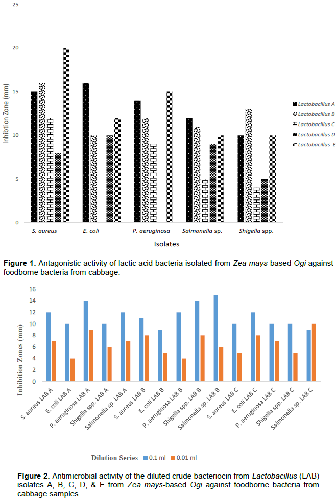
Antagonistic activity of lactic acid bacteria isolated from Zea mays-based Ogi against food-borne bacterial isolates from cabbage
The inhibition zone diameters (IZDs) of Lactobacillus isolates (A, B, C, D, and E) against the foodborne bacteria (s. aureus, e. coli, P. aeruginosa, Salmonella sp., and Shigella spp.) are shown in Figure 1. Lactobacillus isolates A, B, C, D, and E had inhibition zone diameters (IZDs) which ranged from 10 - 15 mm, 10 - 16 mm, 0 - 12 mm, 0 - 10 mm and 10 - 20 mm, respectively against the isolated foodborne bacteria (s. aureus, e. coli, P. aeruginosa, Salmonella sp., and Shigella spp.) (Figure 1).
Determination of the antimicrobial activity of the diluted crude bacteriocin from Lactobacillus isolates A, B, C, D, and E obtained from Zea mays-based Ogi against foodborne bacteria from cabbage
Results showed that 0.1 aliquot of the crude bacteriocin from Lactobacillus isolate A exhibited IZDs of 12, 10, 14, 10, and 12 mm against S. aureus, E. coli, P. aeruginosa, Shigella spp., and Salmonella sp. respectively while at 0.01 aliquot, IZDs of 7, 4, 9, 6, and 7 mm were recorded against S. aureus, E. coli, P. aeruginosa, Shigella spp., and Salmonella sp. respectively (Figure 2).
At 0.1 aliquot of the crude bacteriocin from Lactobacillus isolate B, inhibition zone diameters (IZDs) of 11, 9, 12, 14, and 15 mm were recorded against S. aureus, E. coli, P. aeruginosa, Shigella spp., and Salmonella sp. respectively, while at 0.01 aliquot, an inhibition zone diameter of 8, 5, 4, 8, and 6 mm were recorded against S. aureus, E. coli, P. aeruginosa, Shigella spp, and Salmonella sp. respectively (Figure 2).
At 0.1 aliquot of the crude bacteriocin from Lactobacillus isolate C, IZDs of 10, 12, 10, 10, and 9 mm were recorded against S. aureus, E. coli, P. aeruginosa, Shigella spp., and Salmonella sp. respectively, while at 0.01 aliquot, an IZD of 5, 8, 7, 5, and 10 mm were recorded against S. aureus, E. coli, P. aeruginosa, Shigella spp., and Salmonella sp. respectively (Figure 2).
Bacteriocin crude extract were not recovered from Lactobacillus isolate D and E.
Effect of pH on stability of bacteriocin
The results of the effects of pH on the stability of bacteriocin produced by LAB isolated from Z. mays-based Ogi against foodborne bacteria isolated from cabbage samples revealed that the inhibition zone for all bacterial isolates decreased as the pH increased, ranging from IZD of 21 to 0 mm (Table 3).
Lactic acid bacteria (LAB) are important organisms recognized for their fermentative ability as well as their health and nutritional benefits. They produce various compounds such as bacteriocins bacteriostatic or bacteriocidal proteins during lactic acid fermentation. A large number of bacteriocins from lactic acid bacteria have been characterized until today, and many different studies have indicated the potential usefulness of bacteriocin as food preservative.
This present study was designed to evaluate the effect of bacteriocins from lactic acid bacteria (LAB) in Z. mays-based Ogi on some bacterial pathogens contaminating cabbage in Abakaliki, Nigeria. This study has shown that bacteriocin has antimicrobial activity against Gram-positive (S. aureus) and Gram-negative (E. coli, P. aeruginosa, Salmonella sp., and Shigella spp.) foodborne bacteria contaminating cabbage.
In this study, a total of five Lactobacillus species were isolated from Z. mays-based Ogi-based. These results are similar to the work of Ohenhen et al. (2015) who isolated five different species of Lactobacillus isolates from fermented Ogi samples. Gram-positive (S. aureus) and Gram-negative (E. coli, P. aeruginosa, Salmonella sp., and Shigella spp.) bacterial pathogens were also isolated from the contaminated cabbage collected from Abakpa Main Market. This is also in agreement with the report of Sujeet and Vipin (2017), who observed the presence of the same foodborne bacteria in cabbage and other salad vegetables. The bacteria found are mostly enteric, which suggest a possible feacal-oral transmission. They have also been implicated as common foodborne pathogens causing gastrointestinal illnesses such as diarrhoea (Clevland et al., 2001). It is a well-known fact that food is a valuable source of nutrients for microbes to grow; and as these organisms grow on the food, they may cause spoilage such as bad taste, unpleasant smell, and poor appearance (Pal, 2013). The isolated LAB from Z. mays-based Ogi showed antimicrobial activity against foodborne bacteria isolated from cabbage. The antagonistic activity of LAB isolated from Z. mays based Ogi against foodborne bacteria revealed that even though Lactobacillus isolates A, B, and E had antimicrobial effect on all the indicator microorganisms, the effect on S. aureus was best; evident in the larger zones of inhibition. This could be a result of bacteriocin exhibiting bactericidal activity against closely related producers’ strains. This is in agreement with the work of Tannock (2004) who stated that the inhibitory action of LAB is mainly due to the accumulation of main primary metabolites. He also stated that LAB are capable of producing antimicrobial compounds such as formic acid, benzoic acid, hydrogen peroxide, and bacteriocins. The reason for the high inhibition zone diameter in S. aureus could be as a result of the cell wall structure and the physiological characteristics of the organisms (Tannock, 2004). It was reported that Gram-negative bacteria tend to resist antimicrobial compounds as a result of their complex cell wall composition (Pal et al., 2015). This is in contrast with the work of Ohenhen et al. (2015) who observed highest and lowest zone of inhibitions for E. coli and S. aureus respectively by Lactobacillus plantarum. Other Lactobacillus isolates; C, and D were less effective against the foodborne bacteria isolated from cabbage. The antimicrobial activity of the diluted bacteriocin from Lactobacillus isolate A, Lactobacillus isolate B, and Lactobacillus isolate C obtained from Z. mays showed that all the isolates have antimicrobial effect on the indicator microorganisms at 0.1 ml dilution while at 0.01 ml dilution, it recorded lower inhibition zone. This implies that optimal inhibition zone occurs at 0.1 ml. This result is in agreement with the findings of Kwon et al. (2002) who reported that bacteriocins had broad spectrum of activities against fish pathogens. It has also been reported that certain LAB bacteriocins, especially the class 2 bacteriocin, pediocin, can inhibit Gram-negative bacteria such as Shigella spp., Salmonella sp., and Pseudomonas species.
Hwanhlen et al. (2011) also reported that lactic acid was able to cause sub-lethal injury to E. coli. Similar properties have also been assigned to acetic acid (Hwanhlen et al., 2011). Because bacteriocin does not act equally against target species, many researchers have examined the activity of bacteriocin on specific bacteria species and strains (Castro et al., 2011). Indirect evidence suggests that such injury involve disruption of the lipopolysaccharide (LPSP) layer (Castro et al., 2011). Our study also showed that the crude bacteriocin extract partially purified from LAB displayed a very high antimicrobial activity at the pH 2, compared to the antimicrobial activity displayed at pH 6 and 7. No antimicrobial activity was observed in the diluted crude bacteriocin extract at pH 8. Lactobacillus isolates have tolerance to low pH. This observation is similar to that reported by Ogunbanwo et al. (2003) who indicated that purified bacteriocin extract recovered from L. plantarum was more active at pH 2 and 6, than at pH 10 and 12. The increased sensitivity of S. aureus to the pH amended crude bacteriocin extract could be as a result of the cell wall and physiology of the bacterium. However, Gram-positive bacteria only have a thick-like cell wall, made of peptidoglycan which constructs about 90% of the cell wall (Omar et al., 2006). This observation is in agreement with a report by Rammelsberg and Radler (1990) who observed that antimicrobial activity of purified bacteriocin extracted from L. parecasei subsp. tolerans was more active against S. aureus and Listeria monocytogenes than E. coli. The diluted crude bacteriocin extract at pH 8 exhibited less antimicrobial activity against the indicator organisms. It is possible that a high pH value had a negative effect on the antibacterial activity of the extract. Hence, further studies are needed in the extraction, characterization, and purification of bacteriocin from LAB with a view to utilizing them as alternatives to harmful chemical preservatives and as potential sources of probiotics and antimicrobial agents.
This study has shown that bacteriocin from lactic acid bacteria (LAB) has antimicrobial activities against one Gram-positive (S. aureus) and four Gram-negative (E. coli, P. aeruginosa, Salmonella sp., and Shigella spp.) foodborne bacteria isolated from cabbage. The bacteriocins produced by the LAB isolates in this study were noted to have maximum activity at pH 2. This study show the possibility that bacteriocins could be used as bio-preservatives in acidic food products like fruits juices instead of chemical preservatives which may have adverse effects on human body after consumption.
The authors have not declared any conflict of interests.
REFERENCES
|
Aderiye BI, Laleye SA, Odeyemi A (2007). Thypolipidemic Effect of Lactobacillus and Streptococcus Species from some Nigerian Fermented Foods. Research Journal of Microbiology 2(6):538-544.
Crossref
|
|
|
|
Castro MP, Palarecino NZ, Herman C, Garro AO, Campos CA (2011). Lactic Acid Bacteria Isolated from Artisanal Dry Sausage: Characterization of Antimicrobial Compounds and Study of the Factor Affecting Bacteriocin Production. Meat Sciences 87:321-329.
Crossref
|
|
|
|
|
Chilaka CA, Boevre MD, Atanda OO, Saeger SD (2016). Occurrence of Fusarium Mycotoxins in Cereal Crops and Processed Products (Ogi) from Nigeria. Toxins 47:23-26.
Crossref
|
|
|
|
|
Cleveland J, Montville TJ, Nes IF, Chikindas ML (2001). Bacteriocins: Safe, Natural Antimicrobials for Food Preservation. International Journal of Food Microbiology 71:1-20.
Crossref
|
|
|
|
|
Daba H, Pandean S, Gosselin JF, Simard RE, Huang J, Lacroix C (1991). Detection and Activity of Bacteriocin Produced by Leuconostoc mesenteriodes. Journal of Applied Environmental Microbiology 57:3450-3455.
Crossref
|
|
|
|
|
Harley JP, Prescott LM (2002). Laboratory Exercises in Microbiology. 5th Edition Mc Graw Hill, New York, pp. 65-64.
|
|
|
|
|
Hensyl, W. R., Williams and Wilkins (1994). Bergey's Manual of Systematic Bacteriology, 9th Edition, pp. 45-47.
|
|
|
|
|
Hill D, Sugrue I, Arendt E, Hill C, Stanton C, Ross PR (2017). Recent Advances in Microbial Fermentation for Dairy and Health. F1000 Research 6.
Crossref
|
|
|
|
|
Holt JG, Krig NR, Statey JT, Williams ST (1994). Bergey's Manual of Determinative Bacteriology 9th Edition, Preston Street, Baltmore, Maryland 21202 USA, pp. 528-540.
|
|
|
|
|
Hwanhlen N, Buradaleng S, Waltanachant S, Benjakal, Tani A, Maneerat SS (2011). Isolation and Screening of Lactic Acid Bacteria from Thai Traditional Fermented Fish (plasom) and Production of Plasmon from Selected Strain. Food Control 22:401-407.
Crossref
|
|
|
|
|
Jeeravatnam k, Jamuna M, Bawa SA (2005). Biological Preservation of Food-Bacterocins of Lactic Acid Bacteria. Indian Journal of Biotechnology 4:446-454.
|
|
|
|
|
Khalid K (2011). An Overview of Lactic Acid Bacteria. International Journal of Biosciences 3:1-13.
|
|
|
|
|
Kwon DY, Koo MS, Ryoo CR, Kang CH, Min KH, Kim WJ (2002). Bacteriocin produced by pediococcus sp in kimchi and its characteristics. Journal of Microbiology and Biotechnology 12:96-105.
|
|
|
|
|
Mokoena MP (2017). Lactic Acid Bacteria and Their Bacteriocins: Classi?cation, Biosynthesis and Applications against Uropathogens. Molecules 2:45-47.
Crossref
|
|
|
|
|
Ogunbanwo ST, Sanni AI, Onilude AA (2003). Characterization of Bacteriocin Produced by Lactobacillus plantarum F1 and Lactobacillus brevis OG1. African Journal of resistant Staphylococcus aureus: An Emerging Threat. Lancet Infectious Diseases 5:275-286.
|
|
|
|
|
Ohenhen RE, Isibor JO, Emonfonmwan G, Enabulele SA (2015). Effect of pH and Storage Temperature on Antimicrobial Activity of Bacteriocin Produced by Lactic Acid Bacteria Isolated from Ogi. British Microbiology Research Journal 9(3):1-9.
Crossref
|
|
|
|
|
Omar NB, Abriouel H, Lucas R, Martinez-Canamero M, Guyot J, Galvez A (2006). Isolation of bacteriocinogenic Lactobacillus plantarum Strains from Ben Salga, a Traditional Fermented Gruel from Burkina Faso. International Journal of Food Microbiology 112:44-50.
Crossref
|
|
|
|
|
Oyetayo VO, Osho B (2004). Assessment of Probiotic Properties of a Strain of Lactobacillus plantarum Isolated from Fermenting Corn Slurry. Journal of Food Agriculture Environment 2(1):132-134.
|
|
|
|
|
Pal M (2013). Food spoilage. Ph.D., Lecture Notes. Addis Abba University College of veterinary Medicine, Debre Zeit Ethiopia, pp 1-9.
|
|
|
|
|
Pal M, Gebretensay A, Shiberu T, Mukarim MA, Karanfil O (2015). The Role of Bacteriocin as Food Preservative. College of Veterinary Medicine, Addis Ababa University.
|
|
|
|
|
Rammelsberg M, Radler F (1990). Antibacterial Polypeptides of Lactobacillus species. Journal of Applied Bacteriology 69:177-184.
Crossref
|
|
|
|
|
Sharma K (2009). Manual of Microbiology. Ane Books. Pvt. Ltd.
|
|
|
|
|
Sujeet KM, Vipin K (2017). A Study on Prevalence of Microbial Contamination on the Surface of Raw Salad Vegetable. International Journal of Food Microbiology 6:411-418.
|
|
|
|
|
Tannock GW (2004). A Special Fondness for Lactobacilli. Journal of Applied Environmental Microbiology 70: 3189-3194.
Crossref
|
|